REVIEW ARTICLE
- Advance in Ultrasound Super-resolution Imaging, Cell Manipulation and Inter-brain Communication
- Zheng Hairong, Meng Long, Li Fei, Niu Lili, Qiu Weibao, Ma Teng, Liu Chengbo, Zhu Xuefeng, Wan Liwen, Cai Feiyan
- 2025, 9 (4): 307-325. DOI:10.26599/AUDT.2025.250100
- Abstract ( 10 ) HTML ( 2 ) PDF ( 9993KB ) ( 4 )
-
Ultrasound medicine is an interdisciplinary field that integrates ultrasonics and medicine, encompassing the applications of ultrasound in medical diagnosis, therapy, and basic research. While classical acoustic theories and technologies have reached a developmental bottleneck, their convergence with physics, artificial intelligence (AI), and related advanced technologies has spawned a dynamic research landscape defined by ultra-microscale precision and extreme interdisciplinarity. This paper presents a comprehensive systematic review of sound field modulation theories and their cutting-edge advances in ultra-microscale and highly interdisciplinary biological research. Leveraging acoustic metamaterials, microbubble dynamics, and acoustic streaming coupling effects, breakthroughs have been achieved in deep subwavelength diffraction imaging and precise nanoscale/microscale manipulation at extreme deep subwavelength resolutions. These innovations are fueling biophysical revolutions—including mechanical loading of biomolecules and regulation of ion channel proteins—while enabling breakthroughs in emerging technologies such as sonogenetics and non-invasive ultrasound-based brain-computer interfaces (BCIs). In the future, acoustics is poised to generate disruptive technologies in areas such as artificial structures and devices, non-invasive BCIs, cell and molecular regulation, micro- and nano-imaging/manipulation, and targeted drug delivery. Its unique characteristics—wavelength tunability and cross-scale integration—will continue to drive the deep fusion of physics, biology, and information science, fostering unexploited interdisciplinary synergy.
- Artificial Intelligence in Ultrasound Diagnosis of Liver Nodules: A Comprehensive Review of B-Mode and Contrast-enhanced Applications
- Yu Xiao jie, Song Zheng lai, Chang Xue yong, Yu Jie, Liang Ping
- 2025, 9 (4): 326-346. DOI:10.26599/AUDT.2025.250098
- Abstract ( 13 ) HTML ( 0 ) PDF ( 514KB ) ( 2 )
-
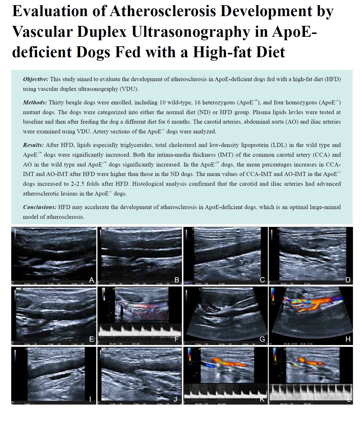
Ultrasound is one of the most commonly used imaging modalities for the screening and diagnosis of liver nodules. However, its diagnostic accuracy is highly dependent on operator expertise, and atypical or small lesions are prone to missed diagnosis or misdiagnosis. In recent years, artificial intelligence (AI) has achieved remarkable progress in medical image analysis, offering novel solutions to improve the objectivity, accuracy, and efficiency of liver ultrasound diagnosis. This review systematically summarizes the current status and advances of AI in the ultrasound diagnosis of liver nodules, with a focus on B-mode and contrast-enhanced ultrasound (CEUS). We detail AI applications in automatic nodule detection and localization, benign–malignant differentiation, multi-class classification (e.g., hepatocellular carcinoma [HCC], cholangiocarcinoma [CCA], hemangioma [HH], metastasis [HM]), and prediction of key pathological biomarkers (e.g., microvascular invasion [MVI], pathological grading, Ki-67, vessels encapsulating tumor clusters [VETC]), analyzes the current research status and summarizes the main limitations of existing studies. By reviewing methodological characteristics such as cohort size, validation strategies, and machine learning algorithms, this paper provides insights into future research directions and promotes the development of clinically translatable AI models, with the ultimate goal of advancing standardization and broad clinical adoption of AI-assisted diagnosis in liver ultrasound.
- Applications of Ultrasound Localization Microscopy in Abdominal Imaging
- Hou Wenfei, Chen Wanting, Liu Huazhen, Tang Jiajia, Yang Meng
- 2025, 9 (4): 347-356. DOI:10.26599/AUDT.2025.250097
- Abstract ( 7 ) HTML ( 3 ) PDF ( 2931KB ) ( 3 )
-
Ultrasound localization microscopy (ULM) is an ultrasound technique capable of overcoming the acoustic diffraction limit to achieve super resolution imaging of microvasculature, simultaneously balancing imaging depth and resolution. Abdominal organs are rich in microvasculature, and pathological processes in these organs are often accompanied by microvascular alterations, such as in tumors, chronic liver and kidney diseases, and allograft. Therefore, for abdominal organs, ULM represents a promising tool for aiding disease diagnosis and monitoring. Currently, an increasing number of studies are exploring the preclinical and clinical applications of ULM in both healthy and diseased abdominal organs. This paper aims to provide a systematic review of ULM applications in abdominal organs, while briefly discussing its limitations and future prospects.
- Contrast-enhanced Ultrasound LI-RADS for Nonradiation Treatment Response Assessment in Liver Tumor: A Pictorial Review Based on LR-TR v2024
- Xu Junmei, Tahmasebi Aylin, Mohammed Amr, Pour Bahareh Kian, Liu Ji-Bin, Eisenbrey John R.
- 2025, 9 (4): 357-374. DOI:10.26599/AUDT.2025.250102
- Abstract ( 5 ) HTML ( 0 ) PDF ( 23720KB ) ( 2 )
-
This pictorial review summarizes the Contrast-enhanced ultrasound (CEUS) Liver Imaging Reporting and Data System (LI-RADS) Treatment Response Algorithm (LR-TR, v2024) for response assessment after nonradiation locoregional therapies (NLT). The NLT covered by LR-TR v2024 includes embolization procedures such as conventional transarterial chemoembolization (cTACE), drug-eluting bead TACE (DEB-TACE), and bland transarterial embolization (TAE), as well as ablation techniques such as radiofrequency ablation (RFA), microwave ablation (MWA), and percutaneous ethanol injection (PEI). The algorithm independently evaluates intralesional and perilesional viability, using arterial-phase enhancement as the dominant criteria for intralesional evaluation and multiphasic enhancement (arterial, portal, and late phases) for perilesional evaluation. The results are then integrated into three standardized response categories, including LR-TR Viable, Equivocal, or Nonviable. Evidence from multicenter studies in hepatocellular carcinoma (HCC) indicates that CEUS LR-TR v2024 provides high reliability and strong reproducibility in detecting residual viable tumor following NLT. This review provides representative imaging features and interpretation tips to familiarize physicians with CEUS LR-TR v2024, aiming to improve accuracy in treatment response assessment (TRA) in HCC and facilitate timely therapeutic adjustments that ultimately benefits patients.
- The Evolving Application of Ultrasound in the Precision Management of Small Hepatocellular Carcinoma
- Guan Xin, Hu Xinyuan, Han Hong, Zhang Dezhi, Xu Huixiong
- 2025, 9 (4): 375-387. DOI:10.26599/AUDT.2025.250094
- Abstract ( 7 ) HTML ( 1 ) PDF ( 1950KB ) ( 1 )
-
Early detection of hepatocellular carcinoma (HCC) is crucial, as patient outcomes depend largely on clinical stage and treatment options at diagnosis. Currently most clinical guidelines emphasize non-invasive cross-sectional imaging for HCC diagnosis and evaluation, yet the full potential of ultrasound is often underexplored. With the development of emerging ultrasound techniques and diagnostic algorithms in recent years, the diagnostic ability of ultrasound for HCC smaller than 3 cm has been substantially improved and is increasingly emphasized in the management of small HCC. Following sequential clinical scenarios, this review discusses the state-of-the-art of ultrasound-related techniques and algorithms for preoperative screening and diagnosis, intraoperative guidance and monitoring as well as posttreatment efficacy evaluation of small HCC. By highlighting how advanced ultrasound complements and enhances standard practices, this review aims to promote ultrasound as an indispensable, versatile, and dynamic tool in the precision management of small HCC.
- Multimodal Ultrasound Radiomics in Liver Disease: Current Status and Future Directions
- Zhong Xian, Xie Xiaoyan
- 2025, 9 (4): 388-408. DOI:10.26599/AUDT.2025.250101
- Abstract ( 5 ) HTML ( 0 ) PDF ( 1118KB ) ( 1 )
-
Multimodal ultrasound, including B-mode imaging, contrast-enhanced ultrasound (CEUS), and ultrasound-based elastography, has demonstrated significant value in evaluating both diffuse liver diseases such as fibrosis and steatosis, and focal liver lesions such as hepatocellular carcinoma (HCC). Radiomics, including both handcrafted radiomics and deep learning approaches, has emerged as a promising strategy to enhance ultrasound-based liver disease assessment. Recent studies have applied radiomics across multimodal ultrasound, achieving notable success in grading fatty liver disease, staging fibrosis, and improving diagnosis, risk stratification, and prognostic prediction in HCC. Multimodal ultrasound provides complementary information on liver morphology, perfusion, and stiffness, while fusion strategies further enhance diagnostic accuracy and robustness. Future efforts should focus on standardized, large-scale multicenter validation, methodological improvements in multimodal integration, and the incorporation of explainable artificial intelligence to support clinical translation. Ultimately, despite ongoing challenges related to data heterogeneity, reproducibility, interpretability, and clinical validation, multimodal ultrasound radiomics holds strong promise for noninvasive, individualized, and clinically meaningful liver disease management.
- Explainable Artificial Intelligence in Echocardiography
- Hu Xuelin, Zhu Ye, Zhang Zisang, Quan Yuanting, Chen Wenwen, Chen Leichong, Xu Guangyu, Qin Luning, Xie Mingxing, Zhang Li
- 2025, 9 (4): 409-425. DOI:10.26599/AUDT.2025.250089
- Abstract ( 11 ) HTML ( 1 ) PDF ( 1465KB ) ( 4 )
-
Recent advancements in artificial intelligence (AI) have generated novel opportunities and challenges in ultrasound imaging. Deep learning algorithms exhibit significant potential in analyzing echocardiographic images, encompassing tasks such as view classification, quantification of cardiac function, and the diagnosis and risk assessment of cardiac diseases. The “black box” nature of AI models limits their clinical applications. Adopting explainable artificial intelligence (XAI) methods is crucial for improving the transparency and understanding of model predictions. This paper reviews the progress of AI applications in echocardiography, with a particular emphasis on XAI as a technical solution to enhance the transparency of model decision-making and its benefits compared to traditional AI models. This review outlines recent advancements in XAI applications for echocardiography and their clinical implications.
- Pulse Field Ablation in Oncology: Current Progress and Future Directions
- Xie Liting, Zhang Chengyue, Lou Wenjing, Xu Fan, Ma Wenyuan, Jiang Tian’an
- 2025, 9 (4): 426-436. DOI:10.26599/AUDT.2025.250099
- Abstract ( 8 ) HTML ( 0 ) PDF ( 1332KB ) ( 2 )
-
Pulsed field ablation (PFA), a non-thermal ablation technique that induces cell death via irreversible electroporation, has emerged as a promising therapeutic strategy for treating tumors located in anatomically complex regions. This review comprehensively examines recent developments in PFA, including its mechanisms foundations, established clinical applications in pancreatic, prostate and liver cancer, and expanding utility with other tumors, etc. Furthermore, we discuss synergistic approaches combining PFA with chemotherapy, immunotherapy, surgery and radiotherapy to augment therapeutic outcomes. In addition, technological innovations—such as robotic assistance, magnetic anchoring, and artificial intelligence—that improve precision and reproducibility are also explored. Despite encouraging clinical results, broader implementation of PFA necessitates higher-quality evidence from large-scale randomized trials and standardized treatment protocols. This review highlights the transformative potential of PFA in oncology while addressing contemporary challenges and future research directions to facilitate clinical translation.
- Volumetric Imaging with 2D Array Ultrasound Transducers for Clinical Applications: A Review
- Hou Shilin, Bao Guocui, Sun Zhe, Li Guo, Zhang Bo, Dai Jiyan
- 2025, 9 (4): 437-448. DOI:10.26599/AUDT.2025.250093
- Abstract ( 14 ) HTML ( 1 ) PDF ( 2710KB ) ( 9 )
-
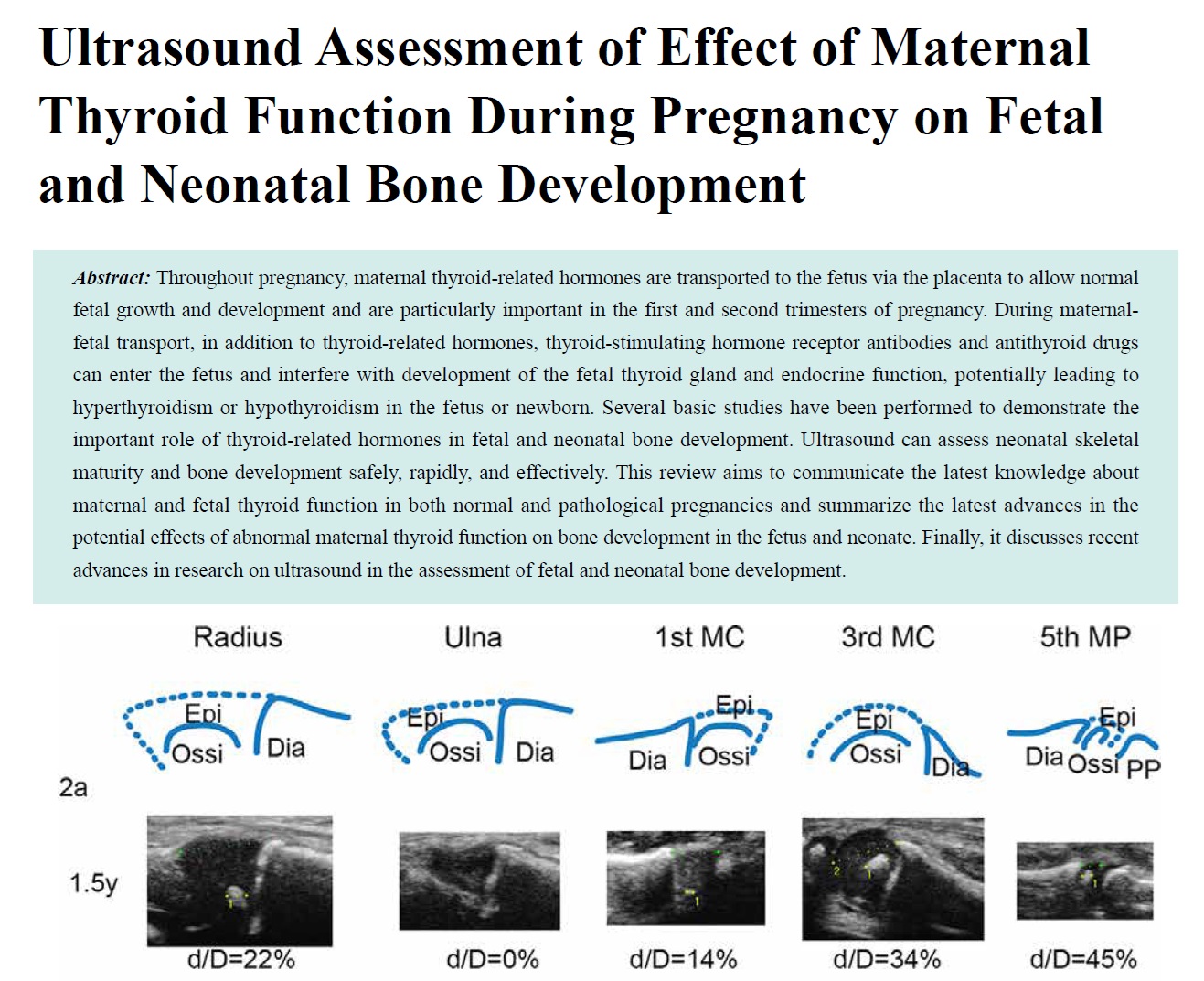
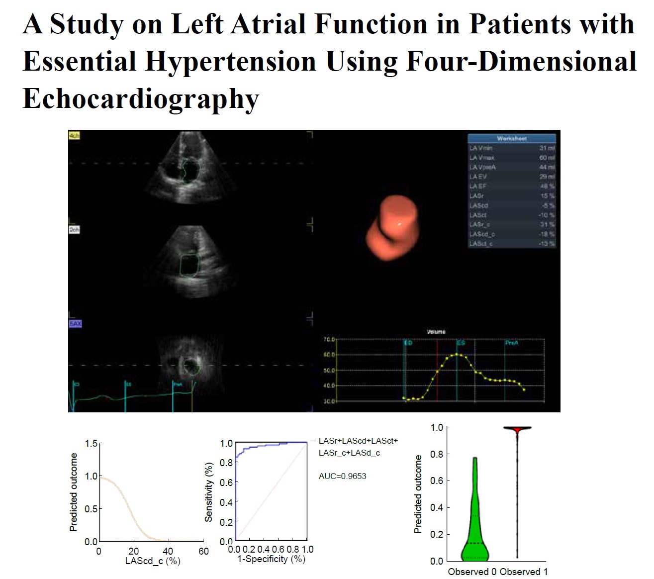
Recent advancements in ultrasound technology have revolutionized both medical imaging and therapeutic applications. Among these, volumetric imaging using two-dimensional (2D) array ultrasound transducers has emerged as a powerful tool, enabling real-time three-dimensional (3D) visualization, which is also referred to as four-dimensional (4D) imaging. 4D ultrasound imaging represents the most advanced diagnostic technique in ultrasound and is considered one of the most essential tools for medical diagnostics, particularly for assessing blood flow in micro-sized blood vessels. Due to its real-time and volumetric imaging capabilities, 4D imaging offers a unique advantage for the early diagnosis of cardiovascular and cerebrovascular diseases. The row-column-addressed (RCA) array is a novel 2D ultrasound transducer designed for ultrafast 3D ultrasonic imaging. Compared to traditional fully-sampled 2D matrix arrays, RCA transducers reduce the number of electronic channels from M×N to M+N, thereby significantly lowering hardware costs and manufacturing complexity. This review explores the design, fabrication, and clinical applications of 2D arrays, including both fully-sampled 2D arrays and RCA arrays. We discuss their roles in cardiology, brain imaging, and interventional procedures, while also addressing current challenges and future developments in the field.
- Current Applications of Artificial Intelligence in Obstetric Ultrasound
- Li Yanran, Cui Yuanjie, Wu Qingqing, Zhang Na
- 2025, 9 (4): 449-456. DOI:10.26599/AUDT.2025.250095
- Abstract ( 7 ) HTML ( 0 ) PDF ( 568KB ) ( 2 )
-
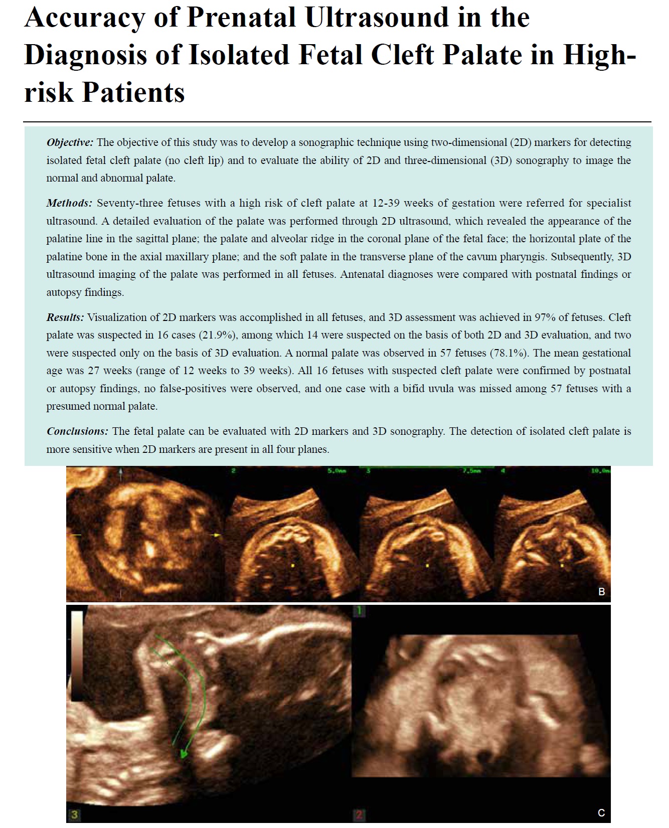
Artificial Intelligence (AI) technology has made remarkable progress in fetal ultrasound examinations, particularly excelling in fetal growth monitoring, organ function assessment, and early disease diagnosis. By automating the analysis of fetal ultrasound images, AI can accurately measure fetal biometric parameters and assist in diagnosing issues such as fetal growth restriction and organ developmental abnormalities. It demonstrates significant application potential in evaluating multiple organ systems including the fetal lungs, nervous system, cardiovascular system, and placenta, substantially enhancing the efficiency and accuracy of prenatal screening. This paper aims to review the current status of AI applications in obstetric ultrasound, while also exploring its limitations and future prospects.
- Advances and Applications in Dermatological Ultrasound
- Xiang Xi, Yang Yujia, Wang Liyun, Qiu Li
- 2025, 9 (4): 457-466. DOI:10.26599/AUDT.2025.250105
- Abstract ( 7 ) HTML ( 1 ) PDF ( 4581KB ) ( 2 )
-
In recent years, dermatological ultrasound has made significant breakthroughs in the research and application of dermatology by virtue of its advantages of non-invasive, real-time imaging and high resolution. The increase of ultrasonic probe frequency makes it possible to visualize the skin structure. Different frequencies of high frequency or ultra-high-frequency ultrasound can realize different degrees of presentation of the fine structure of epidermis, dermis and subcutaneous layer. The wide range of application of shear wave elastography provides a new ultrasonic evaluation index for skin diseases represented by scleroderma. This review provides a detailed summary of the latest application progress and research frontiers of ultrasound in different skin diseases. It includes the diagnosis and efficacy evaluation of scleroderma, the diagnosis and activity evaluation of psoriasis, the diagnosis and follow-up of nevus punctatum, the differentiation of benign and malignant skin tumors, the evaluation of pathological scar, the application in the field of aesthetic medicine, and the evaluation of other common skin diseases. In the future, dermatological ultrasound is expected to play a more important role in accurate classification of diseases and prognosis prediction with the further innovation of equipment and the deep exploration of skin diseases.
- Research Progress and Clinical Translation of Photoacoustic–ultrasound Fusion Imaging in Breast Cancer Diagnosis and Therapy
- Zhang Xiaoqian, Zhang Jingwen, Dong Yijie, Zhou Jianqiao
- 2025, 9 (4): 467-482. DOI:10.26599/AUDT.2025.250103
- Abstract ( 8 ) HTML ( 0 ) PDF ( 1475KB ) ( 3 )
-
Photoacoustic-ultrasound (PA/US) fusion imaging is an emerging dual-modality technique that integrates the high optical contrast of photoacoustic imaging (PAI, also referred to as optoacoustic imaging or photoacoustic tomography) with the anatomical resolution of US. This review summarizes the principles, technical advances, and clinical applications of PA/US in breast cancer diagnosis and therapy. Recent studies demonstrate that PA/US markedly improves diagnostic specificity while maintaining high sensitivity, particularly in differentiating benign from malignant lesions, predicting molecular subtypes, and monitoring therapeutic response. Radiomics and artificial intelligence further enhance the interpretive power of PA/US, enabling functional phenotyping, Ki-67 expression prediction, and axillary lymph node (ALN) metastasis risk assessment. Moreover, multicenter clinical trials, such as the PIONEER study, have validated the clinical feasibility of PA/US, reducing unnecessary biopsies and refining BI-RADS categorization. Despite challenges in system standardization, quantitative accuracy, and large-scale validation, PA/US holds promise as the “fourth major breast imaging modality,” complementing mammography, US, and MRI. With continued progress in AI integration, standardized protocols, and policy recognition, PA/US is expected to achieve routine clinical implementation in the next 5-10 years, supporting individualized breast cancer diagnosis and precision oncology.
- Artificial Intelligence in Ultrasound Imaging: A Review of Progress from Machine Learning to Large Language Model
- Jin Tong, Yu Xiaohu, Ai Zheng, Guo Hongcheng
- 2025, 9 (4): 483-496. DOI:10.26599/AUDT.2025.250104
- Abstract ( 9 ) HTML ( 1 ) PDF ( 607KB ) ( 1 )
-
Biomedical ultrasound imaging, as one of the most common, safe, and cost-effective modalities in clinical diagnosis, witnesses remarkable progress with the integration of artificial intelligence (AI). Early studies based on traditional machine learning (ML) rely on handcrafted features and classical classifiers to achieve automatic recognition and quantitative analysis of ultrasound images. However, such methods are limited in feature representation capacity and generalizability. With the advent of deep learning (DL), convolutional neural networks (CNNs), recurrent neural networks (RNNs), and attention-based architectures are widely applied to tasks such as segmentation, detection, and lesion classification, significantly improving diagnostic accuracy and robustness. More recently, large language models (LLMs) and multimodal foundation models open new avenues for intelligent ultrasound analysis. These models not only integrate imaging and textual information to support automated report generation and cross-modal reasoning but also offer enhanced interpretability and greater potential for clinical adoption. In this review, we provide a systematic review of the evolution of AI in ultrasound image analysis, spanning from traditional ML to deep learning and LLMs, outlining a complete trajectory of methodological advances.

