Advanced Ultrasound in Diagnosis and Therapy ›› 2024, Vol. 8 ›› Issue (4): 172-182.doi: 10.37015/AUDT.2024.240064
• Review Articles • Previous Articles Next Articles
Yang Yuna,b,c,1, Zhang Xina,b,c,1, Zhang Ruizea,b,c, Jiang Jingronga,b,c, Xie Yujia,b,c, Fang Lingyuna,b,c, Zhang Jinga,b,c, Xie Mingxinga,b,c,*( ), Wang Jinga,b,c,*()
Received:2024-10-13
Accepted:2024-12-01
Online:2024-12-30
Published:2024-11-12
Contact:
Xie Mingxing, Wang Jing,
E-mail:xiemx@hust.edu.cn;jingwang2004@hust.edu.cn
About author:First author contact:1 Yun Yang and Xin Zhang contributed equally to this work.
Yang Yun, Zhang Xin, Zhang Ruize, Jiang Jingrong, Xie Yuji, Fang Lingyun, Zhang Jing, Xie Mingxing, Wang Jing. Current Status and Progress in Arterial Stiffness Evaluation: A Comprehensive Review. Advanced Ultrasound in Diagnosis and Therapy, 2024, 8(4): 172-182.
Table 1
Overview of ultrasound techniques in arterial stiffness assessment"
| Parameters | Measurement method | Interpretation | Advantages | Disadvantages | Considerations |
|---|---|---|---|---|---|
| PWV | Doppler Ultrasound | Higher PWV values (m/s) indicate greater AS and higher cardiovascular risk | Convenient; allow for simultaneous evaluation of cardiac function and AS | Influenced by hemodynamic factors; requires accurate distance measurement; operator-dependent | Standardized operation needed, avoid influences from blood pressure and heart rate fluctuations |
| IMT | Two-dimensional ultrasound | Thickened IMT indicates atherosclerosis and increased cardiovascular risk | Real-time direct evaluation of arterial structural changes | Depend on image quality and operator experience | Commonly used for carotid artery evaluation |
| Aortic distensibility | M-mode or two-dimensional ultrasound | Change in aortic diameter during the cardiac cycle Lower distensibility is associated with higher AS and cardiovascular risk | Simple | Easily affected by changes in blood pressure | Standardized operation and measurement needed |
| Flow-mediated dilation | Brachial artery ultrasound | The ability of the artery to dilate in response to increased blood flow, reflects endothelial function | Non-invasive assessment of endothelial function | Requires precise operation and timing control | Strict adherence to protocols; avoid external interferences |
| β-stiffness index | M-mode or two-dimensional ultrasound | A higher index indicates higher AS | Assesses local AS; independent of blood pressure | Limited use in large central arteries | Can be influenced by arterial wall thickness and vascular smooth muscle tone |
| Aortic wall Motion velocity | Tissue Doppler Imaging | The lower the velocity, the greater the AS; local evaluations | Quantitative | Requires specialized training | Operator must have specialized knowledge |
| Strain and strain rate | Speckle Tracking Echocardiography | The deformation of arterial walls during the cardiac cycle, lower value indicates stiffer arteries | Sensitive to early changes in AS | Require post-processing workstation and specialized knowledge; high demands on image quality | Image quality and frame rate should be considered to ensure accurate results |
| Ultrafast ultrasound imaging | Ultra-high frame rate ultrasound technology | Real-time assessment of local AS; assess cardiac function, blood flow, and myocardial strain with | Real-time; higher precision and temporal resolution | The high cost and complexity limit its widespread use in routine clinical practice; require specialized training | Regular calibration needed to ensure measurement accuracy |
| Shear wave elastography | Generate shear waves in tissue and measure their propagation speed | Measure local tissue stiffness; higher value indicates greater stiffness | Applicable to local arteries or small areas; independent of blood pressure | Limited to specialized centers with advanced equipment, and operator training is needed | |
| Ultrasound time harmonic elastography | Harmonic vibrations generate time harmonic frequencies | Evaluation of overall AS. Higher value indicates greater AS | Suitable for overall stiffness of large arteries, especially for chronic disease and research | Requires specialized equipment and expertise, and the imaging time is long |
Table 2
Different pulse wave velocity methods for evaluating arterial stiffness"
| Parameters | Measurement method | Interpretation | Advantages | Disadvantages | Considerations |
|---|---|---|---|---|---|
| PWV | Ultrasound | Used in clinical research to reflect AS in different locations | Evaluate regional AS; integration with functional imaging | Depends on measurement sites; operator dependence | Mainly used for research purposes, with fewer clinical applications nowadays |
| cfPWV | Pressure sensors | Reflects the stiffness of large arteries | Gold standard | Dependent on equipment and environmental factors | Repeated measurement; Regular calibration; Standardized measurement paths and calculation methods; quiet environment; Consider Patient's physiological state and medications |
| Brachial-ankle PWV (baPWV) | Pressure sensors | Reflects the arterial stiffness of the limbs | Suitable for large-scale clinical screening | Does not fully represent the stiffness of central arteries | |
| SphygmoCor | Pressure sensors | Widely used non-invasive measurement of AS | Considered the gold standard for measuring cfPWV | High cost; operator training is required | |
| Complior | Piezoelectric sensor measurement | Widely used non-invasive measurement of AS | Provides reliable and repeatable measurements of PWV | High cost; operator training is required |

Figure 1
PWV can be calculated based on path length and transit time through the foot to foot method. PWV, pulse wave velocity; Dcf; distance from common carotid artery to common femoral artery."
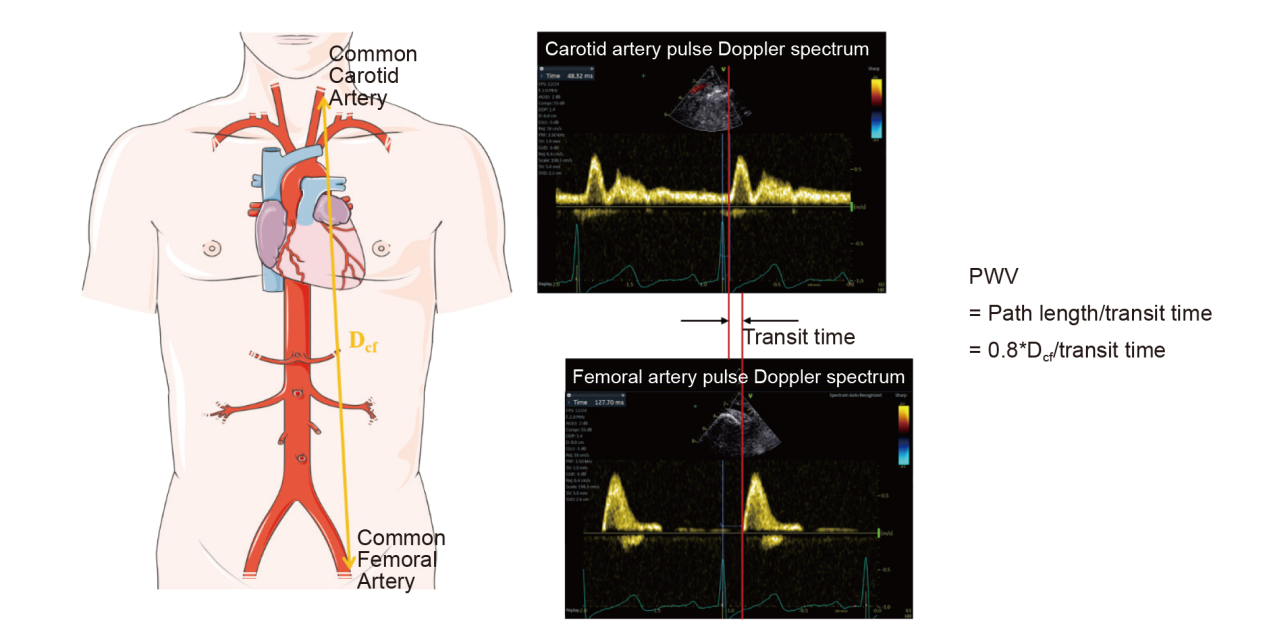
Table 3
Parameters for arterial stiffness evaluation using pressure and waveform analysis-based techniques"
| Parameters | Measurement method | Interpretation | Advantages | Disadvantages | Considerations |
|---|---|---|---|---|---|
| PWV | Pressure sensors | The higher the PWV, the greater the AS | The "gold standard" | Requires specialized equipment, relies on operator experience. | 1. Environmental factors: Measurements should be taken in a quiet environment with suitable temperature. |
| CAVI | Simultaneously measures electrocardiogram, phonocardiogram, and limb blood pressure. | A higher CAVI indicates greater AS | Stable with minimal influence from blood pressure | Certain requirements for the equipment and environment. | 2. Equipment calibration: Equipment should be regularly calibrated and maintained. |
| ABI | Ankle Systolic Pressure / Brachial Systolic Pressure | ABI < 0.9 indicates peripheral artery disease, > 1.4 indicates vascular calcification | Simple, quick, effective for screening peripheral artery disease | Affected by vascular calcification, which may lead to false normal or elevated results | 3. Patient's physiological state: Strenuous exercise, emotional agitation, and intake of coffee or tea before the examination should be avoided. |
| AIx | Pulse waveform analysis | A higher index indicates greater AS and increased central arterial pressure | Can assess central arterial pressure | Affected by factors such as heart rate, age, and height | 5. Consider the effects of medications. |
| Characteristic impedance | Analysis of Aortic Pressure and Flow Waveforms | The higher value indicates the greater the AS | Reflects the instantaneous mechanical properties of the arteries | Complex; requires specialized equipment and trained personnel | 6. Repeat measurements to obtain an average value. |
| Effective arterial elastance | End-systolic pressure/ stroke volume (mmHg/mL) | Higher values indicate increased AS and greater demands on cardiac function | Evaluates ventricular-arterial coupling | Requires invasive measurements or estimations, and the calculations are complex | 7. Need to combine with clinical context. |
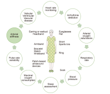
Figure 2
Wearable devices and their uses in cardiovascular symptom."

| [1] |
Mitchell GF, Parise H, Benjamin EJ, Larson MG, Keyes MJ, Vita JA, et al. Changes in arterial stiffness and wave reflection with advancing age in healthy men and women: the Framingham Heart Study. Hypertension 2004; 43:1239-1245.
doi: 10.1161/01.HYP.0000128420.01881.aa pmid: 15123572 |
| [2] |
Safar ME, Asmar R, Benetos A, Blacher J, Boutouyrie P, Lacolley P, et al. Interaction between hypertension and arterial stiffness. Hypertension 2018; 72:796-805.
doi: 10.1161/HYPERTENSIONAHA.118.11212 pmid: 30354723 |
| [3] |
Wu S, Jin C, Li S, Zheng X, Zhang X, Cui L, et al. Aging, arterial stiffness, and blood pressure association in Chinese adults. Hypertension 2019; 73:893-899.
doi: 10.1161/HYPERTENSIONAHA.118.12396 pmid: 30776974 |
| [4] |
Styczynski G, Cienszkowska K, Ludwiczak M, Szmigielski C. Age-related values of aortic pulse wave velocity in healthy subjects measured by Doppler echocardiography. J Hum Hypertens 2021; 35:1081-1087.
doi: 10.1038/s41371-020-00466-4 pmid: 33414505 |
| [5] |
Laurent S, Cockcroft J, Van Bortel L, Boutouyrie P, Giannattasio C, Hayoz D, et al. Expert consensus document on arterial stiffness: methodological issues and clinical applications. Eur Heart J 2006; 27:2588-2605.
doi: 10.1093/eurheartj/ehl254 pmid: 17000623 |
| [6] |
Vlachopoulos C, Aznaouridis K, Stefanadis C. Prediction of cardiovascular events and all-cause mortality with arterial stiffness: a systematic review and meta-analysis. J Am Coll Cardiol 2010; 55:1318-1327.
doi: 10.1016/j.jacc.2009.10.061 pmid: 20338492 |
| [7] |
Ben-Shlomo Y, Spears M, Boustred C, May M, Anderson SG, Benjamin EJ, et al. Aortic pulse wave velocity improves cardiovascular event prediction: an individual participant meta-analysis of prospective observational data from 17,635 subjects. J Am Coll Cardiol 2014; 63:636-646.
doi: S0735-1097(13)05974-3 pmid: 24239664 |
| [8] |
Zanoli L, Briet M, Empana JP, Cunha PG, Mäki-Petäjä KM, Protogerou AD, et al. Vascular consequences of inflammation: a position statement from the ESH Working Group on vascular structure and function and the ARTERY Society. J Hypertens 2020; 38:1682-1698.
doi: 10.1097/HJH.0000000000002508 pmid: 32649623 |
| [9] |
Townsend RR, Wilkinson IB, Schiffrin EL, Avolio AP, Chirinos JA, Cockcroft JR, et al. Recommendations for improving and standardizing vascular research on arterial stiffness: a scientific statement from the American Heart Association. Hypertension 2015; 66:698-722.
doi: 10.1161/HYP.0000000000000033 pmid: 26160955 |
| [10] |
Li X, Cokkinos D, Gadani S, Rafailidis V, Aschwanden M, Levitin A, et al. Advanced ultrasound techniques in arterial diseases. Int J Cardiovasc Imaging 2022; 38:1711-1721.
doi: 10.1007/s10554-022-02558-3 pmid: 35195805 |
| [11] |
Tomiyama H, Yamashina A. Non-invasive vascular function tests: their pathophysiological background and clinical application. Circ J 2010; 74:24-33.
pmid: 19920359 |
| [12] | Cecelja M, Chowienczyk P. Arterial stiffening: causes and consequences. Artery Research 2013; 7:22-27. |
| [13] |
Dumor K, Shoemaker-Moyle M, Nistala R, Whaley-Connell A. Arterial stiffness in hypertension: an update. Curr Hypertens Rep 2018; 20:72.
doi: 10.1007/s11906-018-0867-x pmid: 29974262 |
| [14] | Faqir Muhammad I. Novel biomarkers of cardiometabolic risk in population studies-with a focus on arterial stiffness and diabetes. Lund: Lund University, Faculty of Medicine, 2020. 99p (Lund University, Faculty of Medicine Doctoral Dissertation Series; 2020:99). |
| [15] | Lehmann ED, Gosling RG, Fatemi-Langroudi B, Taylor MG. Non-invasive Doppler ultrasound technique for the in vivo assessment of aortic compliance. J Biomed Eng 1992; 14:250-256. |
| [16] | Kavgacı A, İncedere F, Tunaoğlu S, Karabörk M, Büyükkaragöz B, Leventoğlu E, et al. Comparison of echocardiographic aortic stiffness index measurements and pulse wave velocity measurements in obese and overweight children. Cardiol Young 2024; 34:11-17. |
| [17] | Abhisheka B, Biswas SK, Purkayastha B, Das D, Escargueil A. Recent trend in medical imaging modalities and their applications in disease diagnosis: a review. Multimedia Tools and Applications 2024; 83:43035-43070. |
| [18] | Mushenkova NV, Summerhill VI, Zhang D, Romanenko EB, Grechko AV, Orekhov AN. Current advances in the diagnostic imaging of atherosclerosis: insights into the pathophysiology of vulnerable plaque. Int J Mol Sci 2020;21. |
| [19] |
Wang Z, Wang D, Han M, Ai Y, Zhang X, Yuan L, et al. A novel methodology for semi-automatic measurement of arterial stiffness by doppler ultrasound: clinical feasibility and reproducibility. Ultrasound Med Biol 2021; 47:1725-1736.
doi: 10.1016/j.ultrasmedbio.2021.03.004 pmid: 33858722 |
| [20] | Polak JF, Szklo M, O'Leary DH. Associations of coronary heart disease with common carotid artery near and far wall intima-media thickness: the multi-ethnic study of atherosclerosis. J Am Soc Echocardiogr 2015; 28:1114-1121. |
| [21] |
Coll B, Feinstein SB. Carotid intima-media thickness measurements: techniques and clinical relevance. Curr Atheroscler Rep 2008; 10:444-450.
doi: 10.1007/s11883-008-0068-1 pmid: 18706287 |
| [22] | Fung K, Biasiolli L, Aung N, Hann E, Paiva J, Lukaschuk E, et al. Reference values for aortic distensibility derived from UK Biobank cardiovascular magnetic resonance (CMR) imaging cohort. European Heart Journal - Cardiovascular Imaging 2019;20. |
| [23] | Heiss C, Rodriguez-Mateos A, Bapir M, Skene SS, Sies H, Kelm M. Flow-mediated dilation reference values for evaluation of endothelial function and cardiovascular health. Cardiovasc Res 2023; 119:283-293. |
| [24] | Thijssen DH, Black MA, Pyke KE, Padilla J, Atkinson G, Harris RA, et al. Assessment of flow-mediated dilation in humans: a methodological and physiological guideline. Am J Physiol Heart Circ Physiol 2011; 300:H2-12. |
| [25] |
Ren M, Li X, Xue M. Aortic elasticity evaluated by pulsed tissue doppler imaging of the ascending aorta in different diseases: a systematic review. Angiology 2021; 72:403-410.
doi: 10.1177/0003319721992584 pmid: 33541096 |
| [26] | Chi C, Liu Y, Xu Y, Xu D. Association between arterial stiffness and heart failure with preserved ejection fraction. Front Cardiovasc Med 2021; 8:707162. |
| [27] |
Podgórski M, Grzelak P, Kaczmarska M, Polguj M, Łukaszewski M, Stefańczyk L. Feasibility of two-dimensional speckle tracking in evaluation of arterial stiffness: comparison with pulse wave velocity and conventional sonographic markers of atherosclerosis. Vascular 2018; 26:63-69.
doi: 10.1177/1708538117720047 pmid: 28728481 |
| [28] | Pan FS, Yu L, Luo J, Wu RD, Xu M, Liang JY, et al. Carotid artery stiffness assessment by ultrafast ultrasound imaging: feasibility and potential influencing factors. J Ultrasound Med 2018; 37:2759-2767. |
| [29] |
Marais L, Pernot M, Khettab H, Tanter M, Messas E, Zidi M, et al. Arterial stiffness assessment by shear wave elastography and ultrafast pulse wave imaging: comparison with reference techniques in normotensives and hypertensives. Ultrasound Med Biol 2019; 45:758-772.
doi: S0301-5629(18)30487-3 pmid: 30642659 |
| [30] | Schaafs LA, Tzschätzsch H, Reshetnik A, van der Giet M, Braun J, Hamm B, et al. Ultrasound time-harmonic elastography of the aorta: effect of age and hypertension on aortic stiffness. Invest Radiol 2019; 54:675-680. |
| [31] |
Sigrist RMS, Liau J, Kaffas AE, Chammas MC, Willmann JK. Ultrasound elastography: review of techniques and clinical applications. Theranostics 2017; 7:1303-1329.
doi: 10.7150/thno.18650 pmid: 28435467 |
| [32] |
Reference Values for Arterial Stiffness' Collaboration. Determinants of pulse wave velocity in healthy people and in the presence of cardiovascular risk factors: 'establishing normal and reference values'. Eur Heart J 2010; 31:2338-2350.
doi: 10.1093/eurheartj/ehq165 pmid: 20530030 |
| [33] |
Williams B, Mancia G, Spiering W, Agabiti Rosei E, Azizi M, Burnier M, et al. 2018 ESC/ESH Guidelines for the management of arterial hypertension. Eur Heart J 2018; 39:3021-3104.
doi: 10.1093/eurheartj/ehy339 pmid: 30165516 |
| [34] | Whelton PK, Carey RM, Aronow WS, Casey DE, Jr., Collins KJ, Dennison Himmelfarb C, et al. 2017 ACC/AHA/AAPA/ABC/ACPM/AGS/APhA/ASH/ASPC/NMA/PCNA Guideline for the prevention, detection, evaluation, and management of high blood pressure in adults: executive summary: a report of the american college of cardiology/American heart association task force on clinical practice guidelines. Hypertension 2018; 71:1269-1324. |
| [35] |
Cardoso CRL, Leite NC, Salles GF. Prognostic impact of changes in aortic stiffness for cardiovascular and mortality outcomes in individuals with type 2 diabetes: the Rio de Janeiro cohort study. Cardiovasc Diabetol 2022; 21:76.
doi: 10.1186/s12933-022-01514-8 pmid: 35568947 |
| [36] | Chang G, Hu Y, Ge Q, Chu S, Avolio A, Zuo J. Arterial stiffness as a predictor of the index of atherosclerotic cardiovascular disease in hypertensive patients. Int J Environ Res Public Health 2023;20. |
| [37] |
Townsend RR, Anderson AH, Chirinos JA, Feldman HI, Grunwald JE, Nessel L, et al. Association of pulse wave velocity with chronic kidney disease progression and mortality: findings from the cric study (chronic renal insufficiency cohort). Hypertension 2018; 71:1101-1107.
doi: 10.1161/HYPERTENSIONAHA.117.10648 pmid: 29712736 |
| [38] |
Stone K, Fryer S, Faulkner J, Meyer ML, Heffernan K, Zieff G, et al. The aortic-femoral arterial stiffness gradient demonstrates good between-day reliability. Hypertens Res 2021; 44:1686-1688.
doi: 10.1038/s41440-021-00712-3 pmid: 34548651 |
| [39] | Zheng M, Zhang X, Chen S, Song Y, Zhao Q, Gao X, et al. Arterial stiffness preceding diabetes. Circulation Research 2020; 127:1491-1498. |
| [40] |
Kim ED, Tanaka H, Ballew SH, Sang Y, Heiss G, Coresh J, et al. Associations between kidney disease measures and regional pulse wave velocity in a large community-based cohort: the atherosclerosis risk in communities (aric) study. Am J Kidney Dis 2018; 72:682-690.
doi: S0272-6386(18)30704-2 pmid: 30007506 |
| [41] | Karimpour P, May JM, Kyriacou PA. Photoplethysmography for the assessment of arterial stiffness. Sensors (Basel) 2023;23. |
| [42] | Spronck B, Terentes-Printzios D, Avolio AP, Boutouyrie P, Guala A, Jerončić A, et al. Recommendations for validation of noninvasive arterial pulse wave velocity measurement devices. Hypertension 2024; 81:183-192. |
| [43] |
Milan A, Zocaro G, Leone D, Tosello F, Buraioli I, Schiavone D, et al. Current assessment of pulse wave velocity: comprehensive review of validation studies. J Hypertens 2019; 37:1547-1557.
doi: 10.1097/HJH.0000000000002081 pmid: 30882597 |
| [44] |
Franklin SS, Khan SA, Wong ND, Larson MG, Levy D. Is pulse pressure useful in predicting risk for coronary heart disease? the framingham heart study. Circulation 1999; 100:354-360.
doi: 10.1161/01.cir.100.4.354 pmid: 10421594 |
| [45] |
Saiki A, Ohira M, Yamaguchi T, Nagayama D, Shimizu N, Shirai K, et al. New horizons of arterial stiffness developed using Cardio-Ankle Vascular Index (CAVI). J Atheroscler Thromb 2020; 27:732-748.
doi: 10.5551/jat.RV17043 pmid: 32595186 |
| [46] |
Miyoshi T, Ito H. Arterial stiffness in health and disease: The role of cardio-ankle vascular index. J Cardiol 2021; 78:493-501.
doi: 10.1016/j.jjcc.2021.07.011 pmid: 34393004 |
| [47] | Miyoshi T, Ito H, Shirai K, Horinaka S, Higaki J, Yamamura S, et al. Predictive value of the cardio-ankle vascular index for cardiovascular events in patients at cardiovascular risk. J Am Heart Assoc 2021; 10:e020103. |
| [48] |
Bulbul E, Ozilhan MO, Sezer A, Yetisen M, Ilki FY. Possible clinical benefits of cardio-ankle vascular index measurement in urological diseases. Vasc Health Risk Manag 2023; 19:127-132.
doi: 10.2147/VHRM.S384937 pmid: 36923496 |
| [49] |
Tanaka A, Tomiyama H, Maruhashi T, Matsuzawa Y, Miyoshi T, Kabutoya T, et al. Physiological diagnostic criteria for vascular failure. Hypertension 2018; 72:1060-1071.
doi: 10.1161/HYPERTENSIONAHA.118.11554 pmid: 30354826 |
| [50] |
Giudici A, Khir AW, Reesink KD, Delhaas T, Spronck B. Five years of cardio-ankle vascular index (CAVI) and CAVI0: how close are we to a pressure-independent index of arterial stiffness? J Hypertens 2021; 39:2128-2138.
doi: 10.1097/HJH.0000000000002928 pmid: 34269333 |
| [51] |
Aboyans V, Criqui MH, Abraham P, Allison MA, Creager MA, Diehm C, et al. Measurement and interpretation of the ankle-brachial index: a scientific statement from the American Heart Association. Circulation 2012; 126:2890-909.
doi: 10.1161/CIR.0b013e318276fbcb pmid: 23159553 |
| [52] |
Sonoda H, Nakamura K, Tamakoshi A. Ankle-Brachial Index is a predictor of future incident chronic kidney disease in a general Japanese population. J Atheroscler Thromb 2019; 26:1054-1061.
doi: 10.5551/jat.47779 pmid: 31061261 |
| [53] | Wilkinson IB, Cockcroft JR, Webb DJ. Pulse wave analysis and arterial stiffness. J Cardiovasc Pharmacol 1998; 32:S33-S37. |
| [54] |
Chirinos JA, Kips JG, Roman MJ, Medina-Lezama J, Li Y, Woodiwiss AJ, et al. Ethnic differences in arterial wave reflections and normative equations for augmentation index. Hypertension 2011; 57:1108-1116.
doi: 10.1161/HYPERTENSIONAHA.110.166348 pmid: 21536986 |
| [55] |
DuBose LE, Boles Ponto LL, Moser DJ, Harlynn E, Reierson L, Pierce GL. Higher aortic stiffness is associated with lower global cerebrovascular reserve among older humans. Hypertension 2018; 72:476-482.
doi: 10.1161/HYPERTENSIONAHA.118.11143 pmid: 29915015 |
| [56] |
Jefferson AL, Cambronero FE, Liu D, Moore EE, Neal JE, Terry JG, et al. Higher aortic stiffness is related to lower cerebral blood flow and preserved cerebrovascular reactivity in older adults. Circulation 2018; 138:1951-1962.
doi: 10.1161/CIRCULATIONAHA.118.032410 pmid: 30018169 |
| [57] | Bown CW, Khan OA, Moore EE, Liu D, Pechman KR, Cambronero FE, et al. Elevated aortic pulse wave velocity relates to longitudinal gray and white matter changes. Arterioscler Thromb Vasc Biol 2021; 41:3015-3024. |
| [58] | Vlachopoulos C, Michael O'Rourke, Nichols WW. McDonald's blood flow in arteries Theoretical, Experimental and Clinical Principles. 6th ed. London: CRC Press 2011; 273-310. |
| [59] |
Kelly RP, Ting CT, Yang TM, Liu CP, Maughan WL, Chang MS, et al. Effective arterial elastance as index of arterial vascular load in humans. Circulation 1992; 86:513-521.
doi: 10.1161/01.cir.86.2.513 pmid: 1638719 |
| [60] | Segers P, Stergiopulos N, Westerhof N. Relation of effective arterial elastance to arterial system properties. Am J Physiol Heart Circ Physiol 2002; 282:H1041-H1046. |
| [61] |
Woodrum DA, Herrmann J, Lerman A, Romano AJ, Lerman LO, Ehman RL. Phase-contrast MRI-based elastography technique detects early hypertensive changes in ex vivo porcine aortic wall. J Magn Reson Imaging 2009; 29:583-587.
doi: 10.1002/jmri.21702 pmid: 19243040 |
| [62] | Zhuang B, Sirajuddin A, Zhao S, Lu M. The role of 4D flow MRI for clinical applications in cardiovascular disease: current status and future perspectives. Quant Imaging Med Surg 2021; 11:4193-4210. |
| [63] | Shahzad R, Shankar A, Amier R, Nijveldt R, Westenberg JJM, de Roos A, et al. Quantification of aortic pulse wave velocity from a population based cohort: a fully automatic method. J Cardiovasc Magn Reson 2019; 21:27. |
| [64] | Badji A, Sabra D, Bherer L, Cohen-Adad J, Girouard H, Gauthier CJ. Arterial stiffness and brain integrity: a review of MRI findings. Ageing Res Rev 2019; 53:100907. |
| [65] | Björnfot C, Garpebring A, Qvarlander S, Malm J, Eklund A, Wåhlin A. Assessing cerebral arterial pulse wave velocity using 4D flow MRI. J Cereb Blood Flow Metab 2021; 41: 2769-2777. |
| [66] | Ohyama Y, Redheuil A, Kachenoura N, Ambale Venkatesh B, Lima JAC. Imaging insights on the aorta in aging. Circ Cardiovasc Imaging 2018; 11:e005617. |
| [67] |
Wang DJ, Alger JR, Qiao JX, Hao Q, Hou S, Fiaz R, et al. The value of arterial spin-labeled perfusion imaging in acute ischemic stroke: comparison with dynamic susceptibility contrast-enhanced MRI. Stroke 2012; 43:1018-1024.
doi: 10.1161/STROKEAHA.111.631929 pmid: 22328551 |
| [68] |
Agatston AS, Janowitz WR, Hildner FJ, Zusmer NR, Viamonte M, Jr, Detrano R. Quantification of coronary artery calcium using ultrafast computed tomography. J Am Coll Cardiol 1990; 15:827-832.
doi: 10.1016/0735-1097(90)90282-t pmid: 2407762 |
| [69] | Boas DA, Dunn AK. Laser speckle contrast imaging in biomedical optics. J Biomed Opt 2010; 15:011109. |
| [70] | Tearney GJ, Regar E, Akasaka T, Adriaenssens T, Barlis P, Bezerra HG, et al. Consensus standards for acquisition, measurement, and reporting of intravascular optical coherence tomography studies: a report from the International Working Group for intravascular optical coherence tomography standardization and validation. J Am Coll Cardiol 2012; 59:1058-1072. |
| [71] | Perez MV, Mahaffey KW, Hedlin H, Rumsfeld JS, Garcia A, Ferris T, et al. Large-scale assessment of a smartwatch to identify atrial fibrillation. N Engl J Med 2019; 381:1909-1917. |
| [72] | Williams GJ, Al-Baraikan A, Rademakers FE, Ciravegna F, van de Vosse FN, Lawrie A, et al. Wearable technology and the cardiovascular system: the future of patient assessment. The Lancet Digital Health 2023; 5:e467-e476. |
| [73] | Yao H, Yang W, Cheng W, Tan YJ, See HH, Li S, et al. Near-hysteresis-free soft tactile electronic skins for wearables and reliable machine learning. Proc Natl Acad Sci U S A 2020; 117:25352-25359. |
| [74] |
Hammond-Haley M, Allen C, Han J, Patterson T, Marber M, Redwood S. Utility of wearable physical activity monitors in cardiovascular disease: a systematic review of 11 464 patients and recommendations for optimal use. Eur Heart J Digit Health 2021; 2:231-243.
doi: 10.1093/ehjdh/ztab035 pmid: 36712392 |
| [75] |
Garcia-Carretero R, Vigil-Medina L, Barquero-Perez O, Ramos-Lopez J. Pulse wave velocity and machine learning to predict cardiovascular outcomes in prediabetic and diabetic populations. J Med Syst 2019; 44:16.
doi: 10.1007/s10916-019-1479-y pmid: 31820120 |
| [76] | Huttunen JMJ, Kärkkäinen L, Lindholm H. Pulse transit time estimation of aortic pulse wave velocity and blood pressure using machine learning and simulated training data. PLoS Comput Biol 2019; 15:e1007259. |
| [77] |
Vallée A, Cinaud A, Blachier V, Lelong H, Safar ME, Blacher J. Coronary heart disease diagnosis by artificial neural networks including aortic pulse wave velocity index and clinical parameters. J Hypertens 2019; 37:1682-1688.
doi: 10.1097/HJH.0000000000002075 pmid: 30870247 |
| [78] | Jin W, Chowienczyk P, Alastruey J. Estimating pulse wave velocity from the radial pressure wave using machine learning algorithms. PLoS One 2021; 16:e0245026. |
| [79] |
Dara A, Arvanitaki A, Theodorakopoulou M, Athanasiou C, Pagkopoulou E, Boutou A. Non-invasive assessment of endothelial dysfunction in pulmonary arterial hypertension. Mediterr J Rheumatol 2021; 32:6-14.
doi: 10.31138/mjr.32.1.6 pmid: 34386697 |
| [1] | Chen Anni, Yang Lan, Li Zhenyi, Wang Xinqi, Chen Ya, Jin Lin, Li Zhaojun. Left Ventricular-Arterial Coupling in Cardiovascular Health: Development, Assessment Methods, and Future Directions [J]. Advanced Ultrasound in Diagnosis and Therapy, 2024, 8(4): 159-171. |
| [2] | Zhang Xin, Yang Yun, Zhang Ruize, Zhang Linyue, Xie Yuji, Wu Wenqian, Zhang Jing, Lv Qing, Wang Jing, Xie Mingxing. Noninvasive Evaluation of Left Ventricular-Arterial Coupling: Methodologies and Clinical Relevance [J]. Advanced Ultrasound in Diagnosis and Therapy, 2024, 8(4): 149-158. |
| [3] | Li Zhenyi, Chen Ya, Wang Xinqi, Yang Lan, Chen Anni, Li Zhaojun, Jin Lin. Left and Right Ventricular Interaction: Insight from Echocardiography Imaging [J]. Advanced Ultrasound in Diagnosis and Therapy, 2024, 8(4): 195-204. |
| [4] | Wang Xinqi, Chen Anni, Yang Lan, Chen Ya, Li Zhenyi, Li Zhaojun, Jin Lin. Evaluation Methods and Progress of Right Ventricular-pulmonary Artery Coupling [J]. Advanced Ultrasound in Diagnosis and Therapy, 2024, 8(4): 205-216. |
| [5] | Junrong Hong, MD, Pingyang Zhang, MD, PhD, Mengyao Fei, MD, Lingling Wang, MD. A Study on Left Atrial Function in Patients with Essential Hypertension Using Four-Dimensional Echocardiography [J]. Advanced Ultrasound in Diagnosis and Therapy, 2024, 8(2): 64-73. |
| [6] | Lin Jin, MD, Xinyi Li, BS, Mengjiao Zhang, MS, Xujie Zhang, BS, Chaoyu Xian, BS, Fuyou Liang, PhD, Zhaojun Li, MD. Arterial Stiffness and Cardiovascular Risk: The Role of Brachial Cuff-measured Index [J]. Advanced Ultrasound in Diagnosis and Therapy, 2023, 7(4): 348-355. |
| [7] | Rifei Li, MM, Yuanmei Zhang, MM, Chengkai Zhang, MM, Xuenian Huang, MM, Shangwei Ding, MD. Contrast Echocardiography Evaluation of Microcirculation of Myocardial Infarction Caused by Takotsubo Syndrome: Case Report and Literature Review [J]. Advanced Ultrasound in Diagnosis and Therapy, 2021, 5(3): 258-261. |
| [8] | Jianping Xu, MS, Faping Cui, MS, Shuixiu Dou, MS, Jiafu Ou, MD. Echocardiography of Marfan's Syndrome Patient with New Gene Mutation of FBN1 with 13-year Follow-up [J]. Advanced Ultrasound in Diagnosis and Therapy, 2021, 5(3): 249-253. |
| [9] | Xiaoxue Chen, MD, Shaoling Yang, PhD, Qianqian He, MD, Yin Wang, PhD, Linyan Fan, MD, Fengling Wang, MD, Kun Zhao, MD, Jing Hu, MD. Automated Measurements of Left Ventricular Ejection Fraction and Volumes Using the EchoPAC System [J]. Advanced Ultrasound in Diagnosis and Therapy, 2021, 5(3): 226-235. |
| [10] | Yichen Qu, MD, Ya Yang, MD, Jinjie Xie, MD, Rongjuan Li, MD, Han Zhang, MD, Li Song, MD, Yueli Wang, MD, Jing Li, MD. Incomplete Shone’s Complex with BAV and VSD in Adult Diagnosed by Echocardiography [J]. Advanced Ultrasound in Diagnosis and Therapy, 2021, 5(2): 106-108. |
| [11] | Ting Sun, MD, Guoliang Lu, MD, Jian Fang, MD, Shaobo Xie, MD. Silent Embolization Following Hybrid Device Closure of Atrial Septal Defect [J]. Advanced Ultrasound in Diagnosis and Therapy, 2020, 4(4): 352-353. |
| [12] | Ting Sun, MD, Guoliang Lu, MD, Jian Fang, MD, Shaobo Xie, MD. Transthoracic Echocardiography for Evaluation of an Intrapulmonary Artery Mass [J]. Advanced Ultrasound in Diagnosis and Therapy, 2020, 4(4): 329-334. |
| [13] | Luwen Liu, MS, Shaobo Duan, MD, Yaqiong Li, PhD, Ruiqing Liu, MD, Yuejin Wu, MS, Lianzhong Zhang, MD. Development Status and Prospect of Remote Diagnosis and Treatment of Echocardiography Worldwide [J]. Advanced Ultrasound in Diagnosis and Therapy, 2020, 4(4): 303-307. |
| [14] | Huan Cen, MS, Jinhua Li, MD, Bijing Li, MS, Pengtao Sun, MS. Two- and Three-Dimensional Echocardiography for Primary Cardiac Lymphomas: A Case Report and Literature Review [J]. Advanced Ultrasound in Diagnosis and Therapy, 2020, 4(3): 255-259. |
| [15] | Li Ji, MD, Yuman Li, MD, PhD, Li Zhang, MD, PhD, Yali Yang, MD, PhD, Mingxing Xie, MD, PhD. Aorto-Left Ventricle Fistula in Aortic Valve Endocarditis Found to Mimic Valsalva Sinus Aneurysm Rupture into the Left Ventricle: A case study [J]. Advanced Ultrasound in Diagnosis and Therapy, 2020, 4(1): 18-20. |
| Viewed | ||||||
|
Full text |
|
|||||
|
Abstract |
|
|||||
Share: WeChat
Copyright ©2018 Advanced Ultrasound in Diagnosis and Therapy
|
 Advanced Ultrasound in Diagnosis and Therapy (AUDT)
is licensed under a Creative Commons Attribution 4.0 International License.
Advanced Ultrasound in Diagnosis and Therapy (AUDT)
is licensed under a Creative Commons Attribution 4.0 International License.