Advanced Ultrasound in Diagnosis and Therapy ›› 2024, Vol. 8 ›› Issue (4): 149-158.doi: 10.37015/AUDT.2024.240063
• Review Articles • Previous Articles Next Articles
Zhang Xina,b,c,1, Yang Yuna,b,c,1, Zhang Ruizea,b,c, Zhang Linyuea,b,c, Xie Yujia,b,c, Wu Wenqiana,b,c, Zhang Jinga,b,c, Lv Qinga,b,c, Wang Jinga,b,c,*( ), Xie Mingxinga,b,c,*()
Received:2024-10-13
Accepted:2024-12-02
Online:2024-12-30
Published:2024-11-12
Contact:
Wang Jing, Xie Mingxing,
E-mail:jingwang2004@hust.edu.cn;xiemx@hust.edu.cn
About author:First author contact:1 Xin Zhang and Yun Yang contributed equally to this work.
Zhang Xin, Yang Yun, Zhang Ruize, Zhang Linyue, Xie Yuji, Wu Wenqian, Zhang Jing, Lv Qing, Wang Jing, Xie Mingxing. Noninvasive Evaluation of Left Ventricular-Arterial Coupling: Methodologies and Clinical Relevance. Advanced Ultrasound in Diagnosis and Therapy, 2024, 8(4): 149-158.
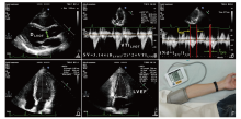
Figure 1
Parameters Required for Measurement in Chen’s Method. (A, B) The left ventricular outflow tract diameter (DLVOT) and the velocity time integral of the LVOT (VTILVOT) were measured to calculate stroke volume (SV). (C) The blood flow spectrum of the LVOT was collected to measure the pre-ejection period (tpre) and ejection time (teje), and their ratio (tNd) was calculated. (D, E) Apical four-chamber and two-chamber views were obtained, and the left ventricular ejection fraction (LVEF) was calculated using the Simpson's biplane method. (F) Left ventricular systolic (SBP) and diastolic pressures (DBP) were measured using an electronic sphygmomanometer. Finally, the parameters were utilized to calculate effective arterial compliance (Ea, Ea= SBP×0.9/SV), end-systolic elastance (Ees), and Ea/Ees."
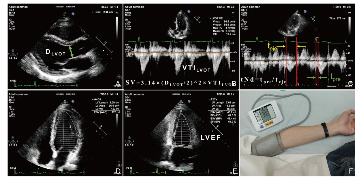
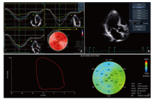
Figure 2
Analysis of echocardiographic myocardial work. (A) Speckle tracking imaging was utilized to analyze the apical four-chamber, two-chamber, and three-chamber echocardiograms in order to obtain the overall longitudinal strain curve of the left ventricle. (B) The apical three-chamber view was employed to determine the timing of the opening and closing of the aortic and mitral valves. Subsequently, valvular timings and non-invasive cuff blood pressure measurements were incorporated with the standard pressure curve built into the software to non-invasively derive the left ventricular pressure curve for specific patients. (C) A pressure-strain loop was constructed with strain on the x-axis and left ventricular systolic pressure on the y-axis to analyze parameters related to myocardial work."

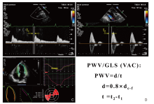
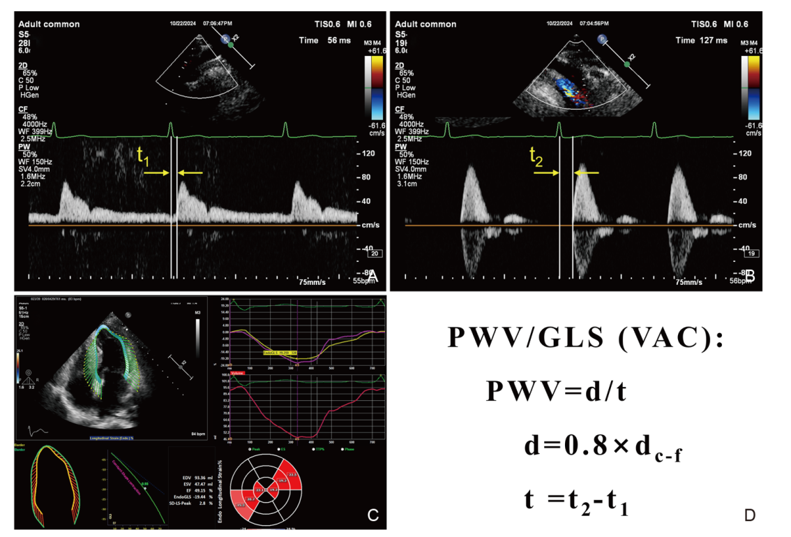
Figure 3
Analysis of the ratio between pulse wave velocity (PWV) and global longitudinal strain (GLS). (A) Blood flow spectrum obtained near the bifurcation of the carotid artery; the time interval between the peak of the ECG R wave and the onset of the carotid artery systolic blood flow spectrum is measured (t1). (B) Blood flow spectrum obtained near the bifurcation of the femoral artery; the time interval between the peak of the ECG R wave and the onset of the femoral artery systolic blood flow spectrum is measured (t2). The distance between the Doppler blood flow spectrum acquisition points of the carotid artery and femoral artery is also measured (dc-f). (C) Assessment of GLS of the left ventricle using speckle tracking imaging. (D) PWV and the PWV/GLS ratio can be calculated using the formula."
| [1] |
Ikonomidis I, Aboyans V, Blacher J, Brodmann M, Brutsaert DL, Chirinos JA, et al. The role of ventricular-arterial coupling in cardiac disease and heart failure: assessment, clinical implications and therapeutic interventions. A consensus document of the European Society of Cardiology Working Group on Aorta & Peripheral Vascular Diseases, European Association of Cardiovascular Imaging, and Heart Failure Association. Eur J Heart Fail 2019; 21:402-424.
doi: 10.1002/ejhf.1436 pmid: 30859669 |
| [2] |
Bastos MB, Burkhoff D, Maly J, Daemen J, den Uil CA, Ameloot K, et al. Invasive left ventricle pressure-volume analysis: overview and practical clinical implications. Eur Heart J 2020; 41:1286-1297.
doi: 10.1093/eurheartj/ehz552 pmid: 31435675 |
| [3] |
Chen CH, Fetics B, Nevo E, Rochitte CE, Chiou KR, Ding PA, et al. Noninvasive single-beat determination of left ventricular end-systolic elastance in humans. J Am Coll Cardiol 2001; 38:2028-2034.
pmid: 11738311 |
| [4] |
Kelly RP, Ting CT, Yang TM, Liu CP, Maughan WL, Chang MS, et al. Effective arterial elastance as index of arterial vascular load in humans. Circulation 1992; 86:513-521.
doi: 10.1161/01.cir.86.2.513 pmid: 1638719 |
| [5] |
Ky B, French B, May Khan A, Plappert T, Wang A, Chirinos JA, et al. Ventricular-arterial coupling, remodeling, and prognosis in chronic heart failure. J Am Coll Cardiol 2013; 62:1165-1172.
doi: 10.1016/j.jacc.2013.03.085 pmid: 23770174 |
| [6] | Fitzpatrick JK, Meyer CS, Schiller NB, Whooley MA, Mishra RK. Ventricular-vascular coupling at rest and after exercise is associated with heart failure hospitalizations in patients with coronary artery disease. J Am Soc Echocardiogr 2018; 31:1212-1220 e1213. |
| [7] | Namasivayam M, Hayward CS, Muller DWM, Jabbour A, Feneley MP. Ventricular-vascular coupling ratio is the ejection fraction in disguise. J Am Soc Echocardiogr 2019;32. |
| [8] | Bertini, Pietro. (2016). iElastance https://itunes.apple.com/us/app/ielastance/id556528864?mt=8. |
| [9] | Gayat E, Mor-Avi V, Weinert L, Yodwut C, Lang RM. Noninvasive quantification of left ventricular elastance and ventricular-arterial coupling using three-dimensional echocardiography and arterial tonometry. Am J Physiol Heart Circ Physiol 2011; 301:H1916-1923. |
| [10] | Oberhoffer FS, Abdul-Khaliq H, Jung A-M, Rohrer TR, Abd El Rahman M. Left ventricular remodelling among Turner syndrome patients: insights from non-invasive 3D echocardiography-derived pressure-volume loop analysis. Clin Res Cardiol 2019; 109:892-903. |
| [11] |
Augustine JA, Lefferts WK, DeBlois JP, Barreira TV, Taylor BA, Liu K, et al. Sex differences in cardiovascular adaptations in recreational marathon runners. Eur J Appl Physiol 2021; 121:3459-3472.
doi: 10.1007/s00421-021-04806-1 pmid: 34515867 |
| [12] | Tona F, Zanatta E, Montisci R, Muraru D, Beccegato E, De Zorzi E, et al. Higher ventricular-arterial coupling derived from three-dimensional echocardiography is associated with a worse clinical outcome in systemic sclerosis. Pharmaceuticals 2021;14. |
| [13] | Scarlatescu AI, Micheu MM, Petre IG, Oprescu N, Mihail AM, Cojocaru ID, et al. Left ventricular-arterial coupling as an independent predictor of adverse events in young patients with st elevation myocardial infarction-a 3d echocardiographic study. Biomedicines 2024;12. |
| [14] | Obokata M, Kurosawa K, Ishida H, Ito K, Ogawa T, Ando Y, et al. Incremental prognostic value of ventricular-arterial coupling over ejection fraction in patients with maintenance hemodialysis. J Am Soc Echocardiogr 2017; 30:444-453.e2. |
| [15] | Smiseth OA, Donal E, Penicka M, Sletten OJ. How to measure left ventricular myocardial work by pressure-strain loops. Eur Heart J Cardiovasc Imaging 2021; 22:259-261. |
| [16] | Roemer S, Jaglan A, Santos D, Umland M, Jain R, Tajik AJ, et al. The utility of myocardial work in clinical practice. J Am Soc Echocardiogr 2021; 34:807-818. |
| [17] |
Russell K, Eriksen M, Aaberge L, Wilhelmsen N, Skulstad H, Remme EW, et al. A novel clinical method for quantification of regional left ventricular pressure-strain loop area: a non-invasive index of myocardial work. Eur Heart J 2012; 33:724-733.
doi: 10.1093/eurheartj/ehs016 pmid: 22315346 |
| [18] | Truong VT, Vo HQ, Ngo TNM, Mazur J, Nguyen TTH, Pham TTM, et al. Normal ranges of global left ventricular myocardial work indices in adults: a meta-analysis. J Am Soc Echocardiogr 2022; 35:369-377.e8. |
| [19] | Papadopoulos K. Özden Tok Ö, Mitrousi K, Ikonomidis I. Myocardial work: methodology and clinical applications. Diagnostics (Basel) 2021;11. |
| [20] |
Galli E, Leclercq C, Hubert A, Bernard A, Smiseth OA, Mabo P, et al. Role of myocardial constructive work in the identification of responders to CRT. Eur Heart J Cardiovasc Imaging 2018; 19:1010-1018.
doi: 10.1093/ehjci/jex191 pmid: 28954293 |
| [21] |
Boe E, Russell K, Eek C, Eriksen M, Remme EW, Smiseth OA, et al. Non-invasive myocardial work index identifies acute coronary occlusion in patients with non-ST-segment elevation-acute coronary syndrome. Eur Heart J Cardiovasc Imaging 2015; 16:1247-1255.
doi: 10.1093/ehjci/jev078 pmid: 25851329 |
| [22] |
Boe E, Skulstad H, Smiseth OA. Myocardial work by echocardiography: a novel method ready for clinical testing. Eur Heart J Cardiovasc Imaging 2019; 20:18-20.
doi: 10.1093/ehjci/jey156 pmid: 30376059 |
| [23] |
Przewlocka-Kosmala M, Marwick TH, Mysiak A, Kosowski W, Kosmala W. Usefulness of myocardial work measurement in the assessment of left ventricular systolic reserve response to spironolactone in heart failure with preserved ejection fraction. Eur Heart J Cardiovasc Imaging 2019; 20:1138-1146.
doi: 10.1093/ehjci/jez027 pmid: 31502637 |
| [24] |
Aalen JM, Donal E, Larsen CK, Duchenne J, Lederlin M, Cvijic M, et al. Imaging predictors of response to cardiac resynchronization therapy: left ventricular work asymmetry by echocardiography and septal viability by cardiac magnetic resonance. Eur Heart J 2020; 41:3813-3823.
doi: 10.1093/eurheartj/ehaa603 pmid: 32918449 |
| [25] | Lustosa RP, van der Bijl P, El Mahdiui M, Montero-Cabezas JM, Kostyukevich MV, Ajmone Marsan N, et al. Noninvasive myocardial work indices 3 months after st-segment elevation myocardial infarction: prevalence and characteristics of patients with postinfarction cardiac remodeling. J Am Soc Echocardiogr 2020; 33:1172-1179. |
| [26] | Sabatino J, De Rosa S, Leo I, Spaccarotella C, Mongiardo A, Polimeni A, et al. Non-invasive myocardial work is reduced during transient acute coronary occlusion. PLoS ONE 2020; 15:e0244397. |
| [27] | Wang CL, Chan YH, Wu VC, Lee HF, Hsiao FC, Chu PH. Incremental prognostic value of global myocardial work over ejection fraction and global longitudinal strain in patients with heart failure and reduced ejection fraction. Eur Heart J Cardiovasc Imaging 2021; 22:348-356. |
| [28] | Jain R, Bajwa T, Roemer S, Huisheree H, Allaqaband SQ, Kroboth S, et al. Myocardial work assessment in severe aortic stenosis undergoing transcatheter aortic valve replacement. Eur Heart J Cardiovasc Imaging 2021; 22:715-721. |
| [29] |
Flachskampf FA, Chandrashekar Y. Myocardial work and work index: related but different for clinical usage. JACC Cardiovasc Imaging 2022; 15:1521-1523.
doi: 10.1016/j.jcmg.2022.07.001 pmid: 35926913 |
| [30] | Sugawara M, Niki K, Ohte N, Okada T, Harada A. Clinical usefulness of wave intensity analysis. Med Biol Eng Comput 2008; 47:197-206. |
| [31] | Parker KH, Jones CJ. Forward and backward running waves in the arteries: analysis using the method of characteristics. J Biomech Eng 1990; 112:322-326. |
| [32] |
Bhuva AN, D'Silva A, Torlasco C, Nadarajan N, Jones S, Boubertakh R, et al. Non-invasive assessment of ventriculo-arterial coupling using aortic wave intensity analysis combining central blood pressure and phase-contrast cardiovascular magnetic resonance. Eur Heart J Cardiovasc Imaging 2020; 21:805-813.
doi: 10.1093/ehjci/jez227 pmid: 31501858 |
| [33] |
Niki K, Sugawara M, Uchida K, Tanaka R, Tanimoto K, Imamura H, et al. A noninvasive method of measuring wave intensity, a new hemodynamic index: application to the carotid artery in patients with mitral regurgitation before and after surgery. Heart Vessels 1999; 14:263-271.
pmid: 10901480 |
| [34] |
Biglino G, Schievano S, Steeden JA, Ntsinjana H, Baker C, Khambadkone S, et al. Reduced ascending aorta distensibility relates to adverse ventricular mechanics in patients with hypoplastic left heart syndrome: noninvasive study using wave intensity analysis. J Thorac Cardiovasc Surg 2012; 144:1307-1313; discussion 1313-1304.
doi: 10.1016/j.jtcvs.2012.08.028 pmid: 23031685 |
| [35] | Vriz O, Zito C, di Bello V, La Carrubba S, Driussi C, Carerj S, et al. Non-invasive one-point carotid wave intensity in a large group of healthy subjects. Heart Vessels 2014; 31:360-369. |
| [36] |
Ikonomidis I, Lekakis J, Papadopoulos C, Triantafyllidi H, Paraskevaidis I, Georgoula G, et al. Incremental value of pulse wave velocity in the determination of coronary microcirculatory dysfunction in never-treated patients with essential hypertension. Am J Hypertens 2008; 21:806-813.
doi: 10.1038/ajh.2008.172 pmid: 18497732 |
| [37] | Mancia G, Fagard R, Narkiewicz K, Redón J, Zanchetti A, Böhm M, et al. 2013 ESH/ESC Guidelines for themanagement of arterial hypertension. J Hypertens 2013; 31:1281-1357. |
| [38] | Smiseth OA, Rider O, Cvijic M, Valkovic L, Remme EW, Voigt JU. Myocardial strain imaging: theory, current practice, and the future. JACC Cardiovasc Imaging 2024. |
| [39] | Ikonomidis I, Katsanos S, Triantafyllidi H, Parissis J, Tzortzis S, Pavlidis G, et al. Pulse wave velocity to global longitudinal strain ratio in hypertension. European Journal of Clinical Investigation 2018;49. |
| [40] | Holm H, Kruger R, Jujic A, Lamiral Z, Uys AS, Girerd N, et al. Ventricular-arterial coupling and cardiovascular risk among young adults: The African-PREDICT study. American Journal of Physiology-Heart and Circulatory Physiology 2023; 325:H362-H371. |
| [41] | Pavlidis G, Tsilivarakis D, Katogiannis K, Vlastos D, Katsanos S, Katsanaki E, et al. Association of aortic stiffness early post myocardial infarction with left ventricular remodelling. European Journal of Clinical Investigation 2023;54. |
| [42] | Wykretowicz A, Schneider A, Krauze T, Szczepanik A, Banaszak A, Minczykowski A, et al. Pulse wave velocity to the global longitudinal strain ratio in survivors of myocardial infarction. European Journal of Clinical Investigation 2019;49. |
| [43] | Seemann F, Arvidsson P, Nordlund D, Kopic S, Carlsson M, Arheden H, et al. Noninvasive quantification of pressure-volume loops from brachial pressure and cardiovascular magnetic resonance. Circulation: Cardiovascular Imaging 2019;12. |
| [44] | Sjöberg P, Liuba P, Arheden H, Heiberg E, Carlsson M. Non-invasive quantification of pressure-volume loops in patients with Fontan circulation. BMC Cardiovasc Disord 2022;22. |
| [45] | Winkler C, Neidlin M, Sonntag SJ, Grünwald A, Groß-Hardt S, Breuer J, et al. Estimation of left ventricular stroke work based on a large cohort of healthy children. Comput Biol Med 2020;123. |
| [46] |
Herberg U, Gatzweiler E, Breuer T, Breuer J. Ventricular pressure-volume loops obtained by 3D real-time echocardiography and mini pressure wire—a feasibility study. Clin Res Cardiol 2013; 102:427-438.
doi: 10.1007/s00392-013-0548-3 pmid: 23397593 |
| [47] | Pironet A, Docherty PD, Dauby PC, Chase JG, Desaive T. Practical identifiability analysis of a minimal cardiovascular system model. Comput Methods Programs Biomed 2019; 171:53-65. |
| [48] | Liu J, Bilgi C, Bregasi A, Mitchell GF, Pahlevan NM. Noninvasive left ventricle pressure-volume loop determination method with cardiac magnetic resonance imaging and carotid tonometry using a physics-informed approach. IEEE J Biomed Health Inform 2024; 28:5487-5496. |
| [49] | Gamarra A, Díez-Villanueva P, Salamanca J, Aguilar R, Mahía P, Alfonso F. Development and clinical application of left ventricular-arterial coupling non-invasive assessment methods. J Cardiovasc Dev Dis 2024;11. |
| [50] |
Vriz O, Fadl Elmula F-EM, Antonini-Canterin F. Noninvasive assessment of ventricular-arterial coupling in heart failure. Heart Failure Clinics 2021; 17:245-254.
doi: 10.1016/j.hfc.2020.12.003 pmid: 33673948 |
| [51] | Kuznetsova T, D'Hooge J, Kloch-Badelek M, Sakiewicz W, Thijs L, Staessen JA. Impact of hypertension on ventricular-arterial coupling and regional myocardial work at rest and during isometric exercise. J Am Soc Echocardiogr 2012; 25:882-890. |
| [52] | Arya N, Schievano S, Caputo M, Taylor AM, Biglino G. Relationship between pulmonary regurgitation and ventriculo-arterial interactions in patients with post-early repair of tetralogy of fallot: insights from wave-intensity analysis. J Clin Med 2022;11. |
| [53] | Shin WJ, Song JG, Jun IG, Moon YJ, Kwon HM, Jung K, et al. Effect of ventriculo-arterial coupling on transplant outcomes in cirrhotics: Analysis of pressure-volume curve relations. J Hepatol 2017; 66:328-337. |
| [54] |
Siniawski H, Lehmkuhl H, Dandel M, Unbehaun A, Kemper D, Weng Y, et al. Prediction of true circulatory decompensation in chronic heart failure for optimal timing of mechanical circulatory support: non-invasive arterial-ventricular coupling. Journal of Functional Biomaterials 2012; 3:100-113.
doi: 10.3390/jfb3010100 pmid: 24956518 |
| [55] |
Narayan HK, French B, Khan AM, Plappert T, Hyman D, Bajulaiye A, et al. Noninvasive measures of ventricular-arterial coupling and circumferential strain predict cancer therapeutics-related cardiac dysfunction. JACC Cardiovasc Imaging 2016; 9:1131-1141.
doi: S1936-878X(16)30035-3 pmid: 27085442 |
| [56] | Biglino G, Giardini A, Ntsinjana HN, Schievano S, Hsia T-Y, Taylor AM. Ventriculoarterial coupling in palliated hypoplastic left heart syndrome: Noninvasive assessment of the effects of surgical arch reconstruction and shunt type. The Journal of Thoracic and Cardiovascular Surgery 2014; 148:1526-1533. |
| [57] |
Zanon F, Aggio S, Baracca E, Pastore G, Corbucci G, Boaretto G, et al. Ventricular-arterial coupling in patients with heart failure treated with cardiac resynchronization therapy: may we predict the long-term clinical response? Eur J Echocardiogr 2009; 10:106-111.
doi: 10.1093/ejechocard/jen184 pmid: 18579495 |
| [58] |
Lam CS, Shah AM, Borlaug BA, Cheng S, Verma A, Izzo J, et al. Effect of antihypertensive therapy on ventricular-arterial mechanics, coupling, and efficiency. Eur Heart J 2013; 34:676-683.
doi: 10.1093/eurheartj/ehs299 pmid: 22963833 |
| [59] |
Hummel SL, Seymour EM, Brook RD, Sheth SS, Ghosh E, Zhu S, et al. Low-sodium DASH diet improves diastolic function and ventricular-arterial coupling in hypertensive heart failure with preserved ejection fraction. Circ Heart Fail 2013; 6:1165-1171.
doi: 10.1161/CIRCHEARTFAILURE.113.000481 pmid: 23985432 |
| [60] |
Berthelot E, Bihry N, Brault-Melin O, Assayag P, Cohen-Solal A, Chemla D, et al. Changes in ventricular-arterial coupling during decongestive therapy in acute heart failure. Eur J Clin Invest 2014; 44:982-988.
doi: 10.1111/eci.12332 pmid: 25186206 |
| [61] |
Lawson MA, Hansen DE, Gupta DK, Bell SP, Adkisson DW, Mallugari RR, et al. Modification of ventriculo-arterial coupling by spironolactone in nonischemic dilated cardiomyopathy. ESC Heart Fail 2021; 8:1156-1166.
doi: 10.1002/ehf2.13161 pmid: 33403831 |
| [62] | Aghezzaf S, Coisne A, Bauters C, Favata F, Delsart P, Coppin A, et al. Feasibility and prognostic significance of ventricular-arterial coupling after myocardial infarction: the RIGID-MI cohort. Eur Heart J Cardiovasc Imaging 2024; 25:668-677. |
| [63] |
Frenneaux M, Williams L. Ventricular-arterial and ventricular-ventricular interactions and their relevance to diastolic filling. Prog Cardiovasc Dis 2007; 49:252-262.
pmid: 17185113 |
| [1] | Li Zhenyi, Chen Ya, Wang Xinqi, Yang Lan, Chen Anni, Li Zhaojun, Jin Lin. Left and Right Ventricular Interaction: Insight from Echocardiography Imaging [J]. Advanced Ultrasound in Diagnosis and Therapy, 2024, 8(4): 195-204. |
| [2] | Yang Yun, Zhang Xin, Zhang Ruize, Jiang Jingrong, Xie Yuji, Fang Lingyun, Zhang Jing, Xie Mingxing, Wang Jing. Current Status and Progress in Arterial Stiffness Evaluation: A Comprehensive Review [J]. Advanced Ultrasound in Diagnosis and Therapy, 2024, 8(4): 172-182. |
| [3] | Chen Anni, Yang Lan, Li Zhenyi, Wang Xinqi, Chen Ya, Jin Lin, Li Zhaojun. Left Ventricular-Arterial Coupling in Cardiovascular Health: Development, Assessment Methods, and Future Directions [J]. Advanced Ultrasound in Diagnosis and Therapy, 2024, 8(4): 159-171. |
| [4] | Wang Xinqi, Chen Anni, Yang Lan, Chen Ya, Li Zhenyi, Li Zhaojun, Jin Lin. Evaluation Methods and Progress of Right Ventricular-pulmonary Artery Coupling [J]. Advanced Ultrasound in Diagnosis and Therapy, 2024, 8(4): 205-216. |
| [5] | Junrong Hong, MD, Pingyang Zhang, MD, PhD, Mengyao Fei, MD, Lingling Wang, MD. A Study on Left Atrial Function in Patients with Essential Hypertension Using Four-Dimensional Echocardiography [J]. Advanced Ultrasound in Diagnosis and Therapy, 2024, 8(2): 64-73. |
| [6] | Rifei Li, MM, Yuanmei Zhang, MM, Chengkai Zhang, MM, Xuenian Huang, MM, Shangwei Ding, MD. Contrast Echocardiography Evaluation of Microcirculation of Myocardial Infarction Caused by Takotsubo Syndrome: Case Report and Literature Review [J]. Advanced Ultrasound in Diagnosis and Therapy, 2021, 5(3): 258-261. |
| [7] | Jianping Xu, MS, Faping Cui, MS, Shuixiu Dou, MS, Jiafu Ou, MD. Echocardiography of Marfan's Syndrome Patient with New Gene Mutation of FBN1 with 13-year Follow-up [J]. Advanced Ultrasound in Diagnosis and Therapy, 2021, 5(3): 249-253. |
| [8] | Xiaoxue Chen, MD, Shaoling Yang, PhD, Qianqian He, MD, Yin Wang, PhD, Linyan Fan, MD, Fengling Wang, MD, Kun Zhao, MD, Jing Hu, MD. Automated Measurements of Left Ventricular Ejection Fraction and Volumes Using the EchoPAC System [J]. Advanced Ultrasound in Diagnosis and Therapy, 2021, 5(3): 226-235. |
| [9] | Yichen Qu, MD, Ya Yang, MD, Jinjie Xie, MD, Rongjuan Li, MD, Han Zhang, MD, Li Song, MD, Yueli Wang, MD, Jing Li, MD. Incomplete Shone’s Complex with BAV and VSD in Adult Diagnosed by Echocardiography [J]. Advanced Ultrasound in Diagnosis and Therapy, 2021, 5(2): 106-108. |
| [10] | Ting Sun, MD, Guoliang Lu, MD, Jian Fang, MD, Shaobo Xie, MD. Silent Embolization Following Hybrid Device Closure of Atrial Septal Defect [J]. Advanced Ultrasound in Diagnosis and Therapy, 2020, 4(4): 352-353. |
| [11] | Ting Sun, MD, Guoliang Lu, MD, Jian Fang, MD, Shaobo Xie, MD. Transthoracic Echocardiography for Evaluation of an Intrapulmonary Artery Mass [J]. Advanced Ultrasound in Diagnosis and Therapy, 2020, 4(4): 329-334. |
| [12] | Luwen Liu, MS, Shaobo Duan, MD, Yaqiong Li, PhD, Ruiqing Liu, MD, Yuejin Wu, MS, Lianzhong Zhang, MD. Development Status and Prospect of Remote Diagnosis and Treatment of Echocardiography Worldwide [J]. Advanced Ultrasound in Diagnosis and Therapy, 2020, 4(4): 303-307. |
| [13] | Huan Cen, MS, Jinhua Li, MD, Bijing Li, MS, Pengtao Sun, MS. Two- and Three-Dimensional Echocardiography for Primary Cardiac Lymphomas: A Case Report and Literature Review [J]. Advanced Ultrasound in Diagnosis and Therapy, 2020, 4(3): 255-259. |
| [14] | Li Ji, MD, Yuman Li, MD, PhD, Li Zhang, MD, PhD, Yali Yang, MD, PhD, Mingxing Xie, MD, PhD. Aorto-Left Ventricle Fistula in Aortic Valve Endocarditis Found to Mimic Valsalva Sinus Aneurysm Rupture into the Left Ventricle: A case study [J]. Advanced Ultrasound in Diagnosis and Therapy, 2020, 4(1): 18-20. |
| [15] | Xingyi Yang, Zhaojun Li, Xianghong Luo. A Lesson from Left Atrial Mass [J]. Advanced Ultrasound in Diagnosis and Therapy, 2018, 2(3): 181-182. |
| Viewed | ||||||
|
Full text |
|
|||||
|
Abstract |
|
|||||
Share: WeChat
Copyright ©2018 Advanced Ultrasound in Diagnosis and Therapy
|
 Advanced Ultrasound in Diagnosis and Therapy (AUDT)
is licensed under a Creative Commons Attribution 4.0 International License.
Advanced Ultrasound in Diagnosis and Therapy (AUDT)
is licensed under a Creative Commons Attribution 4.0 International License.