Advanced Ultrasound in Diagnosis and Therapy ›› 2025, Vol. 9 ›› Issue (3): 290-297.doi: 10.26599/AUDT.2025.240066
• Original Research • Previous Articles Next Articles
Lohith Kumar Bittugondanahalli Prakasha,b,c,*( ), Shivakumar Neeraja, Gaduputi Jahnavia, Kashif Mohammed Sa,d, K Praneethia, Reddy Manda Pranaya, S Sampangi Ramaiaha, Krishnamurthy Umesha, Prabhakar Sumana,e
Received:2024-11-22
Revised:2025-03-02
Accepted:2025-03-12
Online:2025-09-30
Published:2025-10-13
Contact:
Department of Radiology, Division of Clinical Radiology, Christian Medical College, Vellore, Tamil Nadu, India. e-mail:lohithbp@gmail.com(BPL K),
Lohith Kumar Bittugondanahalli Prakash, Shivakumar Neeraj, Gaduputi Jahnavi, Kashif Mohammed S, K Praneethi, Reddy Manda Pranay, S Sampangi Ramaiah, Krishnamurthy Umesh, Prabhakar Suman. Comparative Analysis of Fetal Ventricular Function: AGA vs. SGA Fetuses Using 2D Speckle-Tracking. Advanced Ultrasound in Diagnosis and Therapy, 2025, 9(3): 290-297.
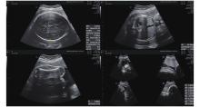
Figure 1
BPD, HC, AC, AFI and FL measured on 2D ultrasonography. BPD and HC measured on an axial plane that traverses the thalami and cavum septum pellucidum. AC is measured at the level of the fetal liver, where the umbilical vein joins the portal vein, and includes the stomach and spine. BPD, biparietal diameter; HC, head circumference; AC, abdominal circumference; FL, femur length. (Images were acquired on E10 Voluson ultrasound machine)"

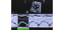
Figure 3
Determination of the cardiac cycle. This figure shows the determination of the right ventricular cardiac cycle to get the annular movement in M-mode by drawing a line perpendicular to the annulus of the right ventricular chamber in the four-chamber view. The cardiac cycle from end-diastole to end-diastole between two troughs is marked on the M-mode graph along with the end-systole. (Images were acquired on E10 Voluson ultrasound machine and analysed using FetalHQ software)"

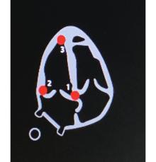
Figure 4
Graphical representation of placement of pointers in the left ventricle. Pointers are placed at the base of the septal wall, the base of the lateral wall, and the apex of the left ventricle. (Images were acquired on E10 Voluson ultrasound machine and analysed using FetalHQ software)"

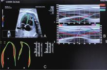
Figure 5
Speckle tracking- Fetal HQ. (A) Marked myocardial region of interest in the left ventricle (LV) and right ventricle (RV); (B) Deformation vectors in the LV and RV; (C) LV and RV global longitudinal strain (GLS) analysis. (Images were acquired on E10 Voluson ultrasound machine and analysed using FetalHQ software)"

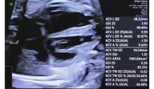
Figure 6
Measurement of the fetal cardiac GSI. A longitudinal line is drawn from the apex to the base of the cardiac outer edge, and a transverse line is drawn from the sidewall of the LV to the sidewall of the RV at the end of the diastole. The GSI can be obtained by dividing the end-diastolic basal–apical length by the end-diastolic transverse width. GSI, global sphericity index; LV, left ventricle; RV, right ventricle. (Images were acquired on E10 Voluson ultrasound machine and analysed using FetalHQ software)"

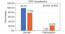
Figure 7
Bar diagram showing CPR Classification. CPR is classified as pathological when the centile for their gestational age is < 5th centile. In the AGA Group, 1.5% had Pathological CPR and in SGA group, 22.2% had pathological CPR, showing statistically significant higher difference in CPR classification between two groups. CPR, cerebro-placental ratio; SGA, small-for-gestational age; AGA, appropriate-for-gestational age."

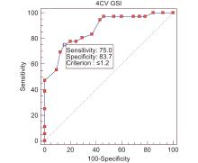
Figure 8
ROC curve showing 4CVGSI in differentiating SGA and AGA. In the study 4CVGSI at ≤ 1.2 had highest validity in predicting SGA. A cutoff of GSI ≤ 1.2 yielded 75% sensitivity and 84.6% specificity, with a positive predictive value (PPV) of 73.0% and a negative predictive value (NPV) of 85.9%."

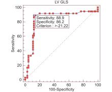
Figure 9
ROC curve showing LV GLS in differentiating SGA and AGA. In the study LV GLS at > −21.19% had highest validity in predicting SGA. A cutoff of LV GLS > −21.19% yielded 88.89% sensitivity and 87.69% specificity, with a positive predictive value (PPV) of 80.0% and a negative predictive value (NPV) of 93.4%."

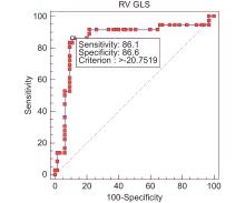
Figure 10
ROC curve showing RV GLS in differentiating SGA and AGA. In the study RV GLS at > −20.59% had highest validity in predicting SGA. A cutoff of RV GLS > −20.59% yielded 86.11% sensitivity and 89.23% specificity, with a positive predictive value (PPV) of 81.6% and a negative predictive value (NPV) of 92.1%."

| [1] | Lees CC, Stampalija T, Baschat A, da Silva Costa F, Ferrazzi E, Figueras F, et al. ISUOG Practice Guidelines: diagnosis and management of small-for-gestational-age fetus and fetal growth restriction. Ultrasound Obstet Gynecol 2020; 56: 298-312. |
| [2] | Mecacci F, Avagliano L, Lisi F, Clemenza S, Serena C, Vannuccini S, et al. Fetal growth restriction: Does an integrated maternal hemodynamic-placental model fit better? Reprod Sci 2021; 28: 2422-2435. |
| [3] | International institute for population sciences (IIPS) and ICF. 2021. National Family Health Survey (NFHS-5), India, 2019-2021: Mizoram. |
| [4] | Gardiner HM. Response of the fetal heart to changes in load: from hyperplasia to heart failure. Heart 2005; 91: 871-873. |
| [5] | de Onis M, Habicht JP. Anthropometric reference data for international use: recommendations from a World Health Organization Expert Committee. Am J Clin Nutr 1996; 64: 650-658. |
| [6] | Physical status: the use and interpretation of anthropometry. Report of a WHO Expert Committee. World Health Organ Tech Rep Ser 1995; 854: 1-452. |
| [7] | Katz J, Wu LA, Mullany LC, Coles CL, Lee AC, Kozuki N, et al. Prevalence of small-for-gestational-age and its mortality risk varies by choice of birth-weight-for-gestation reference population. PLoS One 2014; 9: e92074. |
| [8] | Chew LC, Verma RP. Fetal Growth Restriction. In: StatPearls. Treasure Island (FL): StatPearls Publishing 2022. |
| [9] | Godfrey ME, Messing B, Cohen SM, Valsky DV, Yagel S. Functional assessment of the fetal heart: a review. Ultrasound Obstet Gynecol 2012; 39: 131-144. |
| [10] | van Oostrum NHM, de Vet CM, Clur SB, van der Woude DAA, van den Heuvel ER, Oei SG, et al. Fetal myocardial deformation measured with two-dimensional speckle-tracking echocardiography: longitudinal prospective cohort study of 124 healthy fetuses. Ultrasound Obstet Gynecol 2022; 59: 651-659. |
| [11] | Tan CMJ, Lewandowski AJ. The transitional heart: from early embryonic and fetal development to neonatal life. Fetal Diagn Ther 2020; 47: 373-386. |
| [12] | Senoh D, Hata T, Kitao M. Effect of maternal hyperglycemai on fetal regional circulation in appropriatate for gestational age and small for gestational age fetuses. American Journal of Perinatology 1995; 12: 223-226. |
| [13] | DeVore GR, Cuneo B, Klas B, Satou G, Sklansky M. Comprehensive evaluation of fetal cardiac ventricular widths and ratios using a 24-segment speckle tracking technique. J Ultrasound Med 2019; 38: 1039-1047. |
| [14] | Biswas M, Sudhakar S, Nanda NC, Buckberg G, Pradhan M, Roomi AU, et al. Two- and three-dimensional speckle tracking echocardiography: clinical applications and future directions. Echocardiography 2013; 30: 88-105. |
| [15] | Ringle A, Dornhorst A, Rehman MB, Ruisanchez C, Nihoyannopoulos P. Evolution of subclinical myocardial dysfunction detected by two-dimensional and three-dimensional speckle tracking in asymptomatic type 1 diabetic patients: a long term follow-up study. Echo Res Pract 2017; 4: 73-81. |
| [16] | Claus P, Omar AMS, Pedrizzetti G, Sengupta PP, Nagel E. Tissue tracking technology for assessing cardiac mechanics: principles, normal values, and clinical applications. JACC: Cardiovascular Imaging 2015; 8: 1444-1460. |
| [17] | Li W, Li Z, Liu W, Zhao P, Che G, Wang X, et al. Two-dimensional speckle tracking echocardiography in assessing the subclinical myocardial dysfunction in patients with gestational diabetes mellitus. Cardiovasc Ultrasound 2022; 20: 21. |
| [18] | Liu W, Li W, Li H, Li Z, Zhao P, Guo Z, et al. Two-dimensional speckle tracking echocardiography help identify breast cancer therapeutics-related cardiac dysfunction. BMC Cardiovasc Disord 2022; 22: 548. |
| [19] | DeVore R. Assessment of ventricular contractility in fetuses with an estimated fetal weight less than the tenth centile. Am J Obstet Gynecol 2019; 221: 498.e1-498.e22. |
| [20] | Zhang W, Zhang B, Wu T, Li Y, Qi X, Tian Y, et al. Value of two-dimensional speckle-tracking echocardiography in evaluation of cardiac function in small fetuses. Quant Imaging Med Surg 2024; 14: 8155-8166. |
| [1] | Feng Qing, Yang Huihui, Xu Wanting, He Yu. Application of Two-Dimensional Speckle Tracking Echocardiography in Evaluation of Neonatal Pulmonary Hypertension [J]. Advanced Ultrasound in Diagnosis and Therapy, 2025, 9(3): 254-259. |
| [2] | Elkouahy Fatima Ezzahra, Bennis Ahmed, Merke Nicolas, Ouahid Hajar, Malali Hamid El, Taleb Lhoucine Ben, Mouhsen Azeddine. Advanced Diagnosis of Aortic Stenosis Disease Based on Ultrasound Images: A Novel Artificial Intelligence Approach [J]. Advanced Ultrasound in Diagnosis and Therapy, 2025, 9(3): 298-306. |
| [3] | Qin Shuxuan, He Qing, Wu Zhenni, Lin Yixia, Ji Mengmeng, Zhang Li, Xie Mingxing, Li Yuman. Clinical Utility of Speckle Tracking Echocardiography in Heart Transplantation [J]. Advanced Ultrasound in Diagnosis and Therapy, 2025, 9(2): 103-116. |
| [4] | Yang Lan, Li Zhenyi, Chen Ya, Chen Anni, Wang Xinqi, Jin Lin, Li Zhaojun. Clinical Usefulness of Atrioventricular Coupling in Cardiovascular Disease [J]. Advanced Ultrasound in Diagnosis and Therapy, 2025, 9(1): 1-9. |
| [5] | Yang Yun, Zhang Xin, Zhang Ruize, Jiang Jingrong, Xie Yuji, Fang Lingyun, Zhang Jing, Xie Mingxing, Wang Jing. Current Status and Progress in Arterial Stiffness Evaluation: A Comprehensive Review [J]. Advanced Ultrasound in Diagnosis and Therapy, 2024, 8(4): 172-182. |
| [6] | Chen Anni, Yang Lan, Li Zhenyi, Wang Xinqi, Chen Ya, Jin Lin, Li Zhaojun. Left Ventricular-Arterial Coupling in Cardiovascular Health: Development, Assessment Methods, and Future Directions [J]. Advanced Ultrasound in Diagnosis and Therapy, 2024, 8(4): 159-171. |
| [7] | Zhang Xin, Yang Yun, Zhang Ruize, Zhang Linyue, Xie Yuji, Wu Wenqian, Zhang Jing, Lv Qing, Wang Jing, Xie Mingxing. Noninvasive Evaluation of Left Ventricular-Arterial Coupling: Methodologies and Clinical Relevance [J]. Advanced Ultrasound in Diagnosis and Therapy, 2024, 8(4): 149-158. |
| [8] | Li Zhenyi, Chen Ya, Wang Xinqi, Yang Lan, Chen Anni, Li Zhaojun, Jin Lin. Left and Right Ventricular Interaction: Insight from Echocardiography Imaging [J]. Advanced Ultrasound in Diagnosis and Therapy, 2024, 8(4): 195-204. |
| [9] | Wang Xinqi, Chen Anni, Yang Lan, Chen Ya, Li Zhenyi, Li Zhaojun, Jin Lin. Evaluation Methods and Progress of Right Ventricular-pulmonary Artery Coupling [J]. Advanced Ultrasound in Diagnosis and Therapy, 2024, 8(4): 205-216. |
| [10] | Junrong Hong, MD, Pingyang Zhang, MD, PhD, Mengyao Fei, MD, Lingling Wang, MD. A Study on Left Atrial Function in Patients with Essential Hypertension Using Four-Dimensional Echocardiography [J]. Advanced Ultrasound in Diagnosis and Therapy, 2024, 8(2): 64-73. |
| [11] | Rifei Li, MM, Yuanmei Zhang, MM, Chengkai Zhang, MM, Xuenian Huang, MM, Shangwei Ding, MD. Contrast Echocardiography Evaluation of Microcirculation of Myocardial Infarction Caused by Takotsubo Syndrome: Case Report and Literature Review [J]. Advanced Ultrasound in Diagnosis and Therapy, 2021, 5(3): 258-261. |
| [12] | Jianping Xu, MS, Faping Cui, MS, Shuixiu Dou, MS, Jiafu Ou, MD. Echocardiography of Marfan's Syndrome Patient with New Gene Mutation of FBN1 with 13-year Follow-up [J]. Advanced Ultrasound in Diagnosis and Therapy, 2021, 5(3): 249-253. |
| [13] | Xiaoxue Chen, MD, Shaoling Yang, PhD, Qianqian He, MD, Yin Wang, PhD, Linyan Fan, MD, Fengling Wang, MD, Kun Zhao, MD, Jing Hu, MD. Automated Measurements of Left Ventricular Ejection Fraction and Volumes Using the EchoPAC System [J]. Advanced Ultrasound in Diagnosis and Therapy, 2021, 5(3): 226-235. |
| [14] | Yichen Qu, MD, Ya Yang, MD, Jinjie Xie, MD, Rongjuan Li, MD, Han Zhang, MD, Li Song, MD, Yueli Wang, MD, Jing Li, MD. Incomplete Shone’s Complex with BAV and VSD in Adult Diagnosed by Echocardiography [J]. Advanced Ultrasound in Diagnosis and Therapy, 2021, 5(2): 106-108. |
| [15] | Ting Sun, MD, Guoliang Lu, MD, Jian Fang, MD, Shaobo Xie, MD. Silent Embolization Following Hybrid Device Closure of Atrial Septal Defect [J]. Advanced Ultrasound in Diagnosis and Therapy, 2020, 4(4): 352-353. |
| Viewed | ||||||
|
Full text |
|
|||||
|
Abstract |
|
|||||
Share: WeChat
Copyright ©2018 Advanced Ultrasound in Diagnosis and Therapy
|
 Advanced Ultrasound in Diagnosis and Therapy (AUDT)
is licensed under a Creative Commons Attribution 4.0 International License.
