| [1] |
Osama Mahmoud, BS, Ajay Makkena, BS, Corinne E. Wessner, MS, MBA, RDMS, Ji-Bin Liu, MD, John R. Eisenbrey, PhD, Andrej Lyshchik, MD, PhD.
Contrast-Enhanced Ultrasound LI-RADS: A Pictorial Review
[J]. Advanced Ultrasound in Diagnosis and Therapy, 2023, 7(4): 321-332.
|
| [2] |
Sebastián Eustaquio Martín Pérez, MSc, Raúl Hernández García, PT, Alberto Brito Lorenzo, PT, Carlos Daniel Sabater Cruz, PT, Mario Herrera Pérez, PhD, Fidel Rodríguez Hernández, PhD, Kristin Briem, PhD, Isidro Miguel Martín Pérez, MD.
Ultrasonographic Identification of Muscle Atrophy in Hamstring Muscles after Anterior Cruciate Ligament Repair among Soccer Players: A Case-control Study
[J]. Advanced Ultrasound in Diagnosis and Therapy, 2023, 7(4): 381-389.
|
| [3] |
Lujing Li, MD, Zuofeng Xu, MD.
The Value of VTTQ Combined with B-mode US for Distinguishing Benign from Malignant Breast Masses by Comparing with SE: A Clinical Research
[J]. Advanced Ultrasound in Diagnosis and Therapy, 2023, 7(4): 394-400.
|
| [4] |
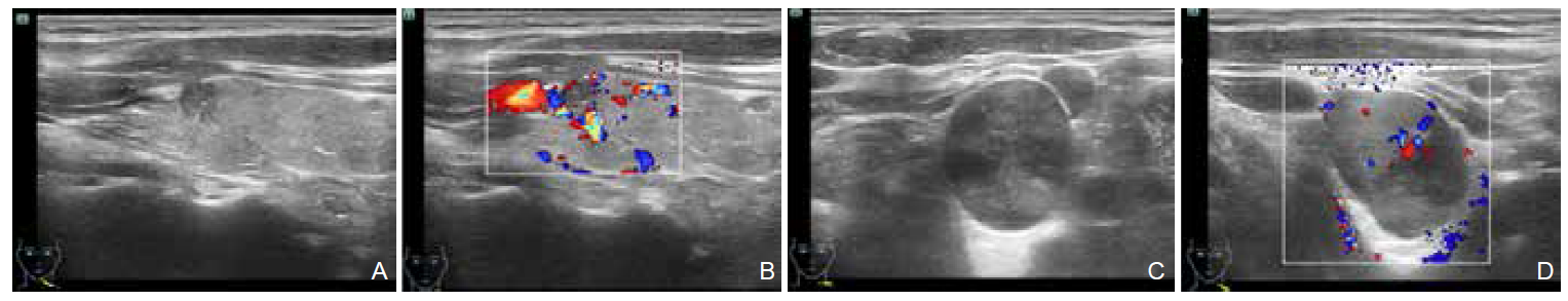
Rodanthi Sfakiotaki, MS, Sergia Liasi, BM, Eleni Papaiakovou, BM, Irene Vraka, PhD, Marina Vakaki, PhD, Chrysoula Koumanidou, PhD.
Juvenile Granulosa Cell Tumor of the Testis: A Preoperative Approach of the Diagnosis with Ultrasound
[J]. Advanced Ultrasound in Diagnosis and Therapy, 2023, 7(4): 409-411.
|
| [5] |
Yiming Li, BM, Jing Xiao, MD, Fang Xie, MD, Yu Lin, BM, Mingbo Zhang, MD, Yukun Luo, MD.
The Value of CEUS in the Diagnosis and Treatment of Thyroid Primary Squamous Cell Carcinoma: A Case Report
[J]. Advanced Ultrasound in Diagnosis and Therapy, 2023, 7(4): 412-415.
|
| [6] |
Nianyu Xue, MM, Shengmin Zhang, BS.
Appendiceal Mucinous Neoplasms Involving the Testis: A Case Report
[J]. Advanced Ultrasound in Diagnosis and Therapy, 2023, 7(4): 420-422.
|
| [7] |
Shuangyu Wu, MM , Xinling Zhang, MD .
Advances and Applications of Transperineal Ultrasound Imaging in Female Pelvic Floor Dysfunction
[J]. Advanced Ultrasound in Diagnosis and Therapy, 2023, 7(3): 235-247.
|
| [8] |
Bo Jiang, MD, Yiman Du, MD, Xiang Fei, MD, Jianing Zhu, MD, Lianhua Zhu, MD, Qiuyang Li, MD, Yukun Luo, MD, PhD.
Ultrasound-Guided Attenuation Parameter May Replace B-mode Ultrasound in Diagnosing Nonalcoholic Fatty Liver Disease
[J]. Advanced Ultrasound in Diagnosis and Therapy, 2023, 7(3): 260-266.
|
| [9] |
Tianxiang Li, BS, Fei Ji, BS, Ruina Zhao, MD, Huazhen Liu, MD, Meng Yang, MD.
Advances in the Research of Ultrasound and Artificial Intelligence in Neuromuscular Disease
[J]. Advanced Ultrasound in Diagnosis and Therapy, 2023, 7(2): 122-129.
|
| [10] |
Zheng Zhang, MS, Li Liu, MD, Desheng Sun, MD, Dirong Zhang, MD, Fengbei Kong, MS, Yalin Wu, PhD, Yu Shi, MD.
Application of the Virtual Reality in the Teaching of Ultrasonography
[J]. Advanced Ultrasound in Diagnosis and Therapy, 2023, 7(2): 193-196.
|
| [11] |
Priscilla Machado, MD, Ji-Bin Liu, MD, Flemming Forsberg, PhD.
Sentinel Lymph Node Identification Using Contrast Lymphosonography: A Systematic Review
[J]. Advanced Ultrasound in Diagnosis and Therapy, 2023, 7(1): 1-7.
|
| [12] |
Cuiwei Wang, MD, Xin-Hua Ye, MD.
Sarcomatoid Intrahepatic Cholangiocarcinoma: A Case Report
[J]. Advanced Ultrasound in Diagnosis and Therapy, 2023, 7(1): 38-41.
|
| [13] |
Yixin Zhang, MS, Yuli Zhao, MS, Yuwen Su, MS, Sen Wang, MS, Li Feng, MD.
Prenatal Ultrasound Diagnosis of Giant Cystic Meconium Peritonitis in Fetus Following in Vitro Fertilization: A Case Report
[J]. Advanced Ultrasound in Diagnosis and Therapy, 2023, 7(1): 42-46.
|
| [14] |
Shi Qiao, MM, Shaote Wang, MD, Yao Deng, MD, Feixiang Xiang, MD, Xiaojuan Qin, MD.
Characterization of Pseudomyxoma Peritonei Originating from Ovarian Borderline Mucinous Tumor Using Ultrasonography: A Case Report
[J]. Advanced Ultrasound in Diagnosis and Therapy, 2023, 7(1): 57-59.
|
| [15] |
Qiaoer Gong, BS, Nianyu Xue, MS.
Application and Progress of Ultrasound Technology in Atherosclerosis
[J]. Advanced Ultrasound in Diagnosis and Therapy, 2023, 7(1): 8-15.
|
 )
)
 Advanced Ultrasound in Diagnosis and Therapy (AUDT)
is licensed under a Creative Commons Attribution 4.0 International License.
Advanced Ultrasound in Diagnosis and Therapy (AUDT)
is licensed under a Creative Commons Attribution 4.0 International License.