Advanced Ultrasound in Diagnosis and Therapy ›› 2024, Vol. 8 ›› Issue (4): 217-230.doi: 10.37015/AUDT.2024.240047
• Review Articles • Previous Articles Next Articles
Mohammed Amra, Machado Priscillaa, Siu Xiao Taniaa, Tahmasebi Aylina, Alnoury Mostafaa, Trabulsi Edouardb, J. Halpern Ethana, R. Eisenbrey Johna, Forsberg Flemminga,*( )
)
Received:2024-09-15
Accepted:2024-10-24
Online:2024-12-30
Published:2024-11-12
Contact:
Forsberg Flemming,
E-mail:flemming.forsberg@jefferson.edu
Mohammed Amr, Machado Priscilla, Siu Xiao Tania, Tahmasebi Aylin, Alnoury Mostafa, Trabulsi Edouard, J. Halpern Ethan, R. Eisenbrey John, Forsberg Flemming. Precision Imaging for Prostate Cancer Localization: How Multiparametric Ultrasound Stands Against Multiparametric MRI. Advanced Ultrasound in Diagnosis and Therapy, 2024, 8(4): 217-230.
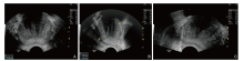
Figure 1
Baseline GrayScale US of the prostate obtaining 3D measurements and systematic biopsy. (A) Transverse diameter; (B) Anteroposterior and sagittal diameters; (C) Core biopsy needle marks."
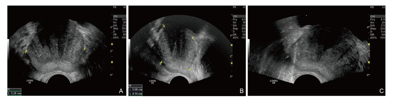

Figure 2
The historical evolution of the histological division of the prostate. (A) The three main methods of dividing the prostate are presented by time, researchers, and core perspectives; (B) Abridged general view of Lowsley’s five-lobe method of prostate dissection; (C) Abridged general view of Franks’ prostate hormone sensitivity dichotomy; (D) Abridged general view of McNeal’s prostate zones theory.. This figure has been reproduced from [Yu et al., Differences in the pathogenetic characteristics of prostate cancer in the transitional and peripheral zones and the possible molecular biological mechanisms, Frontier in Oncology, 2023, Volume 13] under the terms of the Creative Commons Attribution License (CC BY)."
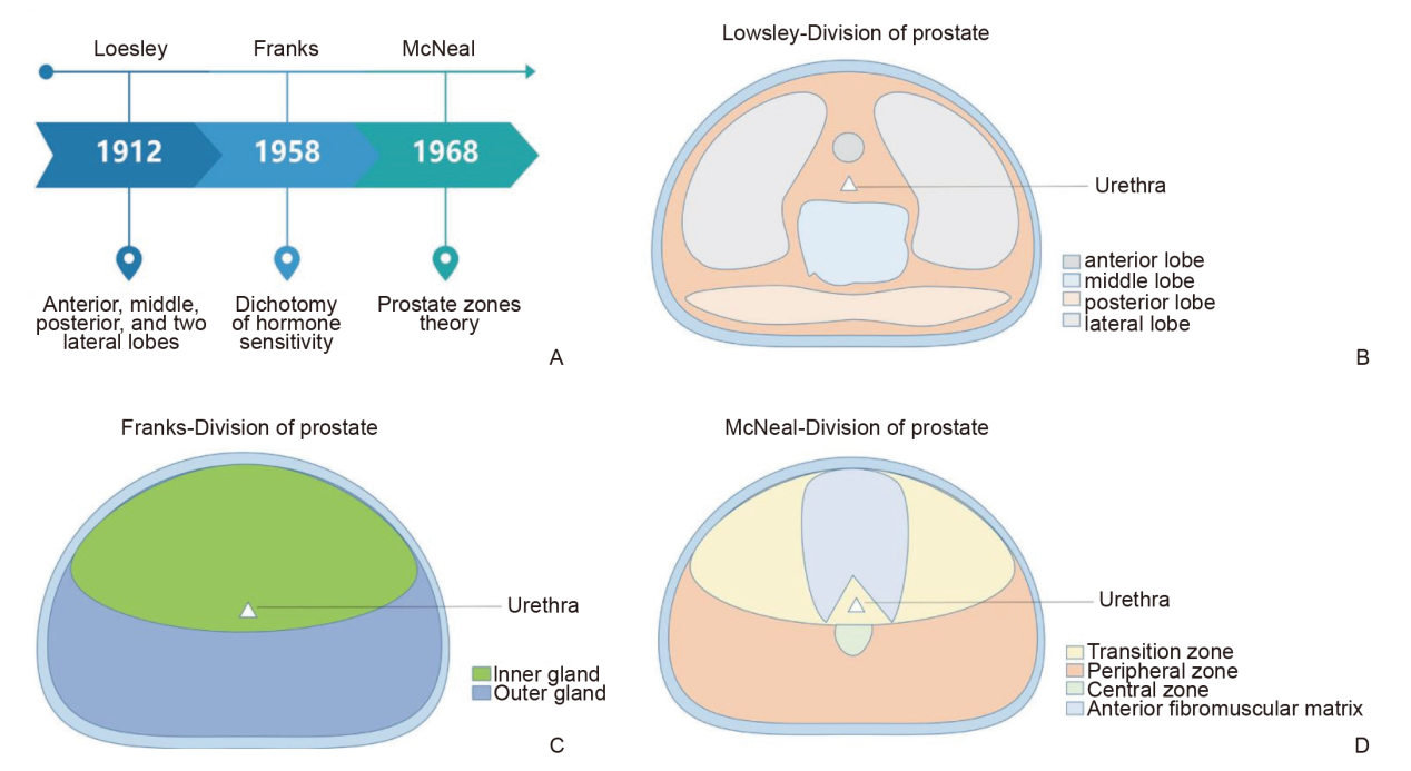
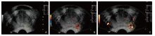
Figure 3
Comparison of GrayScale (GS), Color Doppler Imaging (CDI), and Power Doppler Imaging (PDI) modes. Regions of Interest (ROIs) indicate a suspicious increase in blood flow to the left base of the prostate. (A) GS, inconclusive; (B) CDI, shows increased flow to the left base of the gland; (C) PDI, confirms increased flow to the left base of the gland."
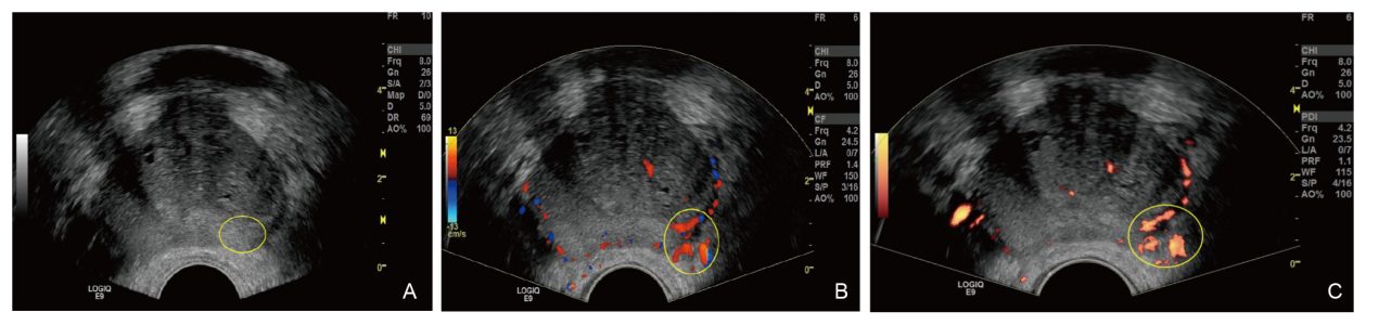
Figure 4
Representative image of dual mode (A) GrayScale and (B) CEUS of a 68-year-old man with PSA of 16.2 ng/ml. (Arrow) points to faster contrast uptake noted in the left base of the prostate with regular uptake to the rest of the prostate."
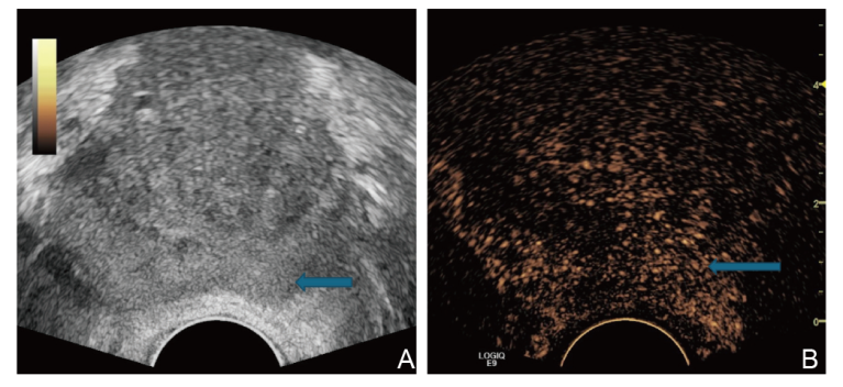

Figure 5
Representative image of dual mode (A) GrayScale and (B) Flash Replenishment Imaging. ROI on flash mode shows irregular vascular pattern and increased contrast uptake in the left base of the prostate compared to the corresponding region in the right base of the gland. ROI, region of interest."
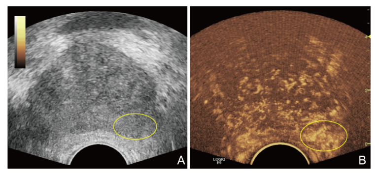
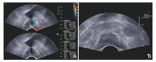
Figure 6
representative figure of dual mode imaging for an 80-year-old man with a focal lesion with hypoechogenicity and elevated stiffness on shear wave elastography. (A) After acquisition of shear wave elastography images, region of interest was placed at and around focal lesion with hypoechogenicity compared with surrounding prostate gland. After three region of interest measurements, mean S and R were 191.2 kPa and 11.2, respectively; (B) Dotted line shows needle guide for real-time biopsy targeted at focal lesion which was confirmed as prostate cancer with Gleason score of 7 at pathology. This figure has been reproduced from [Woo et al., Shear Wave Elastography for Detection of Prostate Cancer, Korean Journal of Radiology, 2015, Volume 15, Pages 346-355] under the terms of the Creative Commons Attribution Non-Commercial License (CC BY NC 3.0)."
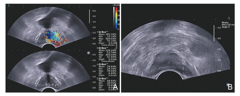
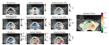
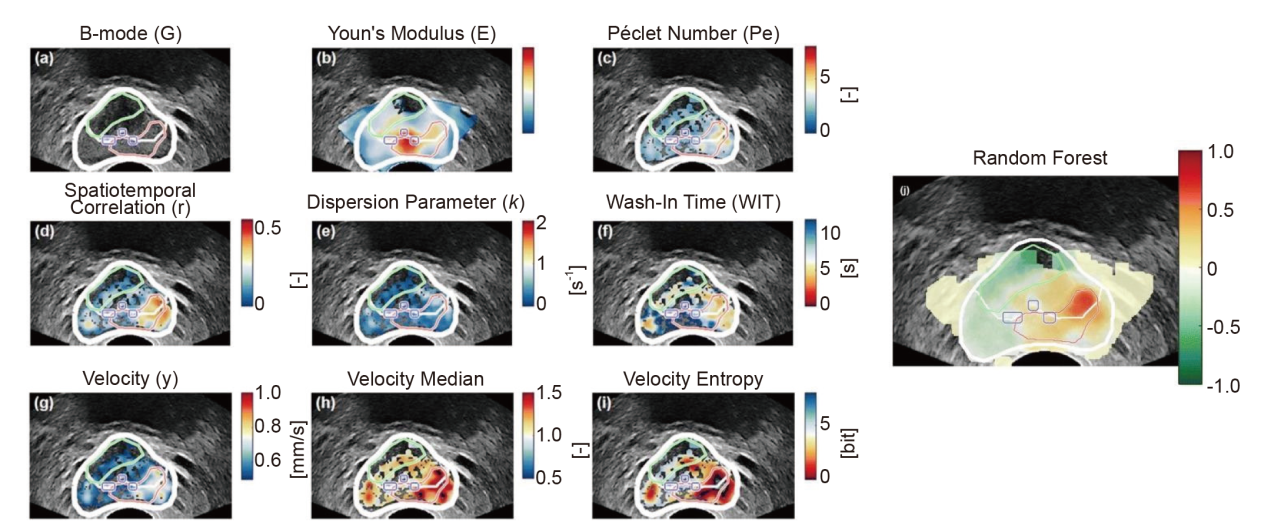
Figure 7
Image plane example, showing the B-mode (A), Young’s modulus (SWE) (B), Péclet number (C), spatiotemporal correlation (D), dispersion-related parameter (E), wash-in time (F), velocity (G), velocity relative to image median (H), 2-mm entropy of velocity (I), and resulting multiparametric map (G). In each map, the prostate and zonal segmentations are depicted in white, the calcifications are encircled in blue, and histopathologically confirmed malignant and benign ROIs are indicated in red and green, respectively. B-mode, brightness mode; SWE, shear wave elastography; ROIs, regions of interest. This figure has been reproduced from [Wildeboer et al., Automated multiparametric localization of prostate cancer based on B-mode, shear-wave elastography, and contrast-enhanced ultrasound radiomics, European Radiology, 2019, Volume 30, Pages 806-815] under the terms of the Creative Commons Attribution 4.0 International License."
| [1] | Key Statistics for Prostate Cancer | Prostate Cancer Facts [Internet]. [cited 2024 Jul 29]. Available from: https://www.cancer.org/cancer/types/prostate-cancer/about/key-statistics.html |
| [2] | Bray F, Laversanne M, Sung H, Ferlay J, Siegel RL, Soerjomataram I, et al. Global cancer statistics 2022: GLOBOCAN estimates of incidence and mortality worldwide for 36 cancers in 185 countries. CA Cancer J Clin 2024; 74:229-263. |
| [3] |
Moe A, Hayne D. Transrectal ultrasound biopsy of the prostate: does it still have a role in prostate cancer diagnosis? Transl Androl Urol 2020; 9:3018-3024.
doi: 10.21037/tau.2019.09.37 pmid: 33457275 |
| [4] |
Bott SRJ, Young MPA, Kellett MJ, Parkinson MC. Contributors to the UCL Hospitals’ Trust Radical Prostatectomy Database. Anterior prostate cancer: is it more difficult to diagnose? BJU Int 2002; 89:886-889.
doi: 10.1046/j.1464-410x.2002.02796.x pmid: 12010233 |
| [5] |
Zhang M, Tang J, Luo Y, Wang Y, Wu M, Memmott B, et al. Diagnostic performance of multiparametric transrectal ultrasound in localized prostate cancer: A comparative study with magnetic resonance imaging. J Ultrasound Med 2019; 38:1823-1830.
doi: 10.1002/jum.14878 pmid: 30561768 |
| [6] | Wei JT, Barocas D, Carlsson S, Coakley F, Eggener S, Etzioni R, et al. Early Detection of Prostate Cancer: AUA/SUO Guideline Part I: Prostate Cancer Screening. J Urol [Internet]. 2023 Jul [cited 2024 Jul 29]; Available from: https://www.auajournals.org/doi/10.1097/JU.0000000000003491 |
| [7] | Turkbey B, Brown AM, Sankineni S, Wood BJ, Pinto PA, Choyke PL. Multiparametric prostate magnetic resonance imaging in the evaluation of prostate cancer. CA Cancer J Clin 2016; 66:326-336. |
| [8] | Spektor M, Mathur M, Weinreb JC. Standards for MRI reporting—the evolution to PI-RADS v 2.0. Transl Androl Urol 2017; 6:355-367. |
| [9] |
Siddiqui MM, Rais-Bahrami S, Truong H, Stamatakis L, Vourganti S, Nix J, et al. Magnetic resonance imaging/ultrasound-fusion biopsy significantly upgrades prostate cancer versus systematic 12-core transrectal ultrasound biopsy. Eur Urol 2013; 64:713-719.
doi: S0302-2838(13)00598-8 pmid: 23787357 |
| [10] |
Siddiqui MM, Rais-Bahrami S, Turkbey B, George AK, Rothwax J, Shakir N, et al. Comparison of MR/ultrasound fusion-Guided biopsy with ultrasound-guided biopsy for the diagnosis of prostate cancer. JAMA 2015; 313:390-397.
doi: 10.1001/jama.2014.17942 pmid: 25626035 |
| [11] |
Rouvière O, Puech P, Renard-Penna R, Claudon M, Roy C, Mège-Lechevallier F, et al. Use of prostate systematic and targeted biopsy on the basis of multiparametric MRI in biopsy-naive patients (MRI-FIRST): a prospective, multicentre, paired diagnostic study. Lancet Oncol 2019; 20:100-109.
doi: S1470-2045(18)30569-2 pmid: 30470502 |
| [12] |
Stabile A, Giganti F, Rosenkrantz AB, Taneja SS, Villeirs G, Gill IS, et al. Multiparametric MRI for prostate cancer diagnosis: current status and future directions. Nat Rev Urol 2020; 17:41-61.
doi: 10.1038/s41585-019-0212-4 pmid: 31316185 |
| [13] |
Rosenkrantz AB, Ginocchio LA, Cornfeld D, Froemming AT, Gupta RT, Turkbey B, et al. Interobserver reproducibility of the PI-RADS version 2 lexicon: A multicenter study of six experienced prostate radiologists. Radiology 2016; 280:793-804.
doi: 10.1148/radiol.2016152542 pmid: 27035179 |
| [14] |
Gupta I, Freid B, Masarapu V, Machado P, Trabulsi E, Wallace K, et al. Transrectal subharmonic ultrasound imaging for prostate cancer detection. Urology 2020; 138:106-112.
doi: S0090-4295(19)31132-X pmid: 31899231 |
| [15] |
Trabulsi EJ, Calio BP, Kamel SI, Gomella LG, Forsberg F, McCue P, et al. Prostate contrast enhanced transrectal ultrasound evaluation of the prostate with whole-mount prostatectomy correlation. Urology 2019; 133:187-191.
doi: S0090-4295(19)30696-X pmid: 31377256 |
| [16] |
Trabulsi EJ, Sackett D, Gomella LG, Halpern EJ. Enhanced transrectal ultrasound modalities in the diagnosis of prostate cancer. Urology 2010; 76:1025-1033.
doi: 10.1016/j.urology.2010.05.022 pmid: 20719368 |
| [17] |
Aigner F, Pallwein L, Schocke M, Andrei L, Junker D, Schäfer G, et al. Comparison of real-time sonoelastography with T2-weighted endorectal magnetic resonance imaging for prostate cancer detection. J Ultrasound Med 2011; 30:643-649.
pmid: 21527612 |
| [18] | Correas JM, Halpern EJ, Barr RG, Ghai S, Walz J, Bodard S, et al. Advanced ultrasound in the diagnosis of prostate cancer. World J Urol 2021; 39:661-676. |
| [19] |
De Coninck V, Braeckman J, Michielsen D. Prostate HistoScanning: a screening tool for prostate cancer? Int J Urol 2013; 20:1184-1190.
doi: 10.1111/iju.12148 pmid: 23594146 |
| [20] | Salib A, Halpern E, Eisenbrey J, Chandrasekar T, Chung PH, Forsberg F, et al. The evolving role of contrast-enhanced ultrasound in urology: a review. World J Urol 2023; 41:673-678. |
| [21] |
Wildeboer RR, Mannaerts CK, van Sloun RJG, Budäus L, Tilki D, Wijkstra H, et al. Automated multiparametric localization of prostate cancer based on B-mode, shear-wave elastography, and contrast-enhanced ultrasound radiomics. Eur Radiol 2020; 30:806-815.
doi: 10.1007/s00330-019-06436-w pmid: 31602512 |
| [22] |
Mannaerts CK, Wildeboer RR, Remmers S, Kajtazovic A, Hagemann J, et al. Multiparametric ultrasound for prostate cancer detection and localization: correlation of B-mode, shear wave elastography and contrast enhanced ultrasound with radical prostatectomy specimens. J Urol 2019; 202:1166-1173.
doi: 10.1097/JU.0000000000000415 pmid: 31246546 |
| [23] |
Li J, Zhu C, Yang S, Mao Z, Lin S, Huang H, et al. Non-invasive diagnosis of prostate cancer and high-grade prostate cancer using multiparametric ultrasonography and serological examination. Ultrasound Med Biol 2024; 50:600-609.
doi: 10.1016/j.ultrasmedbio.2024.01.003 pmid: 38238199 |
| [24] |
Postema A, Mischi M, de la Rosette J, Wijkstra H. Multiparametric ultrasound in the detection of prostate cancer: a systematic review. World J Urol 2015; 33:1651-1659.
doi: 10.1007/s00345-015-1523-6 pmid: 25761736 |
| [25] |
Hodge KK, McNeal JE, Terris MK, Stamey TA. Random systematic versus directed ultrasound guided transrectal core biopsies of the prostate. J Urol 1989; 142:71-74; discussion 74-75.
doi: 10.1016/s0022-5347(17)38664-0 pmid: 2659827 |
| [26] | McNeal JE. The zonal anatomy of the prostate. The Prostate 1981; 2:35-49. |
| [27] |
McNeal JE, Redwine EA, Freiha FS, Stamey TA. Zonal distribution of prostatic adenocarcinoma. Correlation with histologic pattern and direction of spread. Am J Surg Pathol 1988; 12:897-906.
doi: 10.1097/00000478-198812000-00001 pmid: 3202246 |
| [28] | Lee JJ, Thomas IC, Nolley R, Ferrari M, Brooks JD, Leppert JT. Biologic differences between peripheral and transition zone prostate cancer. The Prostate 2015; 75:183-190. |
| [29] |
King CR, Ferrari M, Brooks JD. Prognostic significance of prostate cancer originating from the transition zone. Urol Oncol 2009; 27:592-597.
doi: 10.1016/j.urolonc.2008.05.009 pmid: 18799332 |
| [30] |
Cohen RJ, Shannon BA, Phillips M, Moorin RE, Wheeler TM, Garrett KL. Central zone carcinoma of the prostate gland: A distinct tumor type with poor prognostic features. J Urol 2008; 179:1762-1767.
doi: 10.1016/j.juro.2008.01.017 pmid: 18343454 |
| [31] | Carpagnano FA, Eusebi L, Carriero S, Giannubilo W, Bartelli F, Guglielmi G. Prostate cancer ultrasound: Is still a valid tool? Curr Radiol Rep 2021; 9:7. |
| [32] | Engelbrecht M r. w., Barentsz J o., Jager G j., Van Der Graaf M, Heerschap A, Sedelaar J p. m., et al. Prostate cancer staging using imaging. BJU Int 2000; 86:123-134. |
| [33] |
Heijmink SWTPJ, van Moerkerk H, Kiemeney LALM, Witjes JA, Frauscher F, Barentsz JO. A comparison of the diagnostic performance of systematic versus ultrasound-guided biopsies of prostate cancer. Eur Radiol 2006; 16:927-938.
doi: 10.1007/s00330-005-0035-y pmid: 16391907 |
| [34] |
Salomon L, Colombel M, Patard JJ, Lefrère-Belda MA, Bellot J, Chopin D, et al. Value of ultrasound-guided systematic sextant biopsies in prostate tumor mapping. Eur Urol 1999; 35:289-293.
pmid: 10087390 |
| [35] |
Terris MK, Freiha FS, McNeal JE, Stamey TA. Efficacy of transrectal ultrasound for identification of clinically undetected prostate cancer. J Urol 1991; 146:78-84.
doi: 10.1016/s0022-5347(17)37718-2 pmid: 1711589 |
| [36] |
Onur R, Littrup PJ, Pontes JE, Bianco FJ. Contemporary impact of transrectal ultrasound lesions for prostate cancer detection. J Urol 2004; 172:512-514.
doi: 10.1097/01.ju.0000131621.61732.6b pmid: 15247717 |
| [37] | Halpern EJ, Strup SE. Using gray-scale and color and power Doppler sonography to detect prostatic cancer. AJR Am J Roentgenol 2000; 174:623-627. |
| [38] |
Smeenge M, de la Rosette JJMCH, Wijkstra H. Current status of transrectal ultrasound techniques in prostate cancer. Curr Opin Urol 2012; 22:297-302.
doi: 10.1097/MOU.0b013e3283548154 pmid: 22595778 |
| [39] |
Norberg M, Egevad L, Holmberg L, Sparén P, Norlén BJ, Busch C. The sextant protocol for ultrasound-guided core biopsies of the prostate underestimates the presence of cancer. Urology 1997; 50:562-566.
doi: 10.1016/S0090-4295(97)00306-3 pmid: 9338732 |
| [40] | Chen FK, de Castro Abreu AL, Palmer SL. Utility of ultrasound in the diagnosis, treatment, and follow-up of prostate cancer: state of the art. J Nucl Med 2016; 57:13S-18S. |
| [41] |
Carter HB, Hamper UM, Sheth S, Sanders RC, Epstein JI, Walsh PC. Evaluation of transrectal Ultrasound in the early detection of prostate cancer. J Urol 1989; 142:1008-1010.
doi: 10.1016/s0022-5347(17)38971-1 pmid: 2677409 |
| [42] | Basso Dias A, Ghai S. Micro-ultrasound: current role in prostate cancer diagnosis and future possibilities. Cancers 2023; 15:1280. |
| [43] | Pensa J, Brisbane W, Kinnaird A, Kuppermann D, Hughes G, Ushko D, et al. Evaluation of prostate cancer detection using micro-ultrasound versus MRI through co-registration to whole-mount pathology. Sci Rep 2024; 14:18910. |
| [44] | Eure G, Fanney D, Lin J, Wodlinger B, Ghai S. Comparison of conventional transrectal ultrasound, magnetic resonance imaging, and micro-ultrasound for visualizing prostate cancer in an active surveillance population: A feasibility study. Can Urol Assoc J 2019; 13:E70-77. |
| [45] | You C, Li X, Du Y, Peng L, Wang H, Zhang X, et al. The Microultrasound-guided prostate biopsy in detection of prostate cancer: A systematic review and meta-analysis. J Endourol 2022; 36:394-402. |
| [46] | Sountoulides P, Pyrgidis N, Polyzos SA, Mykoniatis I, Asouhidou E, Papatsoris A, et al. Micro-ultrasound-guided vs multiparametric magnetic resonance imaging-targeted biopsy in the detection of prostate cancer: A systematic review and meta-analysis. J Urol 2021; 205:1254-1262. |
| [47] | Albers P, Bennett J, Evans M, Martin ES, Wang B, Broomfield S, et al. Micro-ultrasound for the detection of clinically significant prostate cancer in biopsy-naive men with negative MRI. Can Urol Assoc J 2024; 18:208-211. |
| [48] | Avolio PP, Lughezzani G, Fasulo V, Maffei D, Sanchez-Salas R, Paciotti M, et al. Assessing the role of high-resolution microultrasound among Naïve patients with negative multiparametric magnetic resonance imaging and a persistently high suspicion of prostate cancer. Eur Urol Open Sci 2022; 47:73-79. |
| [49] | Dias AB, Ghai S. Prostate Cancer Diagnosis with Micro-ultrasound: What We Know now and New Horizons. Radiol Clin North Am 2024; 62:189-197. |
| [50] |
Bigler SA, Deering RE, Brawer MK. Comparison of microscopic vascularity in benign and malignant prostate tissue. Hum Pathol 1993; 24:220-226.
doi: 10.1016/0046-8177(93)90304-y pmid: 8432518 |
| [51] | Bono AV, Celato N, Cova V, Salvadore M, Chinetti S, Novario R. Microvessel density in prostate carcinoma. Prostate Cancer Prostatic Dis 2002; 5:123-127. |
| [52] | Russo G, Mischi M, Scheepens W, De la Rosette JJ, Wijkstra H. Angiogenesis in prostate cancer: onset, progression and imaging. BJU Int 2012; 110:E794-808. |
| [53] |
Holmgren L, O’Reilly MS, Folkman J. Dormancy of micrometastases: balanced proliferation and apoptosis in the presence of angiogenesis suppression. Nat Med 1995; 1:149-153.
doi: 10.1038/nm0295-149 pmid: 7585012 |
| [54] | Ashi K, Kirkham B, Chauhan A, Schultz SM, Brake BJ, Sehgal CM. Quantitative colour Doppler and greyscale ultrasound for evaluating prostate cancer. Ultrasound J Br Med Ultrasound Soc 2021; 29:106-111. |
| [55] | Kelly IM, Lees WR, Rickards D. Prostate cancer and the role of color Doppler US. Radiology 1993; 189:153-156. |
| [56] | Del Rosso A, Di Pierro ED, Masciovecchio S, Galatioto GP, Vicentini C. Does transrectal color Doppler ultrasound improve the diagnosis of prostate cancer? Arch Ital Urol Androl 2012; 84:22-25. |
| [57] |
Taverna G, Morandi G, Seveso M, Giusti G, Benetti A, Colombo P, et al. Colour Doppler and microbubble contrast agent ultrasonography do not improve cancer detection rate in transrectal systematic prostate biopsy sampling. BJU Int 2011; 108:1723-1727.
doi: 10.1111/j.1464-410X.2011.10199.x pmid: 21756276 |
| [58] |
Nelson ED, Slotoroff CB, Gomella LG, Halpern EJ. Targeted biopsy of the prostate: the impact of color Doppler imaging and elastography on prostate cancer detection and Gleason score. Urology 2007; 70:1136-1140.
doi: 10.1016/j.urology.2007.07.067 pmid: 18158034 |
| [59] |
Cornud F, Hamida K, Flam T, Hélénon O, Chrétien Y, Thiounn N, et al. Endorectal color doppler sonography and endorectal MR imaging features of nonpalpable prostate cancer. Am J Roentgenol 2000; 175:1161-1168.
pmid: 11000183 |
| [60] |
Ismail M, Petersen RO, Alexander AA, Newschaffer C, Gomella LG. Color Doppler imaging in predicting the biologic behavior of prostate cancer: correlation with disease-free survival. Urology 1997; 50:906-912.
doi: 10.1016/S0090-4295(97)00403-2 pmid: 9426722 |
| [61] |
Murphy KJ, Rubin JM. Power Doppler: It’s a good thing. Semin Ultrasound CT MR 1997; 18:13-21.
pmid: 9143062 |
| [62] | Dias AB, O’Brien C, Correas JM, Ghai S. Multiparametric ultrasound and micro-ultrasound in prostate cancer: a comprehensive review. Br J Radiol 2022; 95:20210633. |
| [63] |
Sauvain JL, Palascak P, Bourscheid D, Chabi C, Atassi A, Bremon JM, et al. Value of power doppler and 3D vascular sonography as a method for diagnosis and staging of prostate cancer. Eur Urol 2003; 44:21-31.
doi: 10.1016/s0302-2838(03)00204-5 pmid: 12814671 |
| [64] | Zeng S, Wu S, Chen C, Zhu X, Liu Y, Zeng Q, et al. Performance characteristics of 3-D power doppler ultrasound (3-D-PD) with the virtual organ computer-aided analysis (VOCAL) technique in the detection of prostate cancer. Ultrasound Med Biol 2022; 48:91-97. |
| [65] | Halpern EJ, Frauscher F, Forsberg F, Strup SE, Nazarian LN, O’Kane P, et al. High-frequency doppler US of the prostate: Effect of patient position. Radiology 2002; 222:634-639. |
| [66] |
Raine-Fenning NJ, Nordin NM, Ramnarine KV, Campbell BK, Clewes JS, Perkins A, et al. Determining the relationship between three-dimensional power Doppler data and true blood flow characteristics: an in-vitro flow phantom experiment. Ultrasound Obstet Gynecol 2008; 32:540-550.
doi: 10.1002/uog.6110 pmid: 18686275 |
| [67] | Chong WK, Papadopoulou V, Dayton PA. Imaging with ultrasound contrast agents: current status and future. Abdom Radiol 2018; 43:762-772. |
| [68] |
Paefgen V, Doleschel D, Kiessling F. Evolution of contrast agents for ultrasound imaging and ultrasound-mediated drug delivery. Front Pharmacol 2015; 6:197.
doi: 10.3389/fphar.2015.00197 pmid: 26441654 |
| [69] | Calliada F, Campani R, Bottinelli O, Bozzini A, Sommaruga MG. Ultrasound contrast agents: Basic principles. Eur J Radiol 1998; 27:S157-160. |
| [70] |
Wink M, Frauscher F, Cosgrove D, Chapelon JY, Palwein L, Mitterberger M, et al. Contrast-enhanced ultrasound and prostate cancer; A multicentre european research coordination project. Eur Urol 2008; 54:982-993.
doi: 10.1016/j.eururo.2008.06.057 pmid: 18584944 |
| [71] |
Sridharan A, Eisenbrey JR, Forsberg F, Lorenz N, Steffgen L, Ntoulia A. Ultrasound contrast agents: microbubbles made simple for the pediatric radiologist. Pediatr Radiol 2021; 51:2117-2127.
doi: 10.1007/s00247-021-05080-1 pmid: 34117892 |
| [72] |
Smeenge M, Mischi M, Laguna Pes MP, de la Rosette JJMCH, Wijkstra H. Novel contrast-enhanced ultrasound imaging in prostate cancer. World J Urol 2011; 29:581-587.
doi: 10.1007/s00345-011-0747-3 pmid: 21847656 |
| [73] |
Li Y, Tang J, Fei X, Gao Y. Diagnostic performance of contrast enhanced ultrasound in patients with prostate cancer: a meta-analysis. Acad Radiol 2013; 20:156-164.
doi: 10.1016/j.acra.2012.09.018 pmid: 23103186 |
| [74] |
Li H, Xia J, Xie S, Guo Y, Xin M, Li F. Prostate cancer: a comparison of the diagnostic performance of transrectal ultrasound versus contrast enhanced transrectal ultrasound in different clinical characteristics. Int J Clin Exp Med 2015; 8:21428-21434.
pmid: 26885087 |
| [75] |
Frauscher F, Klauser A, Volgger H, Halpern EJ, Pallwein L, Steiner H, et al. Comparison of contrast enhanced color doppler targeted biopsy with conventional systematic biopsy: Impact on prostate cancer detection. J Urol 2002; 167:1648-1652.
pmid: 11912381 |
| [76] | Frausher F, klauser A, Halpern EJ, Horninger W, Bartsch G. Detection of prostate cancer with a microbubble ultrasound contrast agent. The Lancet 2001; 357:1849-1850. |
| [77] | Halpern EJ. Contrast-enhanced ultrasound imaging of prostate cancer. Rev Urol 2006; 8:S29-37. |
| [78] | Emanuel AL, Meijer RI, van Poelgeest E, Spoor P, Serné EH, Eringa EC. Contrast-enhanced ultrasound for quantification of tissue perfusion in humans. Microcirculation 2020; 27:e12588. |
| [79] |
Linden RA, Trabulsi EJ, Forsberg F, Gittens PR, Gomella LG, Halpern EJ. Contrast enhanced ultrasound flash replenishment method for directed prostate biopsies. J Urol 2007; 178:2354-2358.
doi: 10.1016/j.juro.2007.08.022 pmid: 17936814 |
| [80] |
Huang H, Zhu ZQ, Zhou ZG, Chen LS, Zhao M, Zhang Y, et al. Contrast-enhanced transrectal ultrasound for prediction of prostate cancer aggressiveness: The role of normal peripheral zone time-intensity curves. Sci Rep 2016; 6:38643.
doi: 10.1038/srep38643 pmid: 27929134 |
| [81] |
Schalk SG, Huang J, Li J, Demi L, Wijkstra H, Huang P, et al. 3-D Quantitative dynamic contrast ultrasound for prostate cancer localization. Ultrasound Med Biol 2018; 44:807-814.
doi: S0301-5629(17)32469-9 pmid: 29395678 |
| [82] |
Tang MX, Mulvana H, Gauthier T, Lim AKP, Cosgrove DO, Eckersley RJ, et al. Quantitative contrast-enhanced ultrasound imaging: a review of sources of variability. Interface Focus 2011; 1:520-539.
doi: 10.1098/rsfs.2011.0026 pmid: 22866229 |
| [83] |
Mischi M, Kuenen MP, Wijkstra H. Angiogenesis imaging by spatiotemporal analysis of ultrasound contrast agent dispersion kinetics. IEEE Trans Ultrason Ferroelectr Freq Control 2012; 59:621-629.
doi: 10.1109/TUFFC.2012.2241 pmid: 22547274 |
| [84] | Kuenen MPJ, Mischi M, Wijkstra H. Contrast-Ultrasound Diffusion Imaging for Localization of Prostate Cancer. IEEE Trans Med Imaging 2011; 30:1493-1502. |
| [85] |
Mischi M, Kuenen MPJ, Wijkstra H. Angiogenesis imaging by spatiotemporal analysis of ultrasound contrast agent dispersion kinetics. IEEE Trans Ultrason Ferroelectr Freq Control 2012; 59:621-629.
doi: 10.1109/TUFFC.2012.2241 pmid: 22547274 |
| [86] |
Kuenen MPJ, Saidov TA, Wijkstra H, Mischi M. Contrast-ultrasound dispersion imaging for prostate cancer localization by improved spatiotemporal similarity analysis. Ultrasound Med Biol 2013; 39:1631-1641.
doi: 10.1016/j.ultrasmedbio.2013.03.004 pmid: 23791350 |
| [87] | Pieters B, Wijkstra H, van Herk M, Kuipers R, Kaljouw E, de la Rosette J, et al. Contrast-enhanced ultrasound as support for prostate brachytherapy treatment planning. J Contemp Brachytherapy 2012; 4:69-74. |
| [88] | Junker D, De Zordo T, Quentin M, Ladurner M, Bektic J, Horniger W, et al. Real-time elastography of the prostate. BioMed Res Int 2014; 2014:180804. |
| [89] | Krouskop TA, Wheeler TM, Kallel F, Garra BS, Hall T. Elastic moduli of breast and prostate tissues under compression. Ultrason Imaging 1998; 20:260-274. |
| [90] | Tyloch DJ, Tyloch JF, Adamowicz J, Neska-Długosz I, Grzanka D, Van Breda S, et al. Comparison of strain and shear wave elastography in prostate cancer detection. Ultrasound Med Biol 2023; 49:889-900. |
| [91] | Tsutsumi M, Miyagawa T, Matsumura T, Endo T, Kandori S, Shimokama T, et al. Real-time balloon inflation elastography for prostate cancer detection and initial evaluation of clinicopathologic analysis. Am J Roentgenol 2010; 194:W471-476. |
| [92] | Mapes-Gonnella T. The emerging role of elastography in cancer: Diagnostic value in detecting and assessing therapeutic response to treatment. J Diagn Med Sonogr 2014; 30:11-17. |
| [93] |
Zhang B, Ma X, Zhan W, Zhu F, Li M, Huang J, et al. Real-time elastography in the diagnosis of patients suspected of having prostate cancer: a meta-analysis. Ultrasound Med Biol 2014; 40:1400-1407.
doi: 10.1016/j.ultrasmedbio.2014.02.020 pmid: 24785435 |
| [94] |
Sumura M, Shigeno K, Hyuga T, Yoneda T, Shiina H, Igawa M. Initial evaluation of prostate cancer with real-time elastography based on step-section pathologic analysis after radical prostatectomy: A preliminary study. Int J Urol 2007; 14:811-816.
doi: 10.1111/j.1442-2042.2007.01829.x pmid: 17760747 |
| [95] | Almalki YE, Mansour MGED, Ali SA, Basha MAA, Abdelkawi MM, Alduraibi SK, et al. Advanced strain elastography is a reliable approach for prostate cancer detection in patients with elevated PSA levels. Sci Rep 2024; 14:2917. |
| [96] | Correas JM, Tissier AM, Khairoune A, Khoury G, Eiss D, Hélénon O. Ultrasound elastography of the prostate: state of the art. Diagn Interv Imaging 2013; 94:551-560. |
| [97] |
Sang L, Wang X mei, Xu D yang, Cai Y fei. Accuracy of shear wave elastography for the diagnosis of prostate cancer: A meta-analysis. Sci Rep 2017; 7:1949.
doi: 10.1038/s41598-017-02187-0 pmid: 28512326 |
| [98] | Correas JM, Tissier AM, Khairoune A, Vassiliu V, Méjean A, Hélénon O, et al. Prostate cancer: diagnostic performance of real-time shear-wave elastography. Radiology 2015; 275:280-289. |
| [99] | Keskin ET, Kaplanoglu V, Senocak C, Basar H, Bozkurt OF. Transrectal shear wave elastography for detection of prostate cancer. Urol J 2023; 90:230-235. |
| [100] |
Aigner F, Pallwein L, Junker D, Schäfer G, Mikuz G, Pedross F, et al. Value of real-time elastography targeted biopsy for prostate cancer detection in men with prostate specific antigen 1.25 ng/ml or greater and 4.00 ng/ml or less. J Urol 2010; 184:913-917.
doi: 10.1016/j.juro.2010.05.026 pmid: 20643432 |
| [101] |
Barr RG, Cosgrove D, Brock M, Cantisani V, Correas JM, Postema AW, et al. WFUMB guidelines and recommendations on the clinical use of ultrasound elastography: Part 5. Prostate. Ultrasound Med Biol 2017; 43:27-48.
doi: S0301-5629(16)30151-X pmid: 27567060 |
| [102] | Junker D, Schäfer G, Aigner F, Schullian P, Pallwein-Prettner L, Bektic J, et al. Potentials and limitations of real-time elastography for prostate cancer detection: a whole-mount step section analysis. Scientific WorldJournal. 2012; 2012:193213. |
| [103] | Ahmed HU, El-Shater Bosaily A, Brown LC, Gabe R, Kaplan R, Parmar MK, et al. Diagnostic accuracy of multi-parametric MRI and TRUS biopsy in prostate cancer (PROMIS): a paired validating confirmatory study. Lancet Lond Engl 2017; 389:815-822. |
| [104] |
Grey ADR, Scott R, Shah B, Acher P, Liyanage S, Pavlou M, et al. Multiparametric ultrasound versus multiparametric MRI to diagnose prostate cancer (CADMUS): a prospective, multicentre, paired-cohort, confirmatory study. Lancet Oncol 2022; 23:428-438.
doi: 10.1016/S1470-2045(22)00016-X pmid: 35240084 |
| [1] | Yuqing Huang, MD, Cui Lei, BS, Xinyu Zhao, PhD, Jing Xiao, PhD, Xian-Quan Shi, PhD. Identification of Differently Expressed miRNAs and Genes between Benign Prostatic Hyperplasia and Prostate Cancer [J]. Advanced Ultrasound in Diagnosis and Therapy, 2024, 8(1): 22-28. |
| [2] | Xiaojuan Yang, MD, Huihui Yang, MD, Yu He,MD, PhD. Diagnostic Values of CEUS, CECT and CEMRI for Renal Cystic Lesions on the Current Bosniak Criterion-A Meta-analysis [J]. Advanced Ultrasound in Diagnosis and Therapy, 2022, 6(4): 165-173. |
| [3] | Felipe Velasquez-Botero, MD, Ananya Balasubramanya, Ying Tang, MD, Qiang Lu, MD, Ji-Bin Liu, MD, John R. Eisenbrey, PhD. Renal Contrast-enhanced Ultrasound: Clinical Applications and Emerging Research [J]. Advanced Ultrasound in Diagnosis and Therapy, 2022, 6(4): 129-146. |
| [4] | Ruiqing Liu, MD, Yaqiong Li, PhD, Bing Mao, MD, Na Li, PhD, Shaobo Duan, MD, Zhiyang Chang, MS, Ye Zhang, MS, Shuaiyang Wang, MS, Lianzhong Zhang, MD. Focal Ablation Therapy for Prostate Cancer: A Literature Review [J]. Advanced Ultrasound in Diagnosis and Therapy, 2020, 4(4): 308-314. |
| [5] | Lixue Zhai, MD, Xiaojuan Zhang, MD, Yuxiu Gao, MD, Zhaoyan Ding, MD, Haiyang Yu, MD, Cheng Zhao, MD. Progress in Imaging Diagnosis and Image-guided Puncture Biopsy of Prostate Cancer [J]. Advanced Ultrasound in Diagnosis and Therapy, 2019, 3(4): 175-181. |
| [6] | Mengna He, MD, Tian’an Jiang, MD. Acute Transient Thyroid Swelling after Fine-needle Aspiration Biopsy: Case Report and Review of the Literature [J]. Advanced Ultrasound in Diagnosis and Therapy, 2018, 2(3): 178-180. |
| Viewed | ||||||
|
Full text |
|
|||||
|
Abstract |
|
|||||
Share: WeChat
Copyright ©2018 Advanced Ultrasound in Diagnosis and Therapy
|
 Advanced Ultrasound in Diagnosis and Therapy (AUDT)
is licensed under a Creative Commons Attribution 4.0 International License.
Advanced Ultrasound in Diagnosis and Therapy (AUDT)
is licensed under a Creative Commons Attribution 4.0 International License.