
ADVANCED ULTRASOUND IN DIAGNOSIS AND THERAPY >
Comparative Study of CT Characteristics in Imported Cases and Indigenous Cases with COVID-19
Online published: 2020-04-17
Objective: The aims of the current study were to describe the serial CT characteristics of patients infected with COVID-19. In addition, in the light of the CT findings, we tried to determine whether virulence weakens during the transmission with quarantine management.
Methods: Demographics, comorbidity, clinical findings, CT scanning, and scores of the affected lung parenchyma were compared for 131 patients with abnormality on CT images classified as COVID-19 pneumonia and the patients were divided between an imported group (n = 83) and indigenous group (n = 48), according to infected location. Two reviewers scored chest CT examinations for segmental involvement, ground glass opacities, consolidation, and honeycombing opacities. The 55 patients with peak CT "severity score" were selected o make a comparative analysis.
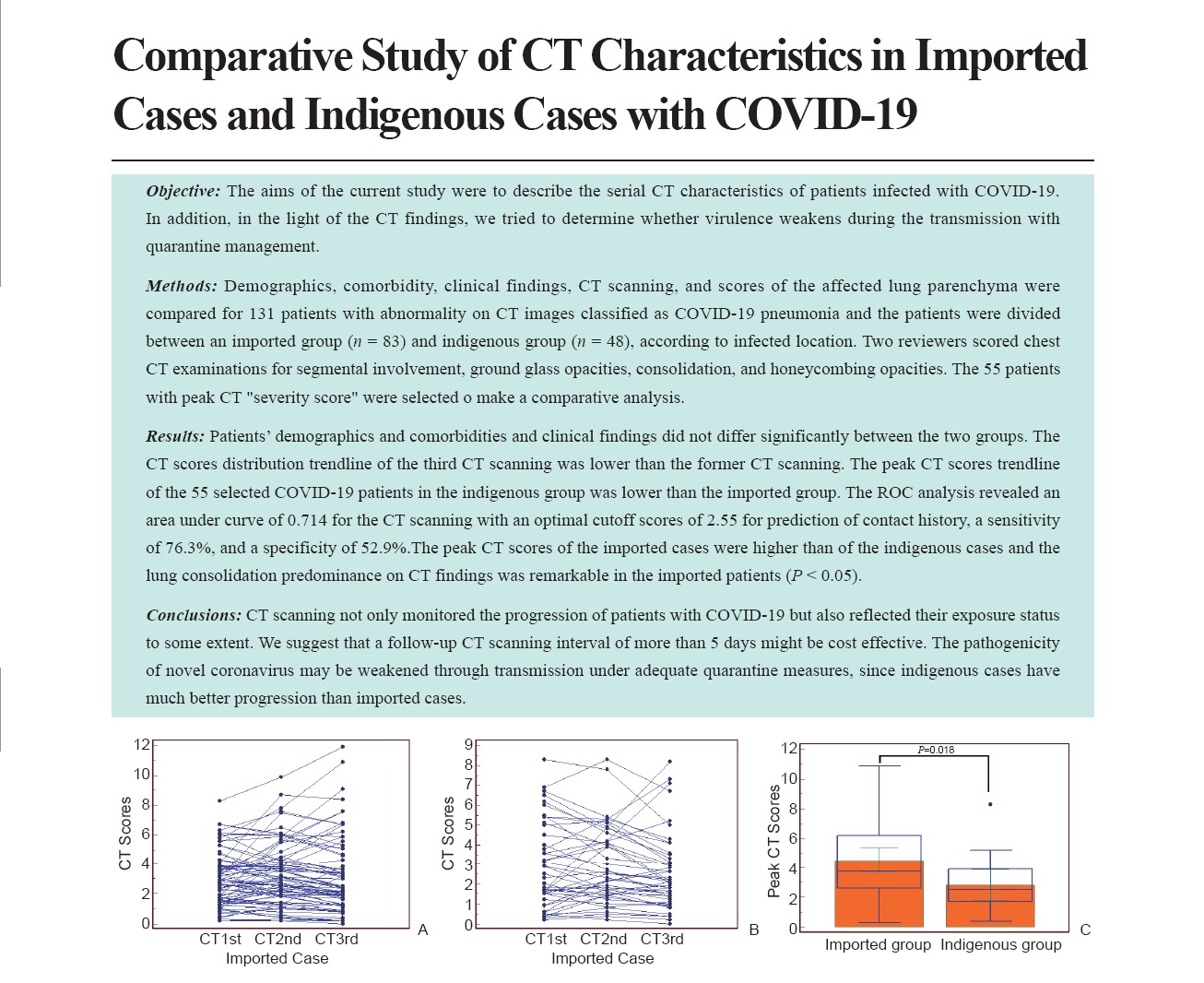
Results: Patients’ demographics and comorbidities and clinical findings did not differ significantly between the two groups. The CT scores distribution trendline of the third CT scanning was lower than the former CT scanning. The peak CT scores trendline of the 55 selected COVID-19 patients in the indigenous group was lower than the imported group. The ROC analysis revealed an area under curve of 0.714 for the CT scanning with an optimal cutoff scores of 2.55 for prediction of contact history, a sensitivity of 76.3%, and a specificity of 52.9%.The peak CT scores of the imported cases were higher than of the indigenous cases and the lung consolidation predominance on CT findings was remarkable in the imported patients (P < 0.05).
Conclusions: CT scanning not only monitored the progression of patients with COVID-19 but also reflected their exposure status to some extent. We suggest that a follow-up CT scanning interval of more than 5 days might be cost effective. The pathogenicity of novel coronavirus may be weakened through transmission under adequate quarantine measures, since indigenous cases have much better progression than imported cases.
Key words: COVID-19; CT; Coronavirus; Pathogenicity
Tan, MD Rong , Li, MD Nina , Liu, MD Ping , Tang, MD Qi , Yu, MD Qizhi . Comparative Study of CT Characteristics in Imported Cases and Indigenous Cases with COVID-19[J]. ADVANCED ULTRASOUND IN DIAGNOSIS AND THERAPY, 2020 , 4(2) : 99 -106 . DOI: 10.37015/AUDT.2020.200016
| [1] | World Health OrganizationM. Coronavirus disease (COVID-19) outbreak. 7 Jan, 2020. Available from: https://www.who.int/emergencies/diseases/novel-coronavirus-2019 |
| [2] | Zhu N, Zhang D, Wang W, Li X, Yang B, Song J, et al. China Novel Coronavirus Investigating and Research Team. A novel coronavirus from patients with pneumonia in China, 2019. N Engl J Med 2020; 382:727-733. |
| [3] | Chen N, Zhou M, Dong X, Qu J, Gong F, Han Y, et al. Epidemiological and clinical characteristics of 99 cases of 2019 novel coronavirus pneumonia in Wuhan, China: a descriptive study. Lancet 2020; 395:507-513. |
| [4] | Li Q, Guan X, Wu P, Wang X, Zhou L, Tong Y, et al. Early transmission dynamics in Wuhan, China, of novel coronavirus-infected pneumonia. N Engl J Med 2020; 382:1199-1207. |
| [5] | Koo HJ, Lim S, Choe J, Choi SH, Sung H, Do KH. Radiographic and CT features of viral pneumonia. Radiographics 2018; 38:719-739. |
| [6] | Bernard-Stoecklin S, Nikolay B, Assiri A, Bin Saeed AA, Ben Embarek PK, El Bushra H, et al. Comparative analysis of eleven healthcare-associated outbreaks of Middle East respiratory syndrome coronavirus (Mers-Cov) from 2015 to 2017. Sci Rep 2019; 9:7385. |
| [7] | Das KM, Lee EY, Enani MA, AlJawder SE, Singh R, Bashir S, et al. CT correlation with outcomes in 15 patients with acute Middle East respiratory syndrome coronavirus. AJR Am J Roentgenol 2015; 204:736-42. |
| [8] | Hansell DM, Bankier AA, MacMahon H, McLoud TC, Müller NL, Remy J. Fleischner Society: glossary of terms for thoracic imaging. Radiology 2008; 246:697-722. |
| [9] | Cha MJ, Chung MJ, Kim K, Lee KS, Kim TJ, Kim TS. Clinical implication of radiographic scores in acute Middle East respiratory syndrome coronavirus pneumonia: Report from a single tertiary-referral center of South Korea. Eur J Radiol 2018; 107:196-202. |
| [10] | Chen J, Pathogenicity and transmissibility of 2019-nCoV—A quick overview and comparison with other emerging viruses. Microbes and Infection.2020. https://doi.org/10.1016/j.micinf. 2020. 01. 004. |
| [11] | Cui J, Li F, Shi ZL. Origin and evolution of pathogenic coronaviruses. Nat Rev Microbiol 2019; 17:181-192. |
| [12] | Ajlan AM, Ahyad RA, Jamjoom LG, Alharthy A, Madani TA. Middle East respiratory syndrome coronavirus (MERS-CoV) infection: Chest CT findings. AJR Am J Roentgenol 2014; 203:782-7. |
| [13] | Kanne JP. Chest CT findings in 2019 novel coronavirus (2019-nCoV) infections from Wuhan, China: Key points for the radiologist. Radiology 2020; 295:16-17. |
| [14] | Chung M, Bernheim A, Mei X, Zhang N, Huang M, Zeng X, Cui J, Xu W, Yang Y, Fayad ZA, Jacobi A, Li K, Li S, Shan H. CT imaging features of 2019 novel coronavirus (2019-nCoV). Radiology 2020; 295:202-207. |
| [15] | Xie X, Zhong Z, Zhao W, Zheng C, Wang F, Liu J. Chest CT for typical 2019-nCoV pneumonia: Relationship to negative RT-PCR testing. Radiology 2020:200343. |
| [16] | Wong KT, Antonio GE, Hui DS, Lee N, Yuen EH, Wu A, et al. Severe acute respiratory syndrome: radiographic appearances and pattern of progression in 138 patients. Radiology 2003; 228:401-6. |
| [17] | Al-Tawfiq JA, Memish ZA. Middle East respiratory syndrome coronavirus: transmission and phylogenetic evolution. Trends Microbiol 2014; 22:573-9. |
| [18] | Memish ZA, Al-Tawfiq JA. Middle East respiratory syndrome coronavirus infection control: the missing piece? Am J Infect Control 2014; 42:1258-60. |
/
| 〈 |
|
〉 |
Share: WeChat
Copyright ©2018 Advanced Ultrasound in Diagnosis and Therapy
|
 Advanced Ultrasound in Diagnosis and Therapy (AUDT)
is licensed under a Creative Commons Attribution 4.0 International License.
Advanced Ultrasound in Diagnosis and Therapy (AUDT)
is licensed under a Creative Commons Attribution 4.0 International License.