
ADVANCED ULTRASOUND IN DIAGNOSIS AND THERAPY >
Histological Reference for Shear Wave Elastography in Liver Fibrosis: Collagen Quantification and Scoring System
Received date: 2019-05-15
Online published: 2019-09-05
Objective: To investigate the significance and value of collagen quantification as an optimized histological standard to evaluate shear wave elastography (SWE) in assessing liver fibrosis.
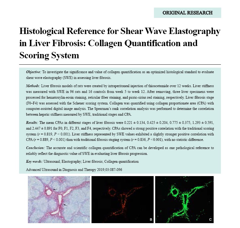
Methods: Liver fibrosis models of rats were created by intraperitoneal injection of thioacetamide over 12 weeks. Liver stiffness was measured with SWE in 96 rats and 16 controls from week 5 to week 12. After removing, three liver specimens were processed for hematoxylin-eosin staining, reticular fiber staining, and picric-sirius red staining, respectively. Liver fibrosis stage (F0-F4) was assessed with the Scheuer scoring system. Collagen was quantified using collagen proportionate area (CPA) with computer-assisted digital image analysis. The Spearman’s rank correlation analysis was performed to determine the correlation between hepatic stiffness measured by SWE, traditional stages and CPA.
Results: The mean CPAs in different stages of liver fibrosis were 0.221 ± 0.134, 0.425 ± 0.204, 0.775 ± 0.375, 1.293 ± 0.591, and 2.447 ± 0.891 for F0, F1, F2, F3, and F4, respectively. CPAs showed a strong positive correlation with the traditional scoring system (r = 0.819, P < 0.001). Liver stiffness represented by SWE values exhibited a slightly stronger positive correlation with CPA (r = 0.889, P < 0.001) than with traditional fibrosis staging system (r = 0.836, P < 0.001), with no statistic difference.
Conclusion: The accurate and scientific collagen quantification of CPA can be developed as one pathological reference to reliably reflect the diagnostic value of SWE in evaluating liver fibrosis progression.
Key words: Ultrasound; Elastography; Liver fibrosis; Collagen quantification
Zhang, MD Yue , Ding, MD Hong , Wu, MD Shengdi , Fan, MD Peili , Li, MD Zheng , Zeng, MD Wenjiao , Wang, MD Wenping . Histological Reference for Shear Wave Elastography in Liver Fibrosis: Collagen Quantification and Scoring System[J]. ADVANCED ULTRASOUND IN DIAGNOSIS AND THERAPY, 2019 , 3(3) : 87 -96 . DOI: 10.37015/AUDT.2019.190815
| [1] | Ferraioli G, Filice C, Castera L, Choi BI, Sporea I, Wilson SR, et al. WFUMB guidelines and recommendations for clinical use of ultrasound elastography: Part 3: liver. Ultrasound Med Biol 2015; 41:1161-79. |
| [2] | Dietrich CF, Bamber J, Berzigotti A, Bota S, Cantisani V, Castera L, et al. EFSUMB guidelines and recommendations on the clinical use of liver ultrasound elastography, update2017 (long version). Ultraschall Med 2017; 38:e16-e47. |
| [3] | Gerber L, Kasper D, Fitting D, Knop V, Vermehren A, Sprinzl K, et al. Assessment of liver fibrosis with 2-D shear wave elastography in comparison to transient elastography and acoustic radiation force impulse imaging in patients with chronic liver disease. Ultrasound Med Biol 2015; 41:2350-9. |
| [4] | Zhuang Y, Ding H, Zhang Y, Sun H, Xu C, Wang W. Two-dimensional shear-wave elastography performance in the noninvasive evaluation of liver fibrosis in patients with chronic hepatitis b: comparison with serum fibrosis indexes. Radiology 2017 6; 283:873-82. |
| [5] | Fraquelli M, Rigamonti C, Casazza G, Donato MF, Ronchi G, Conte D, et al. Etiology-related determinants of liver stiffness values in chronic viral hepatitis B or C. J Hepatol 2011; 54:621-8. |
| [6] | Chen SH, Li YF, Lai HC, Kao JT, Peng CY, Chuang PH, et al. Effects of patient factors on noninvasive liver stiffness measurement using acoustic radiation force impulse elastography in patients with chronic hepatitis C. BMC Gastroenterol 2012; 12:105. |
| [7] | Lucero C, Brown RJ. Noninvasive measures of liver fibrosis and severity of liver disease. Gastroenterol Hepatol (N Y) 2016; 12:33-40. |
| [8] | Germani G, Hytiroglou P, Fotiadu A, Burroughs AK, Dhillon AP. Assessment of fibrosis and cirrhosis in liver biopsies: an update. Semin Liver Dis 2011; 31:82-90. |
| [9] | Germani G, Burroughs AK, Dhillon AP. The relationship between liver disease stage and liver fibrosis: a tangled web. Histopathology 2010; 57:773-84. |
| [10] | Standish RA, Cholongitas E, Dhillon A, Burroughs AK, Dhillon AP. An appraisal of the histopathological assessment of liver fibrosis. Gut 2006; 55:569-78. |
| [11] | Scheuer PJ. Classification of chronic viral hepatitis: a need for reassessment. J Hepatol 1991; 13:372-374. |
| [12] | Berardis S, Dwisthi SP, Najimi M, Sokal EM. Use of mesenchymal stem cells to treat liver fibrosis: current situation and future prospects. World J Gastroenterol 2015; 21:742-58. |
| [13] | Hernandez-Gea V, Friedman SL. Pathogenesis of liver fibrosis. Annu Rev Pathol 2011; 6:425-56. |
| [14] | Kassaye S, Li Y, Huhn G, Peters MG, French AL, Tien PC, et al. Direct and indirect serum markers of liver fibrosis compared with transient elastography among women in the Women's Interagency HIV Study. J AIDS Clin Res 2015; 6:446. |
| [15] | Jeong JY, Kim TY, Sohn JH, Kim Y, Jeong WK, Oh YH, et al. Real time shear wave elastography in chronic liver diseases: accuracy for predicting liver fibrosis, in comparison with serum markers. World J Gastroenterol 2014; 20:13920-9. |
| [16] | Kennedy P, Wagner M, Castéra L, Hong CW, Johnson CL, Sirlin CB, et al. Quantitative elastography methods in liver disease: current evidence and future directions. Radiology 2018 3; 286:738-763. |
| [17] | Knodell RG, Ishak KG, Black WC, Chen TS, Craig R, Kaplowitz N, et al. Formulation and application of a numerical scoring system for assessing histological activity in asymptomatic chronic active hepatitis. Hepatology 1981; 1:431-5. |
| [18] | Anthony PP, Ishak KG, Nayak NC, Poulsen HE, Scheuer PJ, Sobin LH. The morphology of cirrhosis: recommendations on definition, nomenclature, and classification by a working group sponsored by the World Health Organization. J Clin Pathol 1978; 31:395-414. |
| [19] | Bedossa P, Poynard T. An algorithm for the grading of activity in chronic hepatitis C. The METAVIR Cooperative Study Group. Hepatology 1996; 24:289-93. |
| [20] | Ishak K, Baptista A, Bianchi L, Callea F, De Groote J, Gudat F, et al. Histological grading and staging of chronic hepatitis. J Hepatol 1995; 22:696-9. |
| [21] | Goldin RD, Goldin JG, Burt AD, Dhillon PA, Hubscher S, Wyatt J, et al. Intra-observer and inter-observer variation in the histopathological assessment of chronic viral hepatitis. J Hepatol 1996; 25:649-54. |
| [22] | Rammeh S, Khadra HB, Znaidi NS, Romdhane NA, Najjar T, Bouzaidi S, et al. Inter-observer agreement of Ishak and Metavir scores in histological evaluation of chronic viral hepatitis B and C. Ann Biol Clin (Paris) 2014; 72:57-60. |
| [23] | Rosselli M ,MacNaughtan J ,Jalan R ,Pinzani M. Beyond scoring: a modern interpretation of disease progression in chronic liver disease. Gut 2013; 62:1234-41. |
| [24] | Calvaruso V, Dhillon AP, Tsochatzis E, Manousou P, Grillo F, Germani G, et al. Liver collagen proportionate area predicts decompensation in patients with recurrent hepatitis C virus cirrhosis after liver transplantation. J Gastroenterol Hepatol 2012; 27:1227-32. |
| [25] | Manousou P, Burroughs AK, Tsochatzis E, Isgro G, Hall A, Green A, et al. Digital image analysis of collagen assessment of progression of fibrosis in recurrent HCV after liver transplantation. J Hepatol 2013; 58:962-8. |
| [26] | Bihari C, Rastogi A, Sen B, Bhadoria AS, Maiwall R, Sarin SK. Quantitative fibrosis estimation by image analysis predicts development of decompensation, composite events and defines event free survival in chronic hepatitis B patients. Hum Pathol 2016; 55:63-71. |
| [27] | Manousou P, Dhillon AP, Isgro G, Calvaruso V, Luong TV, Tsochatzis E, et al. Digital image analysis of liver collagen predicts clinical outcome of recurrent hepatitis C virus 1 year after liver transplantation. Liver Transpl 2011; 17:178-88. |
| [28] | Farrar CT , DePeralta DK, Day H,Rietz TA, Wei L, Lauwers GY,et al. 3D molecular MR imaging of liver fibrosis and response to rapamycin therapy in a bile duct ligation rat model. J Hepatol 2015; 63:689-96. |
| [29] | Bandula S, Punwani S, Rosenberg WM, Jalan R, Hall AR, Dhillon A, et al. Equilibrium contrast-enhanced CT imaging to evaluate hepatic fibrosis: initial validation by comparison with histopathologic sampling. Radiology 2015; 275:136-43. |
| [30] | Thiele M, Detlefsen S, Sevelsted M?ller L, Madsen BS, Fuglsang Hansen J, Fialla AD, et al. Transient and 2-dimensional shear-wave elastography provide comparable assessment of alcoholic liver fibrosis and cirrhosis. Gastroenterology 2016; 150:123-33. |
| [31] | Thomson W, Kelvin L. Lecture to the Institution of Civil Engineers. Popular Lectures and Addresses May 3; 1883; 1: 80; London: Macmillan and Co.; 1891. |
| [32] | Cong T, Jin XY, Zhao L, Ma L, Li RS, Zhao P, et al. Anti-fibrotic effects of the Masson pine pollen aqueous extract on hepatic fibrosis rat model. Int J Clin Exp Pathol 2015; 8:4651-61. |
| [33] | Gabr SA, Alghadir AH. Prediction of fibrosis in hepatitis C patients: assessment using hydroxyproline and oxidative stress biomarkers. Virus Disease 2014; 25:91-100. |
| [34] | Lareu RR, Zeugolis DI, Abu-Rub M, Pandit A, Raghunath M. Essential modification of the sircol collagen assay for the accurate quantification of collagen content in complex protein solutions. Acta Biomater 2010; 6:3146-51. |
| [35] | Lee HS, Shun CT, Chiou LL, Chen CH, Huang GT, Sheu JC. Hydroxyproline content of needle biopsies as an objective measure of liver fibrosis: emphasis on sampling variability. J Gastroenterol Hepatol 2005; 20:1109-14. |
| [36] | Puchtler H, Meloan SN, Waldrop FS. Are picro-dye reactions for collagens quantitative? Chemical and histochemical considerations. Histochemistry 1988; 88:243-56. |
/
| 〈 |
|
〉 |
Share: WeChat
Copyright ©2018 Advanced Ultrasound in Diagnosis and Therapy
|
 Advanced Ultrasound in Diagnosis and Therapy (AUDT)
is licensed under a Creative Commons Attribution 4.0 International License.
Advanced Ultrasound in Diagnosis and Therapy (AUDT)
is licensed under a Creative Commons Attribution 4.0 International License.