
ADVANCED ULTRASOUND IN DIAGNOSIS AND THERAPY >
Coronary Heart Disease Concomitant with Atherosclerotic Cerebrovascular Disease
Received date: 2019-05-07
Online published: 2019-09-05
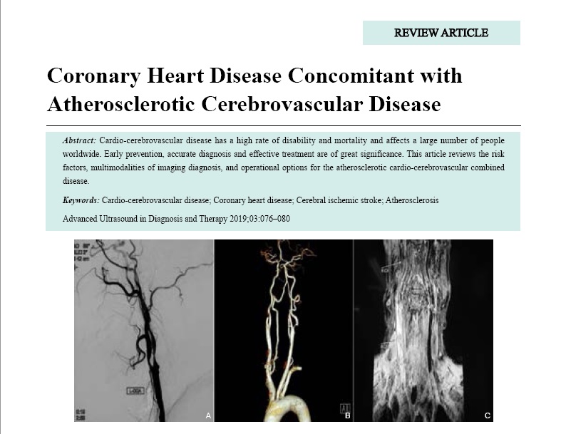
Cardio-cerebrovascular disease has a high rate of disability and mortality and affects a large number of people worldwide. Early prevention, accurate diagnosis and effective treatment are of great significance. This article reviews the risk factors, multimodalities of imaging diagnosis, and operational options for the atherosclerotic cardio-cerebrovascular combined disease.

Liu, MD Yumei , Liu, MD, MS Beibei , Li, MD, PhD Boyu , Hua, MD Yang . Coronary Heart Disease Concomitant with Atherosclerotic Cerebrovascular Disease[J]. ADVANCED ULTRASOUND IN DIAGNOSIS AND THERAPY, 2019 , 3(3) : 76 -80 . DOI: 10.37015/AUDT.2019.190813
| [1] | GBD 2016 DALYs and HALE Collaborators.Global, regional, and national disability-adjusted life-years (DALYs) for 333 diseases and injuries and healthy life expectancy (HALE) for 195 countries and territories, 1990-2016: a systematic analysis for the global burden of disease study 2016. Lancet 2017; 390:1260-344. |
| [2] | Benjamin EJ, Virani SS, Callaway CW, Chamberlain AM, Chang AR, Cheng S, et al. Heart disease and stroke statistics-2018 update: a report from the american heart association. Circulation 2018; 137:e67-e492. |
| [3] | Wang W, Jiang B, Sun H, Ru X, Sun D, Wang L, et al. Prevalence, incidence, and mortality of stroke in China: results from a nationwide population-based survey of 480 687 adults. Circulation 2017; 135:759-71. |
| [4] | Byer E, Ashman R, Toth LA. Electrocardiograms with large, upright T waves and long Q-T intervals. Am Heart J 1947; 33:796-806. |
| [5] | Reeves MJ, Bushnell CD, Howard G, Gargano JW, Duncan PW, Lynch G, et al. Sex differences in stroke: epidemiology, clinical presentation, medical care, and outcomes. Lancet Neurol 2008; 7:915-26. |
| [6] | Collins R, Peto R ,MacMahon S,Hebert P, Fiebach NH, Eberlein KA,et al. Blood pressure, stroke, and coronary heart disease.Part 2, Short-term reductions in blood pressure: overview of randomised drug trials in their epidemiological context. Lancet 1990; 335:827-38. |
| [7] | Shou J, Zhou L, Zhu S, Zhang X. Diabetes is an Independent Risk Factor for Stroke Recurrence in Stroke Patients: A Meta-analysis. J Stroke Cerebrovasc Dis 2015; 24:1961-8. |
| [8] | Peters SA, Singhateh Y, Mackay D, Huxley RR, Woodward M. Total cholesterol as a risk factor for coronary heart disease and stroke in women compared with men: A systematic review and meta-analysis. Atherosclerosis 2016; 248:123-31. |
| [9] | Hoshino T, Sissani L, Labreuche J, Ducrocq G, Lavallee PC, Meseguer E, et al. Prevalence of systemic atherosclerosis burdens and overlapping stroke etiologies and their associations with long-term vascular prognosis in stroke with intracranial atherosclerotic disease. JAMA Neurol 2018; 75:203-11. |
| [10] | Abd-Allah F, Kassem HH, Hashad A, Shamloul RM, Zaki A. Prevalence of intracranial atherosclerosis among patients with coronary artery disease: a 1-year hospital-based study. Eur Neurol 2014; 71:326-30. |
| [11] | Amarenco P, Lavallee PC, Labreuche J, Ducrocq G, Juliard JM, Feldman L, et al. Coronary artery disease and risk of major vascular events after cerebral infarction. Stroke 2013; 44:1505-11. |
| [12] | Kolominsky-Rabas PL, Weber M, Gefeller O, Neundoerfer B, Heuschmann PU. Epidemiology of ischemic stroke subtypes according to TOAST criteria: incidence, recurrence, and long-term survival in ischemic stroke subtypes: a population-based study. Stroke 2001; 32:2735-40. |
| [13] | Liu Y, Hua Y, Feng W, Ovbiagele B. Multimodality ultrasound imaging in stroke: current concepts and future focus. Expert Rev Cardiovasc Ther 2016; 14:1325-33. |
| [14] | Brodoefel H, Reimann A, Burgstahler C, Schumacher F, Herberts T, Tsiflikas I, et al. Noninvasive coronary angiography using 64-slice spiral computed tomography in an unselected patient collective: effect of heart rate, heart rate variability and coronary calcifications on image quality and diagnostic accuracy. Eur J Radiol 2008; 66:134-41. |
| [15] | Mintz GS, Nissen SE, Anderson WD, Bailey SR, Erbel R, Fitzgerald PJ, et al. American college of cardiology clinical expert consensus document on standards for acquisition, measurement and reporting of intravascular ultrasound studies (IVUS). a report of the american college of cardiology task force on clinical expert consensus documents.J Am Coll Cardiol 2001; 37:1478-92. |
| [16] | Terashima M, Kaneda H, Suzuki T. The role of optical coherence tomography in coronary intervention. Korean J Intern Med 2012; 27:1-12. |
| [17] | Chen CJ, Kumar JS, Chen SH, Ding D, Buell TJ, Sur S, et al. Optical coherence tomography: future applications in cerebrovascular imaging. Stroke 2018; 49:1044-50. |
| [18] | Salonen JT, Salonen R. Ultrasound B-mode imaging in observational studies of atherosclerotic progression. Circulation 1993;87:II56-65. |
| [19] | Inaba Y, Chen JA, Bergmann SR. Carotid plaque, compared with carotid intima-media thickness, more accurately predicts coronary artery disease events: a meta-analysis. Atherosclerosis 2012; 220:128-33. |
| [20] | Jashari F, Ibrahimi P, Bajraktari G, Gronlund C, Wester P, Henein MY. Carotid plaque echogenicity predicts cerebrovascular symptoms: a systematic review and meta-analysis. Eur J Neurol 2016; 23:1241-7. |
| [21] | Rafailidis V, Charitanti A, Tegos T, Destanis E, Chryssogonidis I. Contrast-enhanced ultrasound of the carotid system: a review of the current literature. J Ultrasound 2017; 20:97-109. |
| [22] | Yang J, Hua Y, Li X, Gao M, Li Q, Liu B, et al. The assessment of diagnostic accuracy for basilar artery stenosis by transcranial color-coded sonography. Ultrasound Med Biol 2018 May; 44:995-1002. |
| [23] | Valaikiene J, Ryliskyte L, Valaika A, Puronaite R, Dementaviciene J, Vaitkevicius A, et al. A high prevalence of intracranial stenosis in patients with coronary artery disease and the diagnostic value of transcranial duplex sonography. J Stroke Cerebrovasc Dis 2019; 28:1015-21. |
| [24] | Weimar C, Bilbilis K, Rekowski J, Holst T, Beyersdorf F, Breuer M, et al. Safety of simultaneous coronary artery bypass grafting and carotid endarterectomy versus isolated coronary artery bypass grafting: a randomized clinical trial. Stroke 2017; 48:2769-75. |
| [25] | Dick AM, Brothers T, Robison JG, Elliott BM, Kratz JM, Toole JM, et al. Combined carotid endarterectomy and coronary artery bypass grafting versus coronary artery bypass grafting alone: a retrospective review of outcomes at our institution. Vasc Endovascular Surg 2011; 45:130-4. |
| [26] | Illuminati G, Ricco JB, Calio F, Pacile MA, Miraldi F, Frati G, et al. Short-term results of a randomized trial examining timing of carotid endarterectomy in patients with severe asymptomatic unilateral carotid stenosis undergoing coronary artery bypass grafting. J Vasc Surg 2011; 54:993-9. |
| [27] | Dong H, Jiang X, Peng M, Zou Y, Che W, Qian H, et al. The interval between carotid artery stenting and open heart surgery is related to perioperative complications. Catheter Cardiovasc Interv 2016; 87:564-9. |
/
| 〈 |
|
〉 |