
ADVANCED ULTRASOUND IN DIAGNOSIS AND THERAPY >
Morphology and Hemodynamic Characteristics of Internal Jugular Vein Hypoplasia and Relation with Cerebral Venous Sinus Stenosis
Received date: 2019-04-15
Online published: 2019-06-28
Objective: The characteristics of Internal jugular vein (IJV) morphology and hemodynamics of IJV hypoplasia have not been well illustrated.
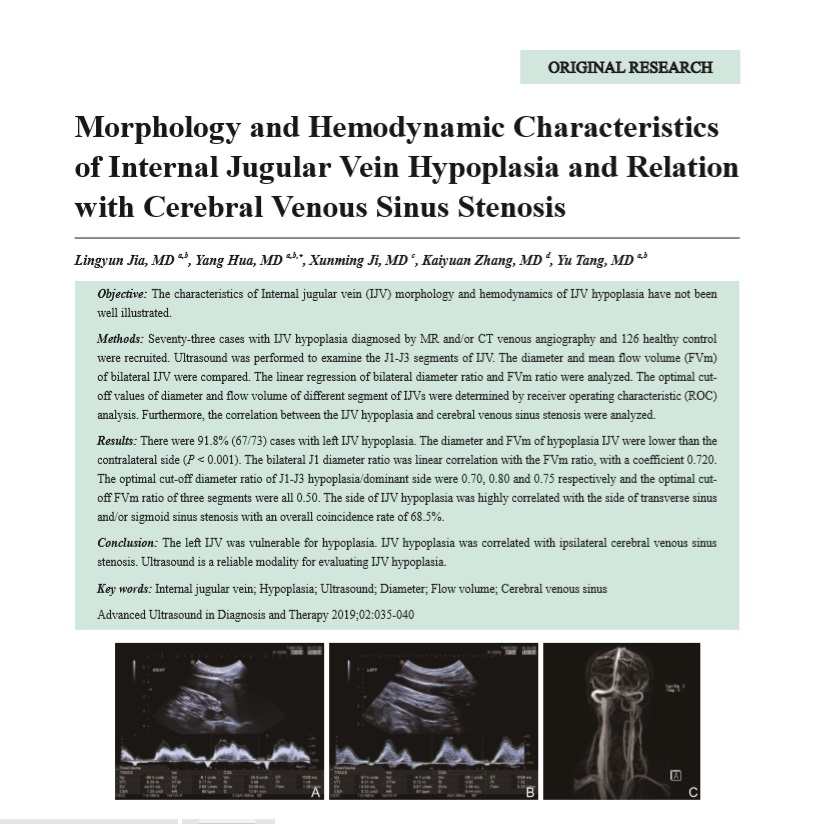
Methods: Seventy-three cases with IJV hypoplasia diagnosed by MR and/or CT venous angiography and 126 healthy control were recruited. Ultrasound was performed to examine the J1-J3 segments of IJV. The diameter and mean flow volume (FVm) of bilateral IJV were compared. The linear regression of bilateral diameter ratio and FVm ratio were analyzed. The optimal cutoff values of diameter and flow volume of different segment of IJVs were determined by receiver operating characteristic (ROC) analysis. Furthermore, the correlation between the IJV hypoplasia and cerebral venous sinus stenosis were analyzed.
Results: There were 91.8% (67/73) cases with left IJV hypoplasia. The diameter and FVm of hypoplasia IJV were lower than the contralateral side (P<0.001). The bilateral J1 diameter ratio was linear correlation with the FVm ratio, with a coefficient 0.720. The optimal cut-off diameter ratio of J1-J3 hypoplasia/dominant side were 0.70, 0.80 and 0.75 respectively and the optimal cutoff FVm ratio of three segments were all 0.50. The side of IJV hypoplasia was highly correlated with the side of transverse sinus and/or sigmoid sinus stenosis with an overall coincidence rate of 68.5%.
Conclusion: The left IJV was vulnerable for hypoplasia. IJV hypoplasia was correlated with ipsilateral cerebral venous sinus stenosis. Ultrasound is a reliable modality for evaluating IJV hypoplasia.
Key words: Internal jugular vein; Hypoplasia; Ultrasound; Diameter; Flow volume; Cerebral venous sinus
Jia, MD Lingyun , Hua, MD Yang , Ji, MD Xunming , Zhang, MD Kaiyuan , Tang, MD Yu . Morphology and Hemodynamic Characteristics of Internal Jugular Vein Hypoplasia and Relation with Cerebral Venous Sinus Stenosis[J]. ADVANCED ULTRASOUND IN DIAGNOSIS AND THERAPY, 2019 , 3(2) : 35 -40 . DOI: 10.37015/AUDT.2019.190807
| [1] | Tong LS, Guo ZN, Ou YB, Yu YN, Zhang XC, Tang J, et al. Cerebral venous collaterals: A new fort for fighting ischemic stroke? Prog Neurobiol 2018; 163-164:172-193. |
| [2] | Jia LY, Hua Y, Ji XM, Liu JT. Correlation analysis of internal jugular vein abnormalities and cerebral venous sinus thrombosis. Chinese Medical Journal 2012; 125: 3671-3674. [in Chinese] |
| [3] | Zhou D, Meng R, Zhang X, Guo L, Li S, Wu W, et al. Intracranial hypertension induced by internal jugular vein stenosis can be resolved by stenting. Eur J Neurol 2018; 25:365-e13. |
| [4] | Han K, Chao AC, Chang FC, Chung CP, Hsu HY, Sheng WY, et al. Obstruction of venous drainage linked to transient global amnesia. PLoS One 2015; 10:e0132893. |
| [5] | Hsu HY, Chao AC, Chen YY, Yang FY, Chung CP, Sheng WY, et al. Reflux of jugular and retrobulbar venous flow in transient monocular blindness. Ann Neurol 2008; 63:247-253. |
| [6] | Sethi SK, Daugherty AM, Gadda G, Utriainen DT, Jiang J, Raz N, et al. Jugular anomalies in multiple sclerosis are associated with increased collateral venous flow. AJNR Am J Neuroradiol 2017; 38:1617-1622. |
| [7] | Dai YF, Li KC, Zhang KY, Liang PP. Administration method of contrast agent for large field of view 3D high-spatial-resolution contrast-enhanced MRV of cerebral venous sinus and jugular veins. Chin J Med Imaging Technol 2014; 30:126-129. [in Chinese] |
| [8] | Zaharchuk G, Fischbein NJ, Rosenberg J, Herfkens RJ, Dake MD. Comparison of MR and contrast venography of the cervical venous system in multiple sclerosis. AJNR Am J Neuroradiol 2011; 32:1482-1489. |
| [9] | Han K, Chao AC, Chang FC, Hsu HY, Chung CP, Sheng WY, et al. Diagnosis of transverse sinus hypoplasia in magnetic resonance venography: new insights based on magnetic resonance imaging in combined dataset of venous outflow impairment case-control studies: post hoc case-control study. Medicine (Baltimore) 2016; 95:e2862. |
| [10] | Laganà MM, Pelizzari L, Scaccianoce E, Dipasquale O, Ricci C, Baglio F, et al. Assessment of internal jugular vein size in healthy subjects with magnetic resonance and semiautomatic processing. Behav Neurol 2016; 2016:9717210. |
| [11] | Chambers B, Chambers J, Churilov L, Cameron H, Macdonell R. Internal jugular and vertebral vein volume flow in patients with clinically isolated syndrome or mild multiple sclerosis and healthy controls: results from a prospective sonographer-blinded study. Phlebology 2014; 29:528-535. |
| [12] | Chung CP, Lin YJ, Chao AC, Lin SJ, Chen YY, Wang YJ, et al. Jugular venous hemodynamic changes with aging. Ultrasound Med Biol 2010; 36:1776-1182. |
| [13] | Freitas CAF, Santos LRMD, Santos AN, Amaral Neto ABD, Brand?o LG. Anatomical study of jugular foramen in the neck. Braz J Otorhinolaryngol 2018; pii: S 1808-(18) 30715-30718. |
| [14] | Saiki K, Tsurumoto T, Okamoto K, Wakebe T. Relation between bilateral differences in internal jugular vein caliber and flow patterns of dural venous sinuses. Anat Sci Int 2013; 88:141-150. |
| [15] | Markey KA, Mollan SP, Jensen RH, Sinclair AJ. Understanding idiopathic intracranial hypertension: mechanisms, management, and future directions. Lancet Neurol 2016; 15:78-91. |
| [16] | West JL, Greeneway GP, Garner RM, Aschenbrenner CA, Singh J, Wolfe SQ, et al. Correlation between angiographic stenosis and physiologic venous sinus outflow obstruction in idiopathic intracranial hypertension. J Neurointerv Surg 2019; 11:90-94 |
| [17] | Chao AC, Han K, Chang FC, Hsu HY, Chung CP, Sheng WY, et al. Ultrasound diagnosis of transverse sinus hypoplasia using flow profiles of the internal jugular vein. PLoS One 2017; 12:e0181119. |
/
| 〈 |
|
〉 |
Share: WeChat
Copyright ©2018 Advanced Ultrasound in Diagnosis and Therapy
|
 Advanced Ultrasound in Diagnosis and Therapy (AUDT)
is licensed under a Creative Commons Attribution 4.0 International License.
Advanced Ultrasound in Diagnosis and Therapy (AUDT)
is licensed under a Creative Commons Attribution 4.0 International License.