
ADVANCED ULTRASOUND IN DIAGNOSIS AND THERAPY >
CT Perfusion Imaging: A Valuable and Feasible Resolution of Pulmonary Nodules
Received date: 2019-01-04
Online published: 2019-06-29
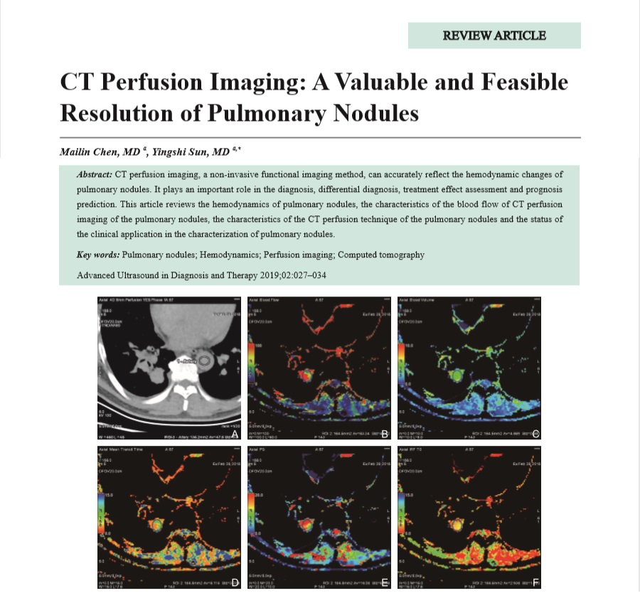
CT perfusion imaging, a non-invasive functional imaging method, can accurately reflect the hemodynamic changes of pulmonary nodules. It plays an important role in the diagnosis, differential diagnosis, treatment effect assessment and prognosis prediction. This article reviews the hemodynamics of pulmonary nodules, the characteristics of the blood flow of CT perfusion imaging of the pulmonary nodules, the characteristics of the CT perfusion technique of the pulmonary nodules and the status of the clinical application in the characterization of pulmonary nodules.
Key words: Pulmonary nodules; Hemodynamics; Perfusion imaging; Computed tomography
Chen, MD Mai-lin , Sun, MD Ying-shi . CT Perfusion Imaging: A Valuable and Feasible Resolution of Pulmonary Nodules[J]. ADVANCED ULTRASOUND IN DIAGNOSIS AND THERAPY, 2019 , 3(2) : 27 -34 . DOI: 10.37015/AUDT.2019.190806
| [1] | Hanahan D, Weinberg RA. Hallmarks of cancer: the next generation. Cell 2011; 14:646-74. |
| [2] | Folkman J. Tumor angiogenesis: therapeutic implications. N Engl J Med 1971; 285:1182-6. |
| [3] | Janning M, Loges S. Anti-Angiogenics: Their Value in Lung Cancer Therapy. Oncol Res Treat 2018; 41:172-180. |
| [4] | Gummadi S, Eisenbrey J, Li JZ, Li ZJ, Forsberg F, Lyshchik A, et al. Advances in modern clinical ultrasound. AUDT 2018; 2:51-63. |
| [5] | Chang YC, Yu CJ, Chen CM, Hu FC, Hsu HH, Tseng WY, et al. Dynamic contrast-enhanced MRI in advanced nonsmall-cell lung cancer patients treated with first-line bevacizumab, gemcitabine, and cisplatin. J Magn Reson Imaging 2012; 36:387-96. |
| [6] | Choyke PL, Dwyer AJ, Knopp MV. Functional tumor imaging with dynamic contrast-enhanced magnetic resonance imaging. J Magn Reson Imaging 2003; 17:509-20. |
| [7] | Pishko GL, Muldoon LL, Pagel MA, Schwartz DL, Neuwelt EA. Vascular endothelial growth factor blockade alters magnetic resonance imaging biomarkers of vascular function and decreases barrier permeability in a rat model of lung cancer brain metastasis. Fluids Barriers CNS 2015; 12:5. |
| [8] | Koenigkam-Santos M, Optazaite E, Sommer G, Safi S, Heussel CP, Kauczor HU, et al. Contrast-enhanced magnetic resonance imaging of pulmonary lesions: description of a technique aiming clinical practice. Eur J Radiol 2015; 84:185-192. |
| [9] | Broncano J, Luna A, Sánchez-González J, Alvarez-Kindelan A, Bhalla S. Functional MR Imaging in Chest Malignancies. Magn Reson Imaging Clin N Am 2016; 24:135-155. |
| [10] | Mirsadraee S, van Beek EJ. Functional imaging: computed tomography and MRI. Clin Chest Med 2015; 36:349-63. |
| [11] | Ohno Y, Nishio M, Koyama H, Fujisawa Y, Yoshikawa T, Matsumoto S, et al. Comparison of quantitatively analyzed dynamic area-detector CT using various mathematic methods with FDG PET/CT in management of solitary pulmonary nodules. AJR Am J Roentgenol 2013; 200:W593-602. |
| [12] | Ohno Y, Nishio M, Koyama H, Seki S, Tsubakimoto M, Fujisawa Y, et al. Solitary pulmonary nodules: Comparison of dynamic first-pass contrast-enhanced perfusion area-detector CT, dynamic first-pass contrast-enhanced MR imaging, and FDG PET/CT. Radiology 2015; 274:563-75. |
| [13] | Ohno Y, Koyama H, Fujisawa Y, Yoshikawa T, Seki S, Sugihara N, et al. Dynamic contrast-enhanced perfusion area detector CT for non-small cell lung cancer patients: Influence of mathematical models on early prediction capabilities for treatment response and recurrence after chemoradiotherapy. Eur J Radiol 2016; 85:176-186. |
| [14] | Ohno Y, Koyama H, Matsumoto K, Onishi Y, Takenaka D, Fujisawa Y, et al. Differentiation of malignant and benign pulmonary nodules with quantitative first-pass 320-detector row perfusion CT versus FDG PET/CT. Radiology 2011; 258:599-609. |
| [15] | Chae EJ, Song JW, Seo JB, Krauss B, Jang YM, Song KS. Clinical utility of dual-energy CT in the evaluation of solitary pulmonary nodules: initial experience. Radiology 2008; 249:671-81. |
| [16] | Purdie TG, Henderson E, Lee TY. Functional CT imaging of angiogenesis in rabbit VX2 soft-tissue tumour. Phys Med Biol 2001; 46:3161-75. |
| [17] | Miles KA. Perfusion CT for the assessment of tumour vascularity: which protocol? Br J Radiol 2003; 76 Spec No 1: S36-42. |
| [18] | Petralia G, Bonello L, Viotti S, Preda L, d'Andrea G, Bellomi M. CT perfusion in oncology: how to do it. Cancer Imaging 2010; 10:8-19. |
| [19] | Kambadakone AR, Sahani DV. Body perfusion CT: technique, clinical applications, and advances. Radiol Clin North Am 2009; 47:161-78. |
| [20] | Miles KA, Lee TY, Goh V, Klotz E, Cuenod C, Bisdas S, et al. Current status and guidelines for the assessment of tumour vascular support with dynamic contrast-enhanced computed tomography. Eur Radiol 2012; 22:1430-41. |
| [21] | Tacelli N, Remy-Jardin M, Copin MC, Scherpereel A, Mensier E, Jaillard S, et al. Assessment of non-small cell lung cancer perfusion: pathologic-CT correlation in 15 patients. Radiology 2010; 257:863-71. |
| [22] | Ebos JM, Kerbel RS. Antiangiogenic therapy: impact on invasion, disease progression, and metastasis. Nat Rev Clin Oncol 2011; 8:210-21. |
| [23] | Figueiras RG, Padhani AR, Goh VJ, Vilanova JC, González SB, Martín CV, et al. Novel oncologic drugs: what they do and how they affect images. Radiographics 2011; 31:2059-91. |
| [24] | Kiessling F, Boese J, Corvinus C, Ederle JR, Zuna I, Schoenberg SO, et al. Perfusion CT in patients with advanced bronchial carcinomas: a novel chance for characterization and treatment monitoring? Eur Radiol 2004; 14:1226-33. |
| [25] | Li Y, Yang ZG, Chen TW, Yu JQ, Sun JY, Chen HJ. First-pass perfusion imaging of solitary pulmonary nodules with 64-detector row CT: comparison of perfusion parameters of malignant and benign lesions. Br J Radiol 2010; 83:785-90. |
| [26] | Sitartchouk I, Roberts HC, Pereira AM, Bayanati H, Waddell T, Roberts TP. Computed tomography perfusion using first pass methods for lung nodule characterization. Invest Radiol 2008; 43:349-58. |
| [27] | Shan F, Zhang Z, Xing W, Qiu J, Yang S, Wang J, et al. Differentiation between malignant and benign solitary pulmonary nodules: use of volume first-pass perfusion and combined with routine computed tomography. Eur J Radiol 2012; 81:3598-605. |
| [28] | Shu SJ, Liu BL, Jiang HJ. Optimization of the scanning technique and diagnosis of pulmonary nodules with first-pass 64-detector-row perfusion VCT. Clin Imaging 2013; 37:256-64. |
| [29] | Ma SH, Le HB, Jia BH, Wang ZX, Xiao ZW, Cheng XL, et al. Peripheral pulmonary nodules: relationship between multi-slice spiral CT perfusion imaging and tumor angiogenesis and VEGF expression. BMC Cancer 2008; 8:186. |
| [30] | Lee YH, Kwon W, Kim MS, Kim YJ, Lee MS, Yong SJ, et al. Lung perfusion CT: the differentiation of cavitary mass. Eur J Radiol 2010; 73:59-65. |
| [31] | Wang G, Zhang C, Li M, Deng K, Li W. Preliminary application of high-definition computed tomographic Gemstone Spectral Imaging in lung cancer. J Comput Assist Tomogr 2014; 38:77-81. |
| [32] | Xiong Z, Liu JK, Hu CP, Zhou H, Zhou ML, Chen W. Role of immature microvessels in assessing the relationship between CT perfusion characteristics and differentiation grade in lung cancer. Arch Med Res 2010; 41:611-7. |
| [33] | Huellner MW, Collen TD, Gut P, Winterhalder R, Pauli C, Diebold J, et al. Multiparametric PET/CT-perfusion does not add significant additional information for initial staging in lung cancer compared with standard PET/CT. EJNMMI Res 2014; 4:6. |
| [34] | Wang J, Wu N, Cham MD, Song Y. Tumor response in patients with advanced non-small cell lung cancer: perfusion CT evaluation of chemotherapy and radiation therapy. AJR Am J Roentgenol 2009; 193:1090-6. |
| [35] | Hegenscheid K, Behrendt N, Rosenberg C, Kuehn JP, Ewert R, Hosten N, et al. Assessing early vascular changes and treatment response after laser-induced thermotherapy of pulmonary metastases with perfusion CT: initial experience. AJR Am J Roentgenol 2010; 194:1116-23. |
| [36] | Fraioli F, Vetere S, Anile M, Venuta F. Computed tomography perfusion: a new method to evaluate response to therapy in lung cancer. J Thorac Oncol 2011; 6:1599-600. |
| [37] | Ng CS, Chandler AG, Wei W, Anderson EF, Herron DH, Charnsangavej C, et al. Reproducibility of perfusion parameters obtained from perfusion CT in lung tumors. AJR Am J Roentgenol 2011; 197:113-21. |
| [38] | Lind JS, Meijerink MR, Dingemans AM, van Kuijk C, Ollers MC, de Ruysscher D, et al. Dynamic contrast-enhanced CT in patients treated with sorafenib and erlotinib for non-small cell lung cancer: a new method of monitoring treatment? Eur Radiol 2010; 20:2890-8. |
| [39] | Bellomi M, Viotti S, Preda L, D'Andrea G, Bonello L, Petralia G. Perfusion CT in solid body-tumours. Part II: Clinical applications and future development. Radiol Med 2010; 115:858-74. |
| [40] | Fraioli F, Anzidei M, Zaccagna F, Mennini ML, Serra G, Gori B, et al. Whole-tumor perfusion CT in patients with advanced lung adenocarcinoma treated with conventional and antiangiogenetic chemotherapy: initial experience. Radiology 2011; 259:574-82. |
| [41] | Fraioli F, Anzidei M, Serra G, Liberali S, Fiorelli A, Zaccagna F, et al. Whole-tumour CT-perfusion of unresectable lung cancer for the monitoring of anti-angiogenetic chemotherapy effects. Br J Radiol 2013; 86(1029):20120174. |
| [42] | Ohno Y, Fujisawa Y, Koyama H, Kishida Y, Seki S, Sugihara N, et al. Dynamic contrast-enhanced perfusion area-detector CT assessed with various mathematical models: Its capability for therapeutic outcome prediction for non-small cell lung cancer patients with chemoradiotherapy as compared with that of FDG-PET/CT. Eur J Radiol 2017; 86:83-91. |
| [43] | Lissoni P, Fugamalli E, Malugani F, Ardizzoia A, Secondino S, Tancini G, et al. Chemotherapy and angiogenesis in advanced cancer: vascular endothelial growth factor (VEGF) decline as predictor of disease control during taxol therapy in metastatic breast cancer. Int J Biol Markers 2000; 15:308-11. |
| [44] | Jain RK. Normalization of tumor vasculature: an emerging concept in antiangiogenic therapy. Science 2005; 307:58-62. |
| [45] | Qiao PG, Zhang HT, Zhou J, Li M, Ma JL, Tian N, et al. Early evaluation of targeted therapy effectiveness in non-small cell lung cancer by dynamic contrast-enhanced CT. Clin Transl Oncol 2016; 18:47-57. |
| [46] | Garcia-Barros M, Paris F, Cordon-Cardo C, Lyden D, Rafii S, Haimovitz-Friedman A, Fuks Z, et al. Tumor response to radiotherapy regulated by endothelial cell apoptosis. Science 2003; 300:1155-9. |
| [47] | Brix G, Lechel U, Nekolla E, Griebel J, Becker C. Radiation protection issues in dynamic contrast-enhanced (perfusion) computed tomography. Eur J Radiol 2015; 84:2347-58. |
/
| 〈 |
|
〉 |
Share: WeChat
Copyright ©2018 Advanced Ultrasound in Diagnosis and Therapy
|
 Advanced Ultrasound in Diagnosis and Therapy (AUDT)
is licensed under a Creative Commons Attribution 4.0 International License.
Advanced Ultrasound in Diagnosis and Therapy (AUDT)
is licensed under a Creative Commons Attribution 4.0 International License.