
ADVANCED ULTRASOUND IN DIAGNOSIS AND THERAPY >
Potential Threat of Tracheal Diverticulum to Thermal Ablation Treatment of Thyroid Nodule
Received date: 2018-10-08
Online published: 2019-03-30
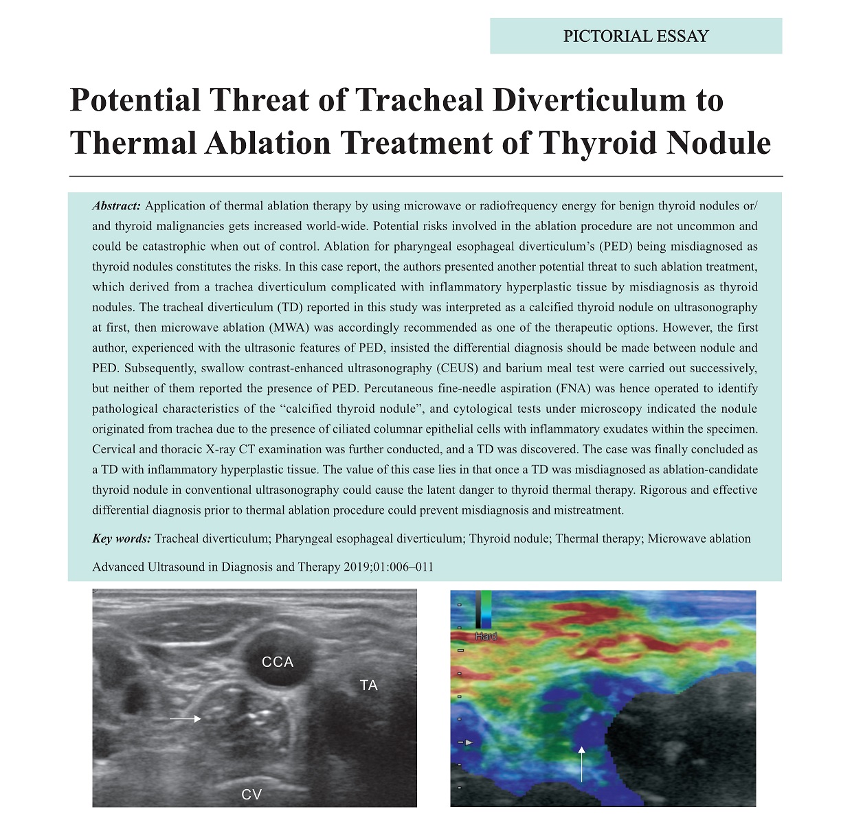
Application of thermal ablation therapy by using microwave or radiofrequency energy for benign thyroid nodules or/ and thyroid malignancies gets increased world-wide. Potential risks involved in the ablation procedure are not uncommon and could be catastrophic when out of control. Ablation for pharyngeal esophageal diverticulum’s (PED) being misdiagnosed as thyroid nodules constitutes the risks. In this case report, the authors presented another potential threat to such ablation treatment, which derived from a trachea diverticulum complicated with inflammatory hyperplastic tissue by misdiagnosis as thyroid nodules. The tracheal diverticulum (TD) reported in this study was interpreted as a calcified thyroid nodule on ultrasonography at first, then microwave ablation (MWA) was accordingly recommended as one of the therapeutic options. However, the first author, experienced with the ultrasonic features of PED, insisted the differential diagnosis should be made between nodule and PED. Subsequently, swallow contrast-enhanced ultrasonography (CEUS) and barium meal test were carried out successively, but neither of them reported the presence of PED. Percutaneous fine-needle aspiration (FNA) was hence operated to identify pathological characteristics of the “calcified thyroid nodule”, and cytological tests under microscopy indicated the nodule originated from trachea due to the presence of ciliated columnar epithelial cells with inflammatory exudates within the specimen. Cervical and thoracic X-ray CT examination was further conducted, and a TD was discovered. The case was finally concluded as a TD with inflammatory hyperplastic tissue. The value of this case lies in that once a TD was misdiagnosed as ablation-candidate thyroid nodule in conventional ultrasonography could cause the latent danger to thyroid thermal therapy. Rigorous and effective differential diagnosis prior to thermal ablation procedure could prevent misdiagnosis and mistreatment.
Zhang, MD Jianquan , Yan, MD Lei , Diao, MD Zongping , Chen, MD Hongqiong , Cheng, MD Jie . Potential Threat of Tracheal Diverticulum to Thermal Ablation Treatment of Thyroid Nodule[J]. ADVANCED ULTRASOUND IN DIAGNOSIS AND THERAPY, 2019 , 3(1) : 6 -11 . DOI: 10.37015/AUDT.2019.190802
| [1] | Tanrivermis Sayit A, Elmali M, Saglam D, Celenk C. The diseases of airway-tracheal diverticulum: a review of the literature. J Thorac Dis 2016; 8:E1163-E1167. |
| [2] | Kurt A, Sayit AT, Ipek A, Tatar IG. A multi detector computed tomography survey of tracheal diverticulum. Eurasian J Med 2013; 45:145-148. |
| [3] | Teng D, Sui G, Liu C, Wang Y, Xia Y, Wang H. Long-term efficacy of ultrasound-guided low power microwave ablation for the treatment of primary papillary thyroid microcarcinoma: a 3-year follow-up study. J Cancer Res Clin Oncol 2018; 144:771-779. |
| [4] | Che Y, Jin S, Shi C, Wang L, Zhang X, Li Y, et al. Treatment of Benign Thyroid Nodules: Comparison of Surgery with Radiofrequency Ablation. AJNR Am J Neuroradiol 2015; 36:1321-1325. |
| [5] | Hamidi O, Callstrom MR, Lee RA, Dean D, Castro MR, Morris JC, et al. Outcomes of Radiofrequency Ablation Therapy for Large Benign Thyroid Nodules: A Mayo Clinic Case Series. Mayo Clin Proc 2018; 93:1018-1025. |
| [6] | Li J, Liu Y, Liu J, Qian L. Ultrasound-guided percutaneous microwave ablation versus surgery for papillary thyroid microcarcinoma. Int J Hyperthermia 2018; 34:653-659. |
| [7] | Kim JH, Baek JH, Sung JY, Min HS, Kim KW, Hah JH, et al. Radiofrequency ablation of low-risk small papillary thyroidcarcinoma: preliminary results for patients ineligible for surgery. Int J Hyperthermia 2017;22; 33:212-219. |
| [8] | Achille G, Castellana M, Russo S, Montepara M, Giagulli VA, Triggiani V. Zenker Diverticulum: A Potential Pitfall in Thyroid Ultrasound Evaluation: A Case Report and Systematic Review of Literature. Endocr Metab Immune Disord Drug Targets 2019; 19:95-99. |
| [9] | Lixin J, Bing H, Zhigang W, Binghui Z. Sonographic diagnosis features of Zenker diverticulum. Eur J Radiol 2011; 80:e13-e19. |
| [10] | Zhang JQ, Yan L, Chen HQ. Microwave ablation of thyroid nodules complicated with Zenker's diverticulum: Case report. Chin J Interv Imaging Ther 2018; 15:48. |
| [11] | Bagheri R, Maddah G, Mashhadi MR, Haghi SZ, Tavassoli A, Ghamari MJ, et al. Esophageal diverticula: Analysis of 25 cases. Asian Cardiovasc Thorac Ann 2014; 22:583-587. |
| [12] | Cui XW, Ignee A, Baum U, Dietrich CF. Feasibility and usefulness of using swallow contrast-enhanced ultrasound to diagnose Zenker's diverticulum: preliminary results. Ultrasound Med Biol 2015; 41:975-981. |
| [13] | Zhang JQ, Chen HQ, Yan L. Potential threat of pharyngo-esophageal diverticulum to thermal ablation treatment of thyroid nodule and rapid differential diagnosis through swallow contrast-enhanced ultrasonography. Chin J Ultrasonogr 2019; 28:35-39. |
| [14] | Zhang JQ. High-frequency contrast-enhanced ultrasound in evaluation of perfusion of superficial lesions: limitations and countermeasures. Acad J Sec Mil Med Univ 2007; 28:1193-1196. |
| [15] | Bushan K, Sharma S, Verma H. A review of thymic tumors. Indian J Surg Oncol 2013; 4:112-116. |
/
| 〈 |
|
〉 |
Share: WeChat
Copyright ©2018 Advanced Ultrasound in Diagnosis and Therapy
|
 Advanced Ultrasound in Diagnosis and Therapy (AUDT)
is licensed under a Creative Commons Attribution 4.0 International License.
Advanced Ultrasound in Diagnosis and Therapy (AUDT)
is licensed under a Creative Commons Attribution 4.0 International License.