
Image Features-based Learning Effectively Improves Inter-Observer Agreement for Beginners in Evaluating Thyroid Nodule with Ultrasound
Received date: 2018-10-10
Online published: 2019-03-30
Objective: Thyroid nodules are a common medical problem in China and in other parts of the world. Many guidelines use ultrasound (US) as the first choice for evaluating thyroid nodules. A major limitation of US is operator dependency, resulting in a variety of discrepancies in diagnosing thyroid nodules in the literatures. Risk stratification of thyroid nodules is based on the patterns of US features in the 2015 American Thyroid Association (ATA) management guidelines for adult patients with thyroid nodules. We hypothesize that special training targeting features recognition may increase the inter-observer agreement.
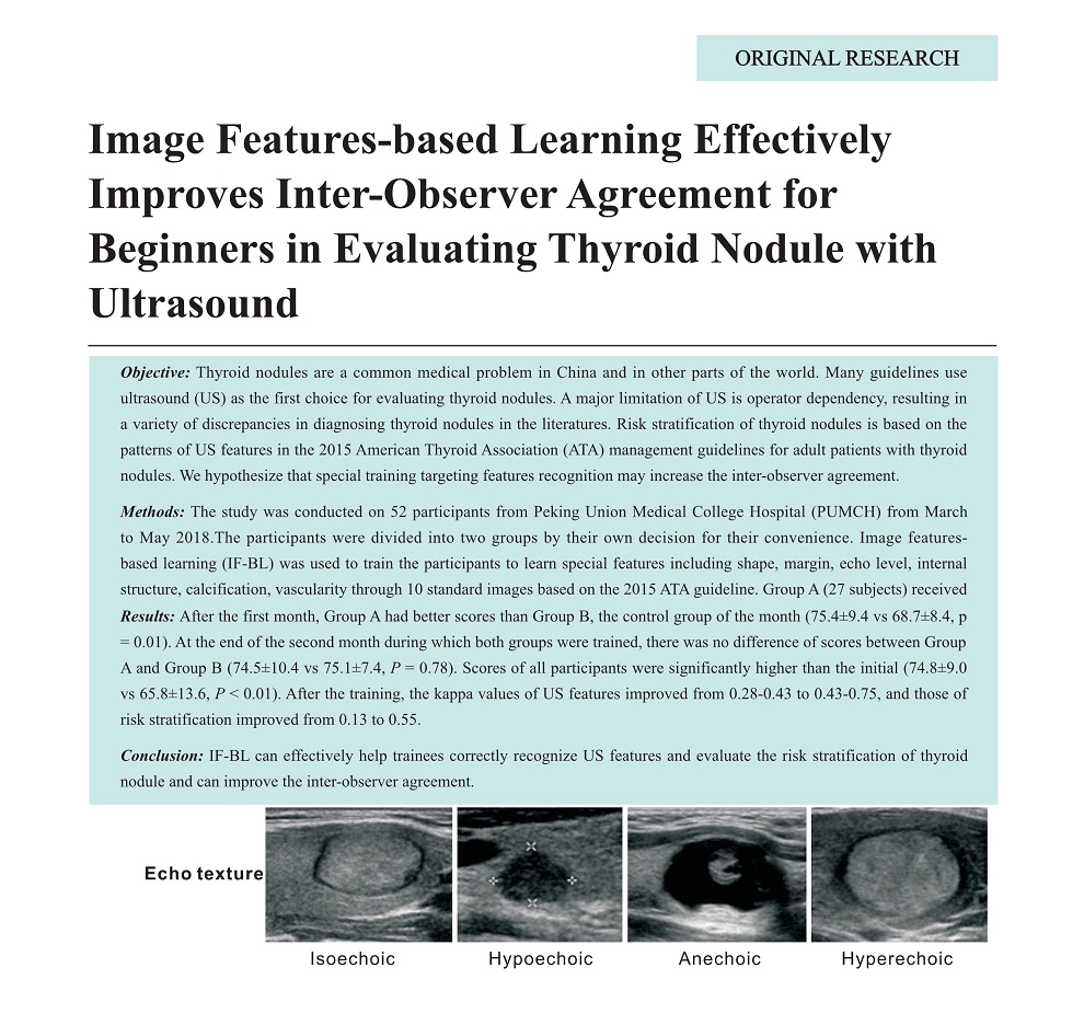
Methods: The study was conducted on 52 participants from Peking Union Medical College Hospital (PUMCH) from March to May 2018.The participants were divided into two groups by their own decision for their convenience. Image featuresbased learning (IF-BL) was used to train the participants to learn special features including shape, margin, echo level, internal structure, calcification, vascularity through 10 standard images based on the 2015 ATA guideline. Group A (27 subjects) received IF-BL during the first month, and Group B (25 subjects) received IF-BL during the second month. All participants evaluated US features and risk stratification in 60 US images of 20 thyroid nodules before and after the training. The test results were graded by a teaching assistant according to the rule of 0.5 points assigned to every feature and 2 points assigned to risk stratification, with a total of 100 points. Inter-observer agreements of US features and risk stratification were assessed and compared before and after the training.
Results: After the first month, Group A had better scores than Group B, the control group of the month (75.4±9.4 vs 68.7±8.4, p = 0.01). At the end of the second month during which both groups were trained, there was no difference of scores between Group A and Group B (74.5±10.4 vs 75.1±7.4, P = 0.78). Scores of all participants were significantly higher than the initial (74.8±9.0 vs 65.8±13.6, P < 0.01). After the training, the kappa values of US features improved from 0.28-0.43 to 0.43-0.75, and those of risk stratification improved from 0.13 to 0.55.
Conclusion: IF-BL can effectively help trainees correctly recognize US features and evaluate the risk stratification of thyroid nodule and can improve the inter-observer agreement.
Key words: Thyroid nodule; Cancer; ATA; Training
Wang, MD Ying , Gao, MD Luying , Jiang, MD Yuxin , Pan, MD Hui , Zhao, MA Jun , Zhou, MA Xin , Wu, MM Qiong , Liu, MM Ruyu , Zhang, MD Bo . Image Features-based Learning Effectively Improves Inter-Observer Agreement for Beginners in Evaluating Thyroid Nodule with Ultrasound[J]. ADVANCED ULTRASOUND IN DIAGNOSIS AND THERAPY, 2019 , 3(1) : 1 -5 . DOI: 10.37015/AUDT.2019.190801
| [1] | Guo H, Sun M, He W, Chen H, Li W, Tang J, et al. The prevalence of thyroid nodules and its relationship with metabolic parameters in a Chinese community-based population aged over 40 years. Endocrine 2014; 45:230-5. |
| [2] | Hegedüs L. Clinical practice. The thyroid nodule. N Engl J Med 2014; 351:1764-71. |
| [3] | Tan GH, Gharib H. Thyroid incidentalomas: management approaches to nonpalpable nodules discovered incidentally on thyroid imaging. Ann Intern Med 1997; 126:226-31. |
| [4] | Leenhardt L, Erdogan MF, Hegedus L, Mandel SJ, Paschke R, Rago T, et al. 2013 European thyroid association guidelines for cervical ultrasound scan and ultrasound-guided techniques in the postoperative management of patients with thyroid cancer. Eur Thyroid J 2013; 2:147-59. |
| [5] | Perros P, Boelaert K, Colley S, Evans C, Evans RM, Gerrard Ba G, et al. Guidelines for the management of thyroid cancer. Clin Endocrinol (Oxf) 2014; 81:1-122. |
| [6] | Shin JH, Baek JH, Chung J, Ha EJ, Kim JH, Lee YH, et al. Ultrasonography diagnosis and imaging-based management of thyroid nodules: revised Korean society of thyroid radiology consensus statement and recommendations. Korean J Radiol 2016; 17:370-95. |
| [7] | Gharib H, Papini E, Paschke R, Duick DS, Valcavi R, Hegedüs L, et al. American Association of Clinical Endocrinologists, Associazione Medici Endocrinologi, and European Thyroid Association Medical guidelines for clinical practice for the diagnosis and management of thyroid nodules: executive summary of recommendations. EndocrPract 2016; 22:622-39. |
| [8] | Haugen BR, Alexander EK, Bible KC, Doherty GM, Mandel SJ, Nikiforov YE, et al. 2015 American Thyroid Association management guidelines for adult patients with thyroid nodules and differentiated thyroid cancer: the American Thyroid Association Guidelines Task Force on Thyroid Nodules and Differentiated Thyroid Cancer. Thyroid 2016; 26:1-133. |
| [9] | Moon WJ, Jung SL, Lee JH, Na DG, Baek JH, Lee YH, et al. Benign and malignant thyroid nodules: US differentiation--multicenter retrospective study. Radiology 2008; 247:762-70. |
| [10] | Park CS, Kim SH, Jung SL, Kang BJ, Kim JY, Choi JJ, et al. Observer variability in the sonographic evaluation of thyroid nodules. J Clin Ultrasound 2010; 38:287-93. |
| [11] | Choi SH, Kim EK, Kwak JY, Kim MJ, Son EJ. Interobserver and intraobserver variations in ultrasound assessment of thyroid nodules. Thyroid 2010; 20:167-72. |
| [12] | Srinivas MN, Amogh VN, Gautam MS, Prathyusha IS, Vikram NR, Retnam MK, et al. A prospective study to evaluate the reliability of thyroid imaging reporting and data system in differentiation between benign and malignant thyroid lesions. J Clin Image Sci 2016; 6:5. |
/
| 〈 |
|
〉 |
Share: WeChat
Copyright ©2018 Advanced Ultrasound in Diagnosis and Therapy
|
 Advanced Ultrasound in Diagnosis and Therapy (AUDT)
is licensed under a Creative Commons Attribution 4.0 International License.
Advanced Ultrasound in Diagnosis and Therapy (AUDT)
is licensed under a Creative Commons Attribution 4.0 International License.