
ADVANCED ULTRASOUND IN DIAGNOSIS AND THERAPY >
The Correlation between Traditional Ultrasound Features and the Expression of Estrogen Receptor, Progesterone Receptor, Human Epidermal Growth Factor Receptor-2, and Ki-67 in Breast Carcinoma
Received date: 2018-10-23
Online published: 2018-12-29
Objective: To evaluate the correlation between traditional ultrasound and the expression of estrogen receptor (ER), progesterone receptor (PR), human epidermal growth factor receptor-2 (Her-2), and Ki-67 expression in breast carcinoma.
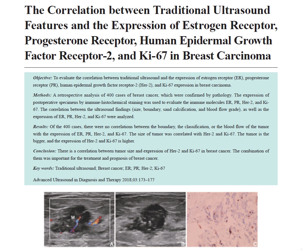
Methods: A retrospective analysis of 400 cases of breast cancer, which were confirmed by pathology. The expression of postoperative specimens by immune-histochemical staining was used to evaluate the immune molecules ER, PR, Her-2, and Ki-67. The correlation between the ultrasound findings (size, boundary, sand calcification, and blood flow grade), as well as the expression of ER, PR, Her-2, and Ki-67 were analyzed.
Results: Of the 400 cases, there were no correlations between the boundary, the classification, or the blood flow of the tumor with the expression of ER, PR, Her-2, and Ki-67. The size of tumor was correlated with Her-2 and Ki-67. The tumor is the bigger, and the expression of Her-2 and Ki-67 is higher.
Conclusion: There is a correlation between tumor size and expression of Her-2 and Ki-67 in breast cancer. The combination of them was important for the treatment and prognosis of breast cancer.
Key words: Traditional ultrasound; Breast cancer; ER; PR; Her-2; Ki-67
Wang, MM Ping , Li Junfan , Yue, MM Wensheng , Li, MM Wenyan , Luo, MM Yuqun . The Correlation between Traditional Ultrasound Features and the Expression of Estrogen Receptor, Progesterone Receptor, Human Epidermal Growth Factor Receptor-2, and Ki-67 in Breast Carcinoma[J]. ADVANCED ULTRASOUND IN DIAGNOSIS AND THERAPY, 2018 , 2(3) : 173 -177 . DOI: 10.37015/AUDT.2018.180820
| [1] | Lialiaris TS, Kouskoukis A, Georqiou G, Tripsianis G, Fiska A, Giatromanolaki A, et al.Expression of 6 common antigenic markers in invasive ductal breast carcinoma: potential clinical implications. Appl Immunohistochem Mol Morphol 2011; 19:106-11. |
| [2] | Park H, Chang SK, Kim JY, Lee BM, Shin HS. Risk factors for distant metastasis as a primary site of treatment failure in early-stage breast cancer. Chonnam Med J 2014; 50:96-101. |
| [3] | Munzone E, Botteri E, Sciandivasci A, Curigliano G, Nolè F, Mastropasqua M, et al.Prognostic value of Ki-67 labeling index in patients with node-negative, triple-negative breast cancer. Breast Cancer Res Treat 2012; 134:277-82. |
| [4] | Rhee J, Han SW, Oh DY, Kim JH, Im SA, Han W, et al.The clinicopathologic characteristics and prognostic significance of triple-negativity in node-negative breast cancer. BMC Cancer 2008; 23:307-14. |
| [5] | Ito Y, Miyauchi A, Inoue H, Fukushima M, Kihara M, Hiqashiyama T, et al.An observational trial for papillary thyroid microcarcinoma in Japanese patients. World J Surg 2010; 34:28-35. |
| [6] | Ayadi L, Khabir A, Amouri H, Karray S, Dammak A, Guermazi M, et al.Correlation of HER-2 over-expression with clinico-pathological parameters in Tunisian breast carcinoma. World J Surg Oncol 2008; 6:112-9. |
| [7] | Francis GD, Dimech M, Giles L, Hopkins A. Frequency and reliability of oestrogen receptor, progesterone receptor and HER2 in breast carcinoma determined by immunohistochemistry in Australasia: results of the RCPA Quality Assurance Program. J Clin Pathol 2007; 60:1277-83. |
| [8] | Liu C, Zhang H, Shuang C, Lu Y, Jin F, Xu H, Lu P. Alterations of ER, PR, HER-2/neu, and P53 protein expression in ductal breast carcinoma and clinical implications. Med Oncol 2010; 27:747-52. |
| [9] | Leong AS, Zhang Z. The changing role of pathology in breast cancer diagnosis and treatment. Pathobiology 2011; 78:99-114. |
| [10] | Goldhirsch A, Ingle JN, Gelber RD, Coates AS, Thürlimann B, Senn HJ, et al. Thresholds for therapies: highlights of the St Gallen International Expert Consensus on the primary therapy of early breast cancer 2009. Ann Oncol 2009; 20:1319-29. |
| [11] | Thakkar JP, Mehta DG. A review of an unfavorable subset of breast cancer: estrogen receptor positive progesterone receptor negative. Oncologist 2011; 16:276-85. |
| [12] | Gutierrez C, Schiff R. HER2: biology, detection, and clinical implications. Arch Pathol Lab Med 2011; 135:55-62. |
| [13] | Camerini A, Donati S, Viacava P, Siclari O, Puccetti C, Tartarelli G, et al. Evaluation of HER2 and p53 expression in predicting response to docetaxel-based first-line chemotherapy in advanced breast cancer. J Exp Clin Cancer Res 2011; 30:38-45. |
| [14] | Nishimura R, Osako T, Okumura Y, Tashima R, Toyozumi Y, Arima N. Changes in the ER, PgR, HER2, P53 and Ki-67 biological markers between primary and recurrent breast cancer: discordance rates and prognosis. World J Surg Oncol 2011; 9:131-7. |
| [15] | Bardou VJ, Arpino G, Elledge RM, Osborne CK, Clark GM. Progesterone receptor status significantly improves outcome prediction over estrogen receptor status alone for adjuvant endocrine therapy in two large breast cancer databases. J Clin Oncol 2003; 21:1973-9. |
| [16] | Hwang SH, Park DJ, Jee YS, Kim HH, Lee HJ, Yang HK, et al.Risk factors for operative complications in elderly patients during laparoscopy-assisted gastrectomy. J Am Coll Surg 2009; 208:186-92. |
| [17] | Paradiso A, Manqia A, Barletta A, Marzullo F, Ventrella V, Racanelli A, et al. Mammography and morphobiologic characteristics of human breast cancer. Tumori 1993; 79:422-6. |
| [18] | Lamb PM, Perry NM, Vinnicombe SJ, Wells CA. Correlation between ultrasound characteristics, mammographic findings and histological grade in patients with invasive ductal carcinoma of the breastl. Clin Radio 2000; 55:40-4. |
| [19] | de Azambuja E, Cardoso F, de Castro G Jr, Colozza M, Mano MS, Durbecq V ,et al. Ki-67 as prognostic marker in early breast cancer: a meta-analysis of published studies involving 12155 patients.Br J Cancer 2007; 96:1504-13. |
| [20] | Prebil LA, Ereman RR, Powell MJ, Jamshidian F, Kerlikowske K, Shepherd JA, et al. First pregnancy events and future breast density: modification by age at first pregnancy and specific VEGF and IGF1R gene variants. Cancer Causes Control 2014; 25:859-68. |
| [21] | Avadi L, Khabir A, Amouri H, Karray S, Dammak A, Guermazi M, et al. Correlation of HER-2 over-expression with clinico-pathological parameters in Tunisian breast carcinoma. World J Surg Oncol 2008; 22:112-8. |
| [22] | Park H, Chang SK, Kim JY, Lee BM, Shin HS. Risk factors for distant metastasis as a primary site of treatment failure in early-stage breast cancer. Chonnam Med J 2014; 50:96-101. |
| [23] | Ivkovic-Kapicl T, Knezevic-Usaj S, Djilas-Ivanovic D, Panjkovic M. Correlation of HER-2/neu protein overexpression with other prognostic and predictive factors in invasive ductal breast cancer. In Vivo 2007; 21:673-8. |
/
| 〈 |
|
〉 |
Share: WeChat
Copyright ©2018 Advanced Ultrasound in Diagnosis and Therapy
|
 Advanced Ultrasound in Diagnosis and Therapy (AUDT)
is licensed under a Creative Commons Attribution 4.0 International License.
Advanced Ultrasound in Diagnosis and Therapy (AUDT)
is licensed under a Creative Commons Attribution 4.0 International License.