
ADVANCED ULTRASOUND IN DIAGNOSIS AND THERAPY >
Analysis of Characteristics Microvessel Density of Thyroid Malignant and Benign Nodules on Contrast-Enhanced Ultrasonography
Received date: 2018-10-29
Online published: 2018-12-29
Objective: To analyze the contrast-enhanced ultrasonography (CEUS) characteristics and microvessel density (MVD) of thyroid carcinomas vs. benign thyroid lesions, and to determine the value of CEUS in the clinical diagnosis of thyroid carcinoma.
Methods: Forty-five patients (34 women, 11 men; mean age, 45.95 ± 12.20 years) with 57 thyroid nodules underwent preoperative CEUS with SonoVue (injected as a bolus via an elbow vein). The CEUS data were analyzed using QLAB quantitative analysis software. The enhancement patterns and quantitative parameters of thyroid carcinoma were evaluated.Additionally, immunohistochemistry was performed to evaluate MVD.
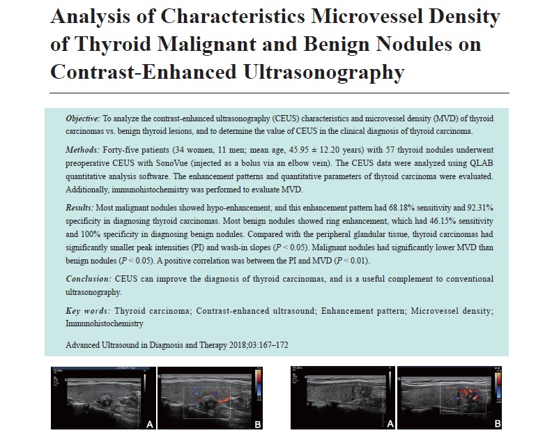
Results: Most malignant nodules showed hypo-enhancement, and this enhancement pattern had 68.18% sensitivity and 92.31% specificity in diagnosing thyroid carcinomas. Most benign nodules showed ring enhancement, which had 46.15% sensitivity and 100% specificity in diagnosing benign nodules. Compared with the peripheral glandular tissue, thyroid carcinomas had significantly smaller peak intensities (PI) and wash-in slopes (P < 0.05). Malignant nodules had significantly lower MVD than benign nodules (P < 0.05). A positive correlation was between the PI and MVD (P < 0.01).
Conclusion: CEUS can improve the diagnosis of thyroid carcinomas, and is a useful complement to conventional ultrasonography.
Liu, MD Jingjing , Liu, MD Liping , Zhang, MD Yanjing , Zhao,MD Yufang , Hao, MD Yanhong , Li, MD Tingting , , Huang, MD Xiaochun . Analysis of Characteristics Microvessel Density of Thyroid Malignant and Benign Nodules on Contrast-Enhanced Ultrasonography[J]. ADVANCED ULTRASOUND IN DIAGNOSIS AND THERAPY, 2018 , 2(3) : 167 -172 . DOI: 10.37015/AUDT.2018.180819
| [1] | Golden SH, Robinson KA, Saldanha I, Anton B, Ladenson PW. Prevalence and incidence of endocrine and metabolic disorders in the United States: a comprehensive review. J Clin Endocrinol Metab 2009; 94:1853-78. |
| [2] | Roti E, degli Uberti EC, Bondanelli M, Braverman LE . Thyroid papillary microcarcinoma: a descriptive and meta analysis study. Eur J Endocrinol 2008; 159:659-73. |
| [3] | Guth S, Theune U, Aberle J, Galach A, Bamberger CM. Very high prevalence of thyroid nodules detected by high frequency (13 MHz) ultrasound examination. Eur J Clin Invest 2009; 39:699-706. |
| [4] | Wiest PW, Hartshorne MF, Inskip PD, Crooks LA, Vela BS, Telepak RJ, et al. Thyroid palpation versus high-resolution thyroid ultrasonography in the detection of nodules. J Ultrasound Med 1998; 17:487-96. |
| [5] | Tomimori E, Pedrinola F, Cavaliere H, Knobel M, Medeiros-Neto G. Prevalence of incidental thyroid disease in a relatively low iodine intake area. Thyroid 1995; 5:273-6. |
| [6] | Brander A, Viikinkoski P, Nickels J, Kivisaari L. Thyroid gland: US screening in a random adult population. Radiology 1991; 181:683-7. |
| [7] | Tunbridge WM, Evered DC, Hall R, Appleton D, Brewis M, Clark F, et al. The spectrum of thyroid disease in a community: the Whickham Survey. Clin Endocrinol (Oxf) 1977; 7:481-93. |
| [8] | Fish SA, Langer JE, Mandel SJ. Sonographic imaging of thyroid nodules and cervical lymph nodes. Endocrinol Metab Clin N Am 2008; 37:401-17. |
| [9] | Burns PN, Wilson SR, Simpson DH. Pulse inversion imaging of liver blood flow: improved method for characterizing focal masses with microbubble contrast.Invest Radiol 2000; 35:58-71. |
| [10] | Burns PN, Wilson SR. Focal liver masses: enhancement patterns on contrast-enhanced images: concordance of US scans with CT scans and MR images. Radiology 2007; 242:162-74. |
| [11] | Minami Y , Kudo M: Ultrasound fusion imaging of hepatocellular carcinoma: A review of current evidence. Dig Dis 2014; 32:690-5. |
| [12] | Yu D, Han Y, Chen T: Contrast-enhanced ultrasound for differentiation of benign and malignant thyroid lesions: Meta.analysis. Otolaryngol Head Neck Surg 2014; 151:909-15. |
| [13] | Jiang J, Huang L, Zhang H, Ma W, Shang X, Zhou Q, et al: Contrast-enhanced sonography of thyroid nodules. J Clin Ultrasound 2015; 43:153-6. |
| [14] | Nemec U, Nemec SF, Novotny C, Weber M, Czerny C, Krestan CR: Quantitative evaluation of contrast-enhanced ultrasound after intravenous administration of a microbubble contrast agent for differentiation of benign and malignant thyroid nodules: Assessment of diagnostic accuracy. Eur Radiol 2012; 22:1357-65. |
| [15] | Agha A, Jung EM, Janke M, Hornung M, Georgieva M, Schlitt HJ, et al, Preoperative diagnosis of thyroid adenomas using high resolution contrast-enhanced ultrasound (CEUS). Clin Hemorheol Microcirc 2013; 55:403-9. |
| [16] | Massimo G, Claudia C, Stefano G, Barbara M, Enzo S, Eleonora M, et al, The use of semi-quantitative ultrasound elastosonography in combination with conventional ultrasonography and contrast-enhanced ultrasonography in the assessment of malignancy risk of thyroid nodules with indeterminate cytology. Thyroid Res 2014; 7:9. |
| [17] | Weidner N. Current pathologic methods for measuring intratumor-al microvessel density within breast carcinoma and other solidtumors. Breast Cancer Res Trest 1995; 36:169-80. |
| [18] | Sener E, Sipal S, Gündo?du C. Comparison of Microvessel Density with Prognostic Factors in Invasive Ductal Carcinomas of the Breast. Turkish Journal of Pathology 2016; 32:164-70. |
| [19] | Moon WJ, Baek JH, Jung SL, Kim DW, Kim EK, Kim JY, et al. Ultrasonography and the ultrasound-based management of thyroid nodules: consensus statement and recommendations. Korean J Radiol 2011; 12:1-14. |
| [20] | Zhang B, Jiang YX, Liu JB, Yang M, Dai Q, Zhu QL, et al. Utility of contrast-enhanced ultrasound for evaluation of thyroid nodules. Thyroid 2010; 20:51-7. |
| [21] | Bartolotta TV, Midiri M, Galia M, Runza G, Attard M, Savoia G, et al. Qualitative and quantitative evaluation of solitary thyroid nodules with contrast enhanced ultrasound: initial results. Eur Radiol 2006; 16:2234-41. |
| [22] | Moon HJ, Kwak JY, Kim MJ, Son EJ, Kim EK. Can vascularity at power Doppler US help predict thyroid malignancy? Radiology 2010; 255:260-9. |
| [23] | Deng J, Zhou P, Tian SM, Zhang L, Li JL, Qian Y. Comparison of Diagnostic Efficacy of Contrast-Enhanced Ultrasound, Acoustic Radiation Force Impulse Imaging, and Their Combined Use in Differentiating Focal Solid Thyroid Nodules. Plos One 2014; 9:e90674. |
| [24] | Strouthos C, Lampaskis M , Sboros V, McNeilly A. & Averkiou M. Indicatordilution models for the quantification of microvascular blood flow with bolus administration of ultrasound contrast agents. IEEE Trans Ultrason Ferroelectr Freq Control 2010; 57:1296-310. |
/
| 〈 |
|
〉 |
Share: WeChat
Copyright ©2018 Advanced Ultrasound in Diagnosis and Therapy
|
 Advanced Ultrasound in Diagnosis and Therapy (AUDT)
is licensed under a Creative Commons Attribution 4.0 International License.
Advanced Ultrasound in Diagnosis and Therapy (AUDT)
is licensed under a Creative Commons Attribution 4.0 International License.