
ADVANCED ULTRASOUND IN DIAGNOSIS AND THERAPY >
Accuracy of Prenatal Ultrasound in the Diagnosis of Isolated Fetal Cleft Palate in High-risk Patients
Received date: 2023-07-24
Accepted date: 2024-05-27
Online published: 2024-07-01
ObjectiveThe objective of this study was to develop a sonographic technique using two-dimensional (2D) markers for detecting isolated fetal cleft palate (no cleft lip) and to evaluate the ability of 2D and three-dimensional (3D) sonography to image the normal and abnormal palate.
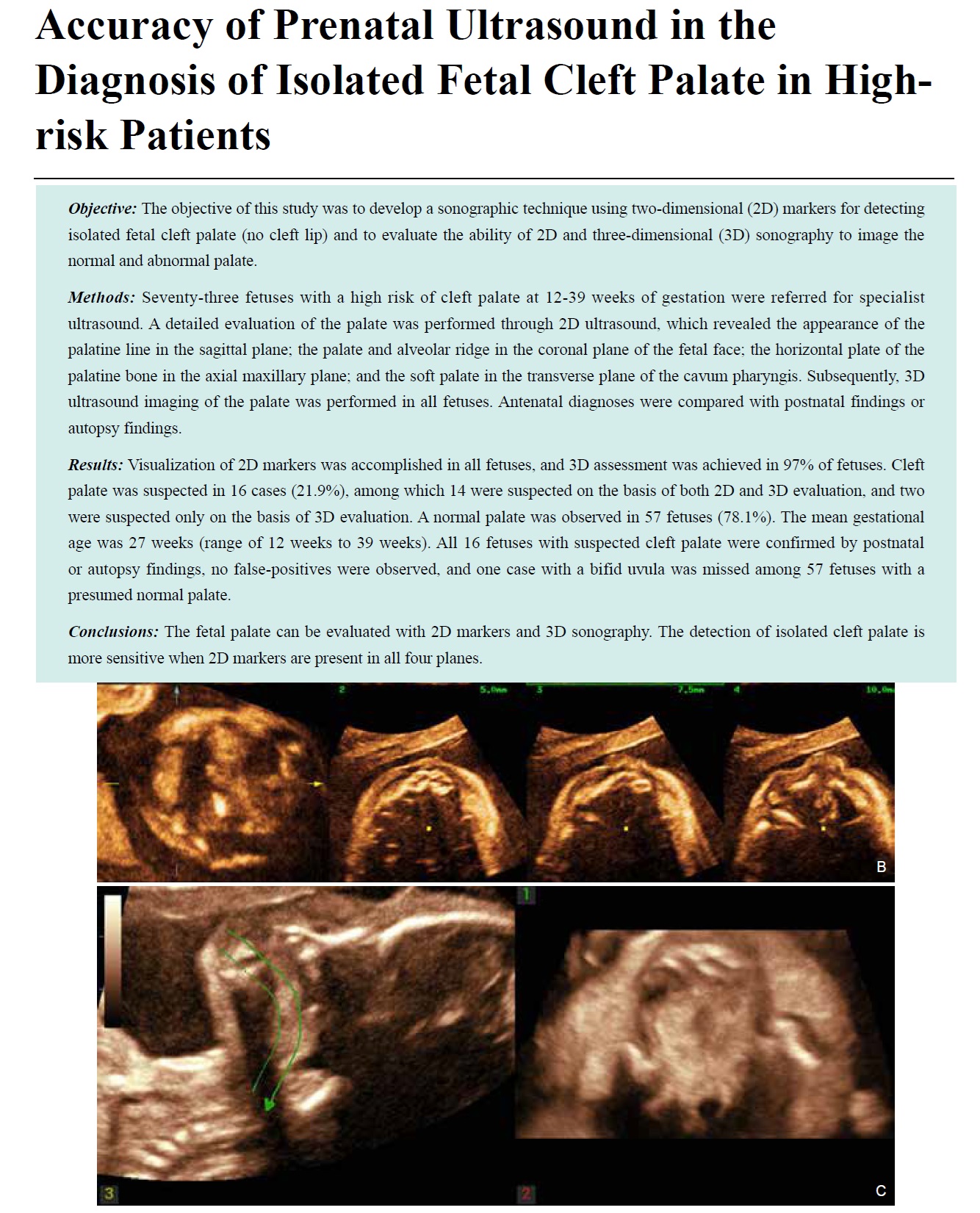
Methods Seventy-three fetuses with a high risk of cleft palate at 12-39 weeks of gestation were referred for specialist ultrasound. A detailed evaluation of the palate was performed through 2D ultrasound, which revealed the appearance of the palatine line in the sagittal plane; the palate and alveolar ridge in the coronal plane of the fetal face; the horizontal plate of the palatine bone in the axial maxillary plane; and the soft palate in the transverse plane of the cavum pharyngis. Subsequently, 3D ultrasound imaging of the palate was performed in all fetuses. Antenatal diagnoses were compared with postnatal findings or autopsy findings.
Results Visualization of 2D markers was accomplished in all fetuses, and 3D assessment was achieved in 97% of fetuses. Cleft palate was suspected in 16 cases (21.9%), among which 14 were suspected on the basis of both 2D and 3D evaluation, and two were suspected only on the basis of 3D evaluation. A normal palate was observed in 57 fetuses (78.1%). The mean gestational age was 27 weeks (range of 12 weeks to 39 weeks). All 16 fetuses with suspected cleft palate were confirmed by postnatal or autopsy findings, no false-positives were observed, and one case with a bifid uvula was missed among 57 fetuses with a presumed normal palate.
Conclusions The fetal palate can be evaluated with 2D markers and 3D sonography. The detection of isolated cleft palate is more sensitive when 2D markers are present in all four planes.
Hongmei Wu, MD , Shuqin Li, MD , Fengfeng Shi, MD , Yuxiu Gao, MD , Jiansheng Li, MD . Accuracy of Prenatal Ultrasound in the Diagnosis of Isolated Fetal Cleft Palate in High-risk Patients[J]. ADVANCED ULTRASOUND IN DIAGNOSIS AND THERAPY, 2024 , 8(2) : 57 -63 . DOI: 10.37015/AUDT.2024.230038
| [1] | Tonni G, Centini G, Rosignoli L. Prenatal screening for fetal face and clefting in a prospective study on low-risk population: can 3- and 4-dimensional ultrasound enhance visualization and d etection rate? Oral Surg Oral Med Oral Pathol Oral Radiol Endod 2005; 100:420-426. |
| [2] | Cunnigham ML. Three-dimensional ultrasonography is superior to 2-dimensional ultrasonography in the detection of orofacial clefts during the second trimester of pregnancy. J Evidence Based Dent Pract 2006; 6:278-279. |
| [3] | Wilhelm L, Borgers H. The “equals sign”: a novel marker in the diagnosis of fetal isolated cleft palate. Ultrasound Obstet Gynecol 2010; 36:439-444. |
| [4] | Sherer DM, Sokolovski M, Santoso PG, Dalloul M, Abulafia O. Nomograms of sonographic measurements throughout gestation of the fetal hard palate width, length and area. Ultrasound Obstet Gynecol 2004; 24:35-41. |
| [5] | Jones MC. Etiology of facial clefts: prospective evaluation of 428 patients. Cleft Palate J 1988; 25:16-20 |
| [6] | Stoll C, Alembik Y, Dott B, Roth MP. Associated malformations in cases with oral clefts. Cleft Palate Craniofac 2000; 37:41-47. |
| [7] | Grandjean H, Larroque D, Levi S. The performance of routine ultrasonographic screening of pregnancies in the Eurofetus Study. Am J Obstet Gynecol 1999; 181:446-454. |
| [8] | Clementi M, Tenconie R, Bianchi F, Stoll C. Evaluation of prenatal diagnosis of cleft lip with or without cleft palate and cleft palate by ultrasound: experience from 20 European registries. EUROSCAN study group. Prenat Diagn 2000; 20:870-875. |
| [9] | Shaikh D, Mercer NS, Sohan K, Kyle P, Soothill P. Prenatal diagnosis of cleft lip and palate. Br J Plastic Surg 2001; 54:288-289. |
| [10] | Offerdal K, Jebens N, Syvertsen T, Blaas HG, Johansen OJ, Eik-Nes SH. Prenatal ultrasound detection of facial clefts: a prospective study of 49 314 deliveries in a non-selected population in Norway. Ultrasound Obstet Gynecol 2008; 31:639-646. |
| [11] | Gillham JC, Anand S, Bullen PJ. Antenatal detection of cleft lip with or without cleft palate: incidence of associated chromosomal and structural anomalies. Ultrasound Obstet Gynecol 2009; 34:410-415. |
| [12] | Rotten D, Levaillant JM. Two- and three-dimensional sonographic assessment of the fetal face. 2. Analysis of cleft lip, alveolus and palate. Ultrasound Obstet Gynecol 2004; 24:402-411. |
| [13] | Sherer DM, Sokolovski M, Santoso PG, Dalloul M, Abulafia O. Nomo-grams of sonographic measurements throughout gestation of the fetal hard palate width, length and area. Ultrasound Obstet Gynecol 2004; 24:35-41. |
| [14] | Tonni G, Centini G, Rosignoli L. Prenatal screening for fetal face and clefting in a prospective study on low-risk population: can 3- and 4-dimensional ultrasound enhance visualization and detection rate? Oral Surg Oral Med Oral Pathol Oral Radiol Endod 2005; 100:420-426. |
| [15] | Clementi M, Tenconi R, Bianchi F, Stoll C. Evaluation of prenatal diagnosis of cleft lip with or without cleft palate by ultrasound: experience from 20 European registries. EUROSCAN Study Group. Prenat Diagn 2000; 20:870-875. |
| [16] | Offerdal K, Jebens N, Syvertsen T, Blaas HG, Johansen OJ, Eik-Nes SH. Prenatal ultrasound detection of facial clefts: a prospective study of 49,314 deliveries in a non-selected population in Norway. Ultrasound Obstet Gynecol 2008; 31:639-646. |
| [17] | Cash C, Set P, Colemann N. The accuracy of antenatal ultrasound in detection of facial clefts in a low-risk screening population. Ultrasound Obstet Gynecol 2001; 18:432-436. |
| [18] | Maarse W, Bergé SJ, Pistorius L. Diagnostic accuracy of transabdominal ultrasound in detecting prenatal cleft lip and palate: a systematic review. Ultrasound Obstet Gynecol 2010; 35:495-502. |
| [19] | Rotten D, Levaillant JM. Two- and three-dimensional sonographic assessment of the fetal face, 2: analysis of cleft lip, alveolus and palate. Ultrasound Obstet Gynecol 2004; 24:402-411. |
| [20] | Campbell S. Prenatal ultrasound examination of the secondary palate. Ultrasound Obstet Gynecol 2007; 29:124-127. |
| [21] | Tonni G, Centini G, Rosignoli L. Prenatal screening for fetal face and clefting in a prospective study on low-risk population: can 3- and 4-dimensional ultrasound enhance visualization and detection rate? Oral Surg Oral Med Oral Pathol Oral Radiol Endod 2005; 100:420-426. |
| [22] | Cunnigham ML. Three-dimensional ultrasonography is superior to 2-dimensional ultrasonography in the detection of orofacial clefts during the second trimester of pregnancy. J Evidence Based Dent Pract 2006; 6:278-279. |
| [23] | Martinez Ten P, Pedregosa JP, Santacruz B, Adiego B, Barrón E, Sepúlveda W. Three-dimensional ultrasound diagnosis of cleft palate: “reverse face”, “flipped face” or “oblique face”—which method is best? Ultrasound Obstet Gynecol 2009; 33:399-406. |
| [24] | Selvaraj Ravi L, Selvaraj D, Nity R, Senthilkumar M. First-Trimester Sonographic Evaluation of Palatine Clefts ANovelDiagnostic Approach. J Ultrasound Med 2017; 36:1397-1414. |
| [25] | Bergé SJ, Plath H, Van de Vondel PT, Appel T, Niederhagen B, J Von Lindern J, et al. Fetal cleft lip and palate: sonographic diagnosis, chromosomal abnormalities, associated anomalies and postnatal outcome in 70 fetuses. Ultrasound Obstet Gynecol 2001; 18:422-431. |
| [26] | Calzolari E, Bianchi F, Rubini M, Ritvanen A, Neville J. Epidemiology of cleft palate in Europe: implications for genetic research. Cleft Palate Craniofac J 2004; 41:244-249. |
| [27] | Ghi T, Perolo A, Banzi C, Contratti G, Valeri B, Savelli L, et al. Two-dimensional ultrasound is accurate in the diagnosis of fetal craniofacial malformation. Ultrasound Obstet Gynecol 2002; 19:543-551. |
| [28] | Rotten D, Levaillant JM. Two- and three-dimensional sonographic assessment of the fetal face. 1. A systematic analysis of the normal face. Ultrasound Obstet Gynecol 2004; 23:224-231. |
| [29] | Rotten D, Levaillant JM, Martinez H, Ducou le Pointe H, Vicaut E. The fetal mandible: a 2D and 3D sonographic approach to the diagnosis of retrognathia and micrognathia. Ultrasound Obstet Gynecol 2002; 19:122-130. |
| [30] | Perrotin F, Poncheville LM, Marret H, Paillet C, Lansac J, Body G. Chromosomal defects and associated malformations in fetal cleft lip with or without cleft palate. Eur J Obstet Gynecol Reprod Biol 2001; 99:19-24. |
| [31] | Campbell S, Lees CC. The three-dimensional reverse face (3DRF) view for the diagnosis of cleft palate. Ultrasound Obstet Gynecol 2003; 22:552-554. |
| [32] | Campbell S. 4D or not 4D: that is the question. Ultrasound Obstet Gynecol 2002; 19:1-4. |
/
| 〈 |
|
〉 |
Share: WeChat
Copyright ©2018 Advanced Ultrasound in Diagnosis and Therapy
|
 Advanced Ultrasound in Diagnosis and Therapy (AUDT)
is licensed under a Creative Commons Attribution 4.0 International License.
Advanced Ultrasound in Diagnosis and Therapy (AUDT)
is licensed under a Creative Commons Attribution 4.0 International License.