
ADVANCED ULTRASOUND IN DIAGNOSIS AND THERAPY >
Multimodal Vascular Ultrasound Findings in A Young Female with Internal Carotid Artery Dissection
Received date: 2021-11-04
Accepted date: 2022-04-22
Online published: 2023-03-30
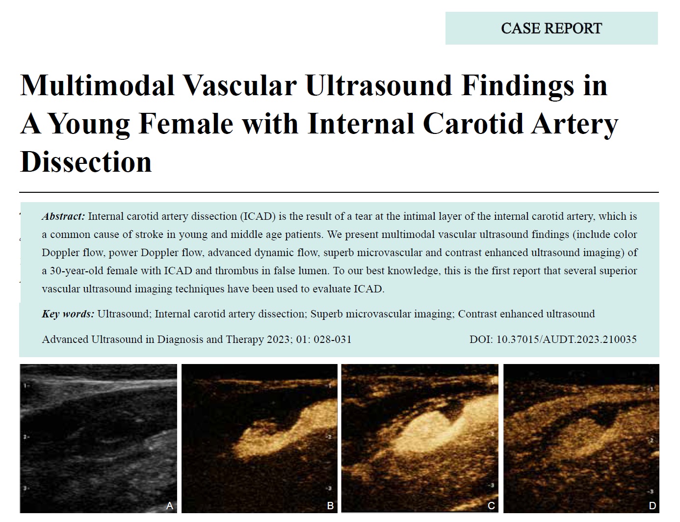
Internal carotid artery dissection (ICAD) is the result of a tear at the intimal layer of the internal carotid artery, which is a common cause of stroke in young and middle age patients. We present multimodal vascular ultrasound findings (include color Doppler flow, power Doppler flow, advanced dynamic flow, superb microvascular and contrast enhanced ultrasound imaging) of a 30-year-old female with ICAD and thrombus in false lumen. To our best knowledge, this is the first report that several superior vascular ultrasound imaging techniques have been used to evaluate ICAD.
Zehui Fu, MD , Chuxue Sun, MD , Haixia Zhou, MD , Xiaowen Lv, MD , Siqi Wang, MD , Hui Chen, MD . Multimodal Vascular Ultrasound Findings in A Young Female with Internal Carotid Artery Dissection[J]. ADVANCED ULTRASOUND IN DIAGNOSIS AND THERAPY, 2023 , 7(1) : 28 -31 . DOI: 10.37015/AUDT.2023.210035
| [1] | Adam G, Darcourt J, Roques M, Ferrier M, Gramada R, Meluchova Z, et al. Standard diffusion-weighted imaging in the brain can detect cervical internal carotid artery dissections. AJNR Am J Neuroradiol 2020; 41:318-322. |
| [2] | Zheng X, Wang Y, Chen G, Ma C, Yan W, Chen M. Diagnosis of ischemic optic neuropathy caused by dissection of the internal carotid artery: a case report. Medicine (Baltimore) 2020; 99:e20034. |
| [3] | Chen L, Chen L, Jiang Y, Hu F, He L, Zheng H. Pulsatile tinnitus was the only manifestation of internal carotid artery dissection. Neurol Sci 2021; 42:1221-1222. |
| [4] | Mes M, Palczewski P, Szczudlik P, Lusakowska A, Maj E, Gawel M. Hypoglossal nerve palsy as an isolated syndrome of internal carotid artery dissection: a review of the literature and a case report. Neurol Neurochir Pol 2018; 52:731-735. |
| [5] | Lee VH, Brown RD, Mandrekar JN, Mokri B. incidence and outcome of cervical artery dissection: a population-based study. Neurology 2006; 67:1809-1812. |
| [6] | Hakimi R, Sivakumar S. Imaging of carotid dissection. Curr Pain Headache Rep 2019; 23:2. |
| [7] | Clevert DA, Sommer WH, Zengel P, Helck A, Reiser M. Imaging of carotid arterial diseases with contrast-enhanced ultrasound (CEUS). Eur J Radiol 2011; 80:68-76. |
| [8] | Brandt T, Hausser I, Orberk E, Grau A, Hartschuh W, Anton-Lamprecht I, et al. Ultrastructural connective tissue abnormalities in patients with spontaneous cervicocerebral artery dissections. Ann Neurol 1998; 44:281-285. |
| [9] | Gould DB, Cunningham K. Internal carotid artery dissection after remote surgery. Iatrogenic complications of anesthesia. Stroke 1994; 25:1276-1278. |
| [10] | Fu Z, Zhang J, Lu Y, Wang S, Mo X, He Y, et al. Clinical applications of superb microvascular imaging in the superficial tissues and organs: a systematic review. Acad Radiol 2021; 28:694-703. |
| [11] | Ashrafi AN, Nassiri N, Gill IS, Gulati M, Park D, de Castro Abreu AL. Contrast-enhanced transrectal ultrasound in focal therapy for prostate cancer. Curr Urol Rep 2018; 19:87. |
| [12] | Holden A, Hope JKA, Osborne M, Moriarty M, Lee K. Value of a contrast agent in equivocal carotid ultrasound studies: pictorial essay. Australas Radiol 2000; 44:253-260. |
| [13] | Ferrer JM, Samsó JJ, Serrando JR, Valenzuela VF, Montoya SB, Docampo MM. Use of ultrasound contrast in the diagnosis of carotid artery occlusion. J Vasc Surg 2000; 31:736-741. |
| [14] | Ventura C, Da Silva E, Cerri G, Puech-Leao P, Tachibana A, Chammas M. Can contrast-enhanced ultrasound with second-generation contrast agents replace computed tomography angiography for distinguishing between occlusion and pseudo-occlusion of the internal carotid artery? Clinics 2015; 70:1-6. |
/
| 〈 |
|
〉 |
Share: WeChat
Copyright ©2018 Advanced Ultrasound in Diagnosis and Therapy
|
 Advanced Ultrasound in Diagnosis and Therapy (AUDT)
is licensed under a Creative Commons Attribution 4.0 International License.
Advanced Ultrasound in Diagnosis and Therapy (AUDT)
is licensed under a Creative Commons Attribution 4.0 International License.