
ADVANCED ULTRASOUND IN DIAGNOSIS AND THERAPY >
Contrast-enhanced Ultrasound of Undifferentiated Embryonal Sarcoma of the Liver in Adult: Two Cases Report and Literature Review
Received date: 2022-03-24
Revised date: 2022-04-14
Accepted date: 2022-04-24
Online published: 2022-10-25
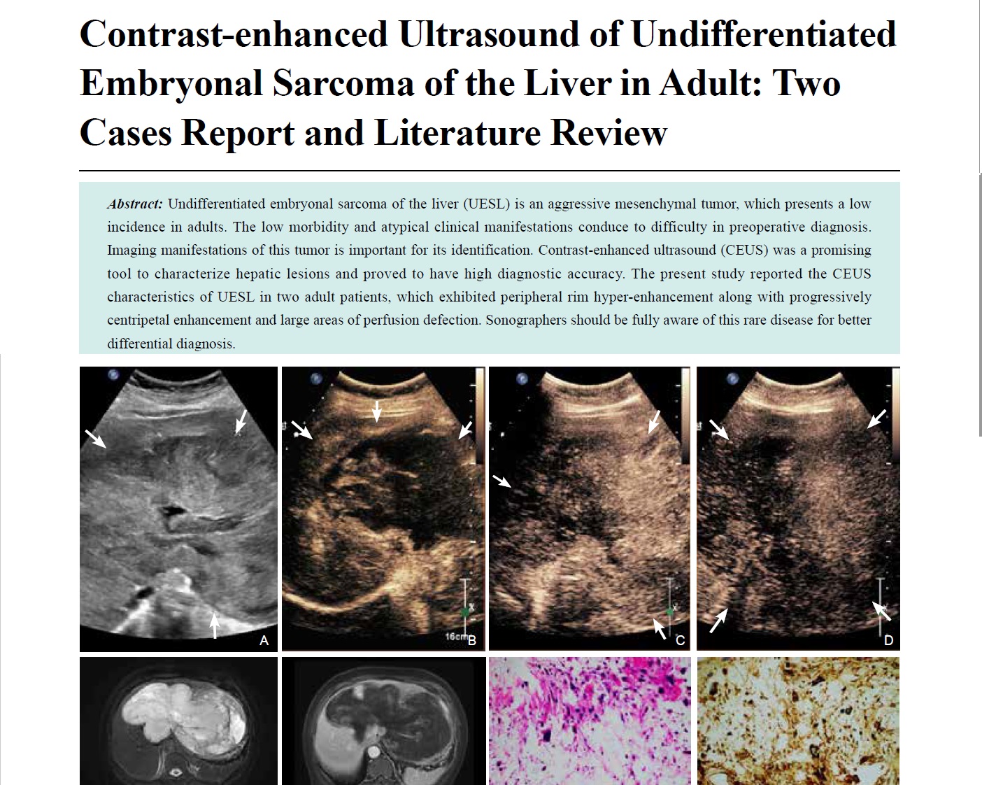
Undifferentiated embryonal sarcoma of the liver (UESL) is an aggressive mesenchymal tumor, which presents a low incidence in adults. The low morbidity and atypical clinical manifestations conduce to difficulty in preoperative diagnosis. Imaging manifestations of this tumor is important for its identification. Contrast-enhanced ultrasound (CEUS) was a promising tool to characterize hepatic lesions and proved to have high diagnostic accuracy. The present study reported the CEUS characteristics of UESL in two adult patients, which exhibited peripheral rim hyper-enhancement along with progressively centripetal enhancement and large areas of perfusion defection. Sonographers should be fully aware of this rare disease for better differential diagnosis.
Yanling Chen, MM , Hantao Wang, MM , Hong Han, PhD , Yi Dong, PhD , Wen-ping Wang, MD . Contrast-enhanced Ultrasound of Undifferentiated Embryonal Sarcoma of the Liver in Adult: Two Cases Report and Literature Review[J]. ADVANCED ULTRASOUND IN DIAGNOSIS AND THERAPY, 2022 , 6(4) : 204 -209 . DOI: 10.37015/AUDT.2022.220014
| [1] | Sy AM, Whitsett M, Li X, Theise ND, Dagher NN, Olsen S. Undifferentiated embryonal sarcoma of the liver: a great masquerade. J Gastrointest Cancer 2019, 50: 1043-1047. |
| [2] | Shu B, Gong L, Huang X, Cao L, Yan Z, Yang S. Undifferentiated embryonal sarcoma of the liver in adults: Retrospective analysis of a case series and systematic revie. Oncol Lett 2020, 20:102. |
| [3] | Wu Z, Wei Y, Cai Z, Zhou Y. Long-term survival outcomes of undifferentiated embryonal sarcoma of the liver: a pooled analysis of 308 patient. ANZ J Surg 2020, 90:1615-1620. |
| [4] | Walther A, Geller J, Coots A, Towbin A, Nathan J, Alonso M, et al. Multimodal therapy including liver transplantation for hepatic undifferentiated embryonal sarcom. Liver Transpl 2014, 20:191-199. |
| [5] | Putra J, Ornvold K. Undifferentiated embryonal sarcoma of the liver: a concise revie. Archives of pathology & laboratory medicine 2015, 139:269-273. |
| [6] | Wu M, Li L, Wang J, Zhang Y, Guo Q, Li X, et al. Contrast-enhanced US for characterization of focal liver lesions: a comprehensive meta-analysi. Eur Radiol 2018, 28:2077-2088. |
| [7] | Mathews J, Duncavage EJ, Pfeifer JD. Characterization of translocations in mesenchymal hamartoma and undifferentiated embryonal sarcoma of the live. Exp Mol Pathol 2013, 95:319-324. |
| [8] | Zhang C, Jia CJ, Xu C, Sheng QJ, Dou XG, Ding Y. Undifferentiated embryonal sarcoma of the liver: Clinical characteristics and outcome. World J Clin Cases 2020, 8:4763-4772. |
| [9] | Wei ZG, Tang LF, Chen ZM, Tang HF, Li MJ. Childhood undifferentiated embryonal liver sarcoma: clinical features and immunohistochemistry analysi. J Pediatr Surg 2008, 43:1912-1919. |
| [10] | Manabe Y, Uojima H, Hidaka H, Shao X, Iwasaki S, Wada N, et al. Undifferentiated embryonal sarcoma of the liver identified after the initial diagnosis of a hepatic cys. Intern Med 2020, 59:2375-2382. |
| [11] | Li Y, Cai Q, Jia N, Chen D, Lu L, Cheng H. Pre-operatively misdiagnosed undifferentiated embryonal sarcoma of the liver: analysis of 16 case. Ann Transl Med 2015, 3:353. |
| [12] | Yu XH, Huang J, Ge NJ, Yang YF, Zhao JY. Recurrent undifferentiated embryonal sarcoma of the liver in adult patient treated by pembrolizumab: A case report. World J Clin Cases 2021, 9:2281-2288. |
| [13] | Gabor F, Franchi-Abella S, Merli L, Adamsbaum C, Pariente D. Imaging features of undifferentiated embryonal sarcoma of the liver: a series of 15 childre. Pediatr Radiol 2016, 46:1694-1704. |
| [14] | Xie ZY, Li LP, Wu WJ, Sun DY, Zhou MH, Zhao YG. Undifferentiated embryonal sarcoma of the liver mistaken for hepatic abscess in an adul. Oncology letters 2014, 8:1184-1186. |
| [15] | Kunze G, Staritz M, Köhler M. Contrast-enhanced ultrasound in different stages of pyogenic liver absces. Ultrasound Med Biol 2015, 41:952-959. |
| [16] | Chen T, Chang X, Lv K, Wang Y, Fu X, Tan L, et al. Contrast-enhanced ultrasound features of intrahepatic cholangiocarcinoma: A new perspectiv. Sci Rep 2019, 9:19363. |
| [17] | Zhao CK, Xu HX, Guo LH, Sun LP, Yu M. A primary hepatic angiosarcoma mimicking intrahepatic cholangiocarcinoma on conventional ultrasound and contrast-enhanced ultrasound: A case report and review of literatures. Clin Hemorheol Microcirc 2017, 66:7-14. |
| [18] | Geyer T, Clevert DA, Schwarz S, Reidler P, Gassenmaier S, Knösel T, et al. Diagnostic value of CEUS prompting liver biopsy: histopathological correlation of hepatic lesions with ambiguous imaging characteristic. Diagnostics (Basel, Switzerland) 2020, 11:35. |
/
| 〈 |
|
〉 |
Share: WeChat
Copyright ©2018 Advanced Ultrasound in Diagnosis and Therapy
|
 Advanced Ultrasound in Diagnosis and Therapy (AUDT)
is licensed under a Creative Commons Attribution 4.0 International License.
Advanced Ultrasound in Diagnosis and Therapy (AUDT)
is licensed under a Creative Commons Attribution 4.0 International License.