
ADVANCED ULTRASOUND IN DIAGNOSIS AND THERAPY >
Ultrasound and Color Doppler Flow Imaging of Paratesticular Leiomyoma
Received date: 2021-08-08
Revised date: 2021-10-05
Online published: 2022-08-08
Objective: In this paper, we retrospectively analyzed the ultrasonographic features of paratesticular leiomyoma to help doctors correctly diagnose the disease before operation and guide surgical treatment.
Methods: From 2013 to 2020, 16 cases of paratesticular leiomyomas confirmed by pathology in our hospital were retrospectively analyzed. The retrospective analysis included the ultrasound and color Doppler flow imaging (CDFI) of paratesticular leiomyoma which were evaluated by two experienced radiologists based on the features of ultrasound images of lesions.
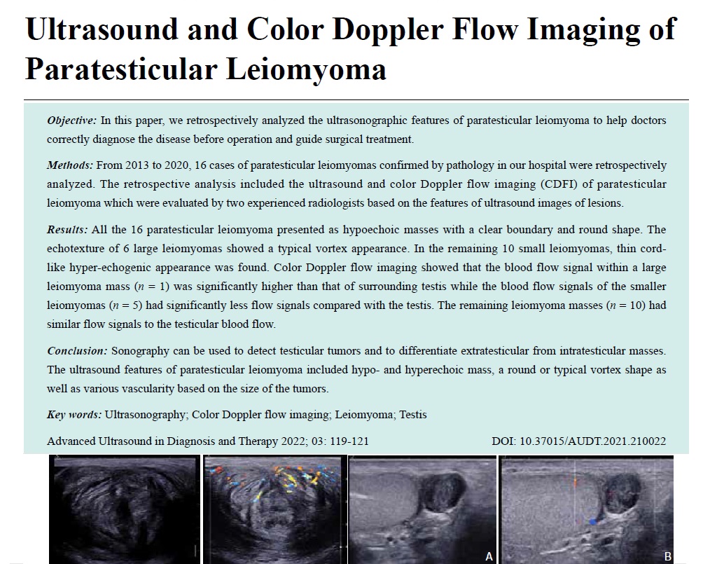
Results: All the 16 paratesticular leiomyoma presented as hypoechoic masses with a clear boundary and round shape. The echotexture of 6 large leiomyomas showed a typical vortex appearance. In the remaining 10 small leiomyomas, thin cord-like hyper-echogenic appearance was found. Color Doppler flow imaging showed that the blood flow signal within a large leiomyoma mass (n = 1) was significantly higher than that of surrounding testis while the blood flow signals of the smaller leiomyomas (n = 5) had significantly less flow signals compared with the testis. The remaining leiomyoma masses (n = 10) had similar flow signals to the testicular blood flow.
Conclusion: Sonography can be used to detect testicular tumors and to differentiate extratesticular from intratesticular masses. The ultrasound features of paratesticular leiomyoma included hypo- and hyperechoic mass, a round or typical vortex shape as well as various vascularity based on the size of the tumors.
Key words: Ultrasonography; Color Doppler flow imaging; Leiomyoma; Testis
Xue, MM Nianyu , Chen, MD Yaya , Wang, BS Guoyao , Zhang, BS Shengmin . Ultrasound and Color Doppler Flow Imaging of Paratesticular Leiomyoma[J]. ADVANCED ULTRASOUND IN DIAGNOSIS AND THERAPY, 2022 , 6(3) : 119 -121 . DOI: 10.37015/AUDT.2021.210022
| [1] | Yamamoto M, Miyake K, Mitsuya H. Intrascrotal extratesticular neurofibroma. Urology 1982, 20: 200-201. |
| [2] | Dougall A J, Wilson R R. Leiomyoma of the vas deferens. Br J Urol 1969, 41: 348-350. |
| [3] | Sean R, James B, Ann C. A rare case of paratesticular leiomyosarcoma. Journal of Surgical Case Reports 2018, 10: 1-2. |
| [4] | Fernandez A, Krishnamoorthy S, Muralitharan S, Johnson T, Ramanan V. Bilateral synchronous paratesticular leiomyoma-a rare entity. J Clin Diagn Res 2017 ; 11: PD05-PD06. |
| [5] | Chen YC, Li MH, Tsai WM. Clinical characteristic of a testis-associated leiomyoma: a case report and literature review. JTUA 2007, 18: 157-160. |
| [6] | Ali Alasmar, Awad Alkaabna, Khalaf Aljader, Nizar Alsaaydah, Oula Waqfi. Bilateral synchronous paratesticular leiomyoma: a case report. JRMS 2014: 21: 60-63. |
| [7] | Tchelepi H, Daneshmand S, Yanle Z, Ralls PW. Sonography of spermatic cord leiomyoma: case report and review of the literature. J Ultrasound Med 2004, 23:569-571. |
| [8] | Vick CW, Bird KI Jr, Rosenfield AT, Viscomi GN, Taylor KJ. Scrotal masses with a uniformly hyperechoic pattern. Radiology 1983, 148:209-211. |
| [9] | Kutchera WA, Bluth EI, Guice SL. Sonographic findings of a spermatic cord lipoma. Case report and review of the literature. J Ultrasound Med 1987, 6:457-460. |
| [10] | Hricak H, Filly RA. Sonography of the scrotum. Invest Radiol 1983; 18:112-121. |
| [11] | Khoubehi B, Mishra V, Ali M, Motiwala H, Karim O. Adult paratesticular tumours. BJU Int 2002; 90:707-715.. |
/
| 〈 |
|
〉 |
Share: WeChat
Copyright ©2018 Advanced Ultrasound in Diagnosis and Therapy
|
 Advanced Ultrasound in Diagnosis and Therapy (AUDT)
is licensed under a Creative Commons Attribution 4.0 International License.
Advanced Ultrasound in Diagnosis and Therapy (AUDT)
is licensed under a Creative Commons Attribution 4.0 International License.