
ADVANCED ULTRASOUND IN DIAGNOSIS AND THERAPY >
Incidental Ultrasound Findings of a Giant Retroperitoneal Schwannoma: A Case Study
Received date: 2021-04-29
Revised date: 2021-06-04
Online published: 2022-06-26
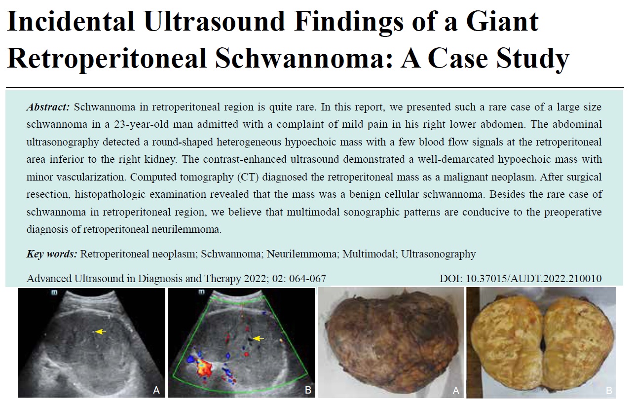
Schwannoma in retroperitoneal region is quite rare. In this report, we presented such a rare case of a large size schwannoma in a 23-year-old man admitted with a complaint of mild pain in his right lower abdomen. The abdominal ultrasonography detected a round-shaped heterogeneous hypoechoic mass with a few blood flow signals at the retroperitoneal area inferior to the right kidney. The contrast-enhanced ultrasound demonstrated a well-demarcated hypoechoic mass with minor vascularization. Computed tomography (CT) diagnosed the retroperitoneal mass as a malignant neoplasm. After surgical resection, histopathologic examination revealed that the mass was a benign cellular schwannoma. Besides the rare case of schwannoma in retroperitoneal region, we believe that multimodal sonographic patterns are conducive to the preoperative diagnosis of retroperitoneal neurilemmoma.
Key words: Retroperitoneal neoplasm; Schwannoma; Neurilemmoma; Multimodal; Ultrasonography
Zhao, MD Jiaqi , Li, MD Weiqing , Ma, MD Xiaolin , Chen, MD Rui , Chen, MD Lin . Incidental Ultrasound Findings of a Giant Retroperitoneal Schwannoma: A Case Study[J]. ADVANCED ULTRASOUND IN DIAGNOSIS AND THERAPY, 2022 , 6(2) : 64 -67 . DOI: 10.37015/AUDT.2022.210010
| [1] | Ratnagiri R, Mallikarjun S. Retroperitoneal ancient schwannoma: two cases and review of literature. J Cancer Res Ther 2014; 10:368-370. |
| [2] | Wong CS, Chu TY, Tam KF. Retroperitoneal schwannoma: a common tumour in an uncommon site. Hong Kong Med J 2010; 16:66-68. |
| [3] | Harada TL, Nagao G, Aoyagi T, Kuroda I, Tokuyama N, Takahashi M, et al. Giant retroperitoneal schwannoma in a 52-year-old man. Radiol Case Rep 2018; 13:810-814. |
| [4] | Kalaycı M, Akyüz U, Demirağ A, Gürses B, Ozkan F, Gökçe O. Retroperitoneal schwannoma: a rare case. Case Rep Gastrointest Med 2011; 2011: 465062. |
| [5] | Xu SY, Sun K, Xie HY, Zhou L, Zheng SS, Wang WL. Hemorrhagic, calcified, and ossified benign retroperitoneal schwannoma: First case report. Medicine (Baltimore) 2016; 95: e4318. |
| [6] | Kinoshita T, Naganuma H, Ishii K, Itoh H. CT features of retroperitoneal neurilemmoma. Eur J Radiol 1998; 27:67-71. |
/
| 〈 |
|
〉 |
Share: WeChat
Copyright ©2018 Advanced Ultrasound in Diagnosis and Therapy
|
 Advanced Ultrasound in Diagnosis and Therapy (AUDT)
is licensed under a Creative Commons Attribution 4.0 International License.
Advanced Ultrasound in Diagnosis and Therapy (AUDT)
is licensed under a Creative Commons Attribution 4.0 International License.