
ADVANCED ULTRASOUND IN DIAGNOSIS AND THERAPY >
Ultrasonographic Features of Intrathyroidal Thymic Carcinoma: Review and Analysis of 10 Cases
Received date: 2022-03-16
Revised date: 2022-04-06
Online published: 2022-06-26
ObjectiveIntrathyroidal thymic carcinoma (ITTC) is a rare epithelial tumor of the thyroid gland. Since ITTC is rare, its imaging findings have not been well defined. In the present study, we studied the US appearance of ITTC by analyzing ten cases retrospectively.
Methods Patients were identified by searching the surgical pathology records at our hospital. There were three male and seven female patients ranging in age from 40 to 79 years. The ultrasound (US) features were evaluated, and the relevant clinical data were combined with the fine needle aspiration (FNA) results from previous publications.
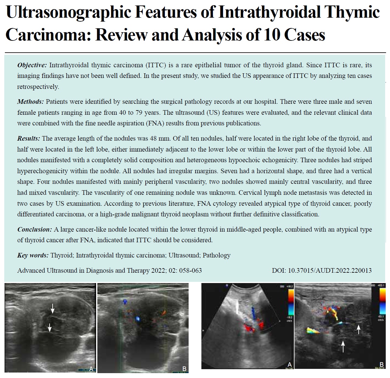
Results The average length of the nodules was 48 mm. Of all ten nodules, half were located in the right lobe of the thyroid, and half were located in the left lobe, either immediately adjacent to the lower lobe or within the lower part of the thyroid lobe. All nodules manifested with a completely solid composition and heterogeneous hypoechoic echogenicity. Three nodules had striped hyperechogenicity within the nodule. All nodules had irregular margins. Seven had a horizontal shape, and three had a vertical shape. Four nodules manifested with mainly peripheral vascularity, two nodules showed mainly central vascularity, and three had mixed vascularity. The vascularity of one remaining nodule was unknown. Cervical lymph node metastasis was detected in two cases by US examination. According to previous literature, FNA cytology revealed atypical type of thyroid cancer, poorly differentiated carcinoma, or a high-grade malignant thyroid neoplasm without further definitive classification.
ConclusionA large cancer-like nodule located within the lower thyroid in middle-aged people, combined with an atypical type of thyroid cancer after FNA, indicated that ITTC should be considered.
Key words: Thyroid; Intrathyroidal thymic carcinoma; Ultrasound; Pathology
Wang, MD Yanhai , Yang, MD Hua , Liu, MD Hanqing , Luo, MD Xiaoli , Liu, BS Luying , Zhou, BS Pingting . Ultrasonographic Features of Intrathyroidal Thymic Carcinoma: Review and Analysis of 10 Cases[J]. ADVANCED ULTRASOUND IN DIAGNOSIS AND THERAPY, 2022 , 6(2) : 58 -63 . DOI: 10.37015/AUDT.2022.220013
| [1] | Miyauchi A, Kuma K, Matsuzuka F, Miyauchi A, Kuma K, Matsuzuka F, et al. Intrathyroidal epithelial thymoma: an entity distinct from squamous cell carcinoma of the thyroid. World J Surg 1985; 9:128-135. |
| [2] | Chan JK, Rosai J. Tumors of the neck showing thymic or related branchial pouch differentiation: a unifying concept. Hum Pathol 1991; 22:349-367. |
| [3] | Zhang Y, Wu QL, Yun JP. Interpretation of the fourth edition of WHO pathological classification of the thyroid tumors in 2017. Zhonghua Er Bi Yan Hou Tou Jing Wai Ke Za Zhi 2018; 53:718-720 [in Chinese]. |
| [4] | Dong W, Zhang P, Li J, Dong W, Zhang P, Li J, et al. Outcome of thyroid carcinoma showing thymus-like differentiation in Ppatients undergoing radical resection. World J Surg 2018; 42:1754-1761. |
| [5] | Fung ACH, Tsang JS, Lang BHH. Thyroid carcinoma showing thymus-like differentiation (CASTLE) with tracheal invasion: a case report. Am J Case Rep 2019; 10:1845-1851. |
| [6] | Inoue Y, Kohi S, Gobara M, Joden F, Yabuki K, Tanoue T, et al. A case of carcinoma showing thymus-like differentiation (CASTLE) of the thyroid. J UOEH 2018; 40:259-266. |
| [7] | Liu Z, Teng XY, Sun DX, Xu WX, Sun SL. Clinical analysis of thyroid carcinoma showing thymus-like differentiation: report of 8 cases. Int Surg 2013; 98:95-100. |
| [8] | Tsutsui H, Hoshi M, Kubota M, Suzuki A, Nakamura N, Usuda J, et al. Management of thyroid carcinoma showing thymus-like differentiation (CASTLE) invading the trachea. Surg Today 2013; 43:1261-1268. |
| [9] | Yamamoto Y, Yamada K, Motoi N, Fujiwara Y, Toda K, Sugitani I, et al. Sonographic findings in three cases of carcinoma showing thymus-like differentiation. J Clin Ultrasound 2013; 41:574-578. |
| [10] | Sun T, Wang Z, Wang J, Wu Y, Li D, Ying H. Outcome of radical resection and postoperative radiotherapy for thyroid carcinoma showing thymus-like differentiation. World J Surg 2011; 35:1840-1846. |
| [11] | Chang S, Joo M, Kim H. Cytologic findings of thyroid carcinoma showing thymus-like differentiation: a case report. Korean J Pathol 2012; 46:30-35. |
| [12] | Ren WH, Dong K, Huang XZ, Zhu YL. Intrathyroidal thymic carcinoma exhibiting neuroendocrine differentiation: case report with cytomorphology, immunocytochemistry, and review of the literature focusing on cytology. Diagn Cytopathol 2019; 47:1197-1202. |
| [13] | Chung SM, Kim KJ, Moon JS, Hong YH, Kang SH. Fever of unknown origin caused by intrathyroidal thymic carcinoma. Korean J Intern Med 2019; 34:683-684. |
| [14] | Tessler FN, Middleton WD, Grant EG, Hoang JK, Berland LL, Teefey SA, et al. ACR thyroid imaging, reporting and data system (TI-RADS): white paper of the ACR TI-RADS committee. J Am Coll Radiol 2017 ; 14: 587-595. |
| [15] | Zhou J, Yin L, Wei X, Zhang S, Song Y, Luo B, et al. 2020 Chinese guidelines for ultrasound malignancy risk stratification of thyroid nodules: the C-TIRADS. Endocrine 2020; 70:256-279. |
| [16] | Ge W, Yao YZ, Chen G, Ding YT. Clinical analysis of 82 cases of carcinoma showing thymus-like differentiation of the thyroid. Oncol Lett 2016; 11:1321-1326. |
| [17] | Liu SM, Lee WH. Carcinoma showing thymus-like differentiation (CASTLE): report of a case specified in the cytomorphology. J Formos Med Assoc 2016; 115:377-379. |
| [18] | Choi KY, Kwon MJ, Ahn HK, Kim JH, Lee DJ. Extrathyroid carcinoma showing thymus-like differentiation (CASTLE): a new case report and review of the therapeutic role of neck dissection and radiotherapy. World J Surg Oncol 2014; 8:247-252. |
| [19] | Zhang G, Liu X, Huang W, Li X, Johnstone M, Deng Y, et al. Carcinoma showing thymus-like elements of the thyroid gland: report of three cases including one case with breast cancer history. Pathol Oncol Res 2015; 21:45-51. |
| [20] | Wharry LI, McCoy KL, Stang MT, Armstrong MJ, LeBeau SO, Tublin ME, et al. Thyroid nodules (≥4 cm): can ultrasound and cytology reliably exclude cancer? World J Surg 2014; 38:614-621. |
| [21] | Kobayashi K, Fujimoto T, Ota H, Hirokawa M, Yabuta T, Masuoka H, et al. Calcifications in thyroid tumors on ultrasonography: calcification types and relationship with histopathological type. Ultrasound Int Open 2018; 4:E45-51. |
| [22] | Yin L, Zhang W, Bai W, He W. Relationship between morphologic characteristics of ultrasonic calcification in thyroid nodules and thyroid carcinoma. Ultrasound Med Biol 2020; 46:20-25. |
| [23] | Mohebbi M, Dehaki MG, Mozaffari M. Comparison between ultrasonographic findings and fine needle aspiration cytology in differentiating malignant and benign thyroid nodules. Eur J Transl Myol 2019; 29:261-267. |
| [24] | Papapostolou KD, Evangelopoulou CC, Ioannidis IA, Kassi GN, Morfas KS, Karaminas NI, et al Taller-than-wide thyroid nodules with microcalcifications are at high risk of malignancy. In Vivo 2020; 34:2101-2105. |
| [25] | Hammad AY, Noureldine SI, Hu T, Ibrahim Y, Masoodi HM, Kandil E. A meta-analysis examining the independent association between thyroid nodule size and malignancy. Gland Surg 2016; 5:312-317. |
| [26] | Mohebati A, Dilorenzo M, Palmer F, Patel SG, Pfister D, Lee N, et al Anaplastic thyroid carcinoma: a 25-year single-institution experience. Ann Surg Oncol 2014; 21:1665-1670. |
| [27] | Xu X, Yang X, Zhao RN, Zhu SL, Zhang XY, Xia Y, et al Comparison of ultrasonic features between anaplastic thyroid carcinoma and papillary thyroid carcinoma. Zhongguo Yi Xue Ke Xue Yuan Xue Bao 2015; 37:71-74 [in Chinese]. |
| [28] | Gu LS, Cui NY, Wang Y, Che SN, Zou SM, He W, et al. Comparison of sonographic characteristics of primary thyroid lymphoma and anaplastic thyroid carcinoma. J Thorac Dis 2017; 9:4774-4784. |
| [29] | Wang Z, Fu B, Xiao Y, Liao J, Xie P. Primary thyroid lymphoma has different sonographic and color Doppler features compared to nodular goiter. J Ultrasound Med 2015; 34:317-323. |
| [30] | Wang Y, Huang L, Lv H, Huang Y, Li D. Primary malignant fibrous histiocytoma of the thyroid: two case reports and review of the literature. J Ultrasound Med 2017; 36:665-669. |
| [31] | Sung JY. Parathyroid ultrasonography: the evolving role of the radiologist. Ultrasonography 2015; 34:268-274. |
| [32] | Fukushima T, Suzuki S, Ohira T, Shimura H, Midorikawa S, Ohtsuru A, et al. Thyroid examination unit of the radiation medical center for the fukushima health management survey. Prevalence of ectopic intrathyroidal thymus in Japan: the Fukushima health management survey. Thyroid 2015; 25:534-537. |
| [33] | Yildiz AE, Elhan AH, Fitoz S. Prevalence and sonographic features of ectopic thyroidal thymus in children: a retrospective analysis. J Clin Ultrasound 2018; 46:375-379. |
/
| 〈 |
|
〉 |
Share: WeChat
Copyright ©2018 Advanced Ultrasound in Diagnosis and Therapy
|
 Advanced Ultrasound in Diagnosis and Therapy (AUDT)
is licensed under a Creative Commons Attribution 4.0 International License.
Advanced Ultrasound in Diagnosis and Therapy (AUDT)
is licensed under a Creative Commons Attribution 4.0 International License.