
ADVANCED ULTRASOUND IN DIAGNOSIS AND THERAPY >
Advances in Modern Clinical Ultrasound
Received date: 2018-06-01
Online published: 2018-08-19
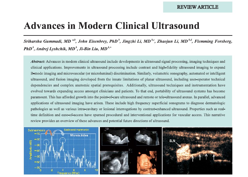
Advances in modern clinical ultrasound include developments in ultrasound signal processing, imaging techniques and clinical applications. Improvements in ultrasound processing include contrast and high-fidelity ultrasound imaging to expand B-mode imaging and microvascular (or microluminal) discrimination. Similarly, volumetric sonography, automated or intelligent ultrasound, and fusion imaging developed from the innate limitations of planar ultrasound, including user-operator technical dependencies and complex anatomic spatial prerequisites. Additionally, ultrasound techniques and instrumentation have evolved towards expanding access amongst clinicians and patients. To that end, portability of ultrasound systems has become paramount. This has afforded growth into the point-of-care ultrasound and remote or tele-ultrasound arenas. In parallel, advanced applications of ultrasound imaging have arisen. These include high frequency superficial sonograms to diagnose dermatologic pathologies as well as various intra-cavitary or lesional interrogations by contrast-enhanced ultrasound. Properties such as realtime definition and ease-of-access have spurned procedural and interventional applications for vascular access. This narrative review provides an overview of these advances and potential future directions of ultrasound.
Gummadi, MD Sriharsha , Eisenbrey, PhD John , Li, MD Jingzhi , Li, MD Zhaojun , Forsberg, PhD Flemming , Lyshchik, MD Andrej , Liu, MD Ji-Bin . Advances in Modern Clinical Ultrasound[J]. ADVANCED ULTRASOUND IN DIAGNOSIS AND THERAPY, 2018 , 2(2) : 51 -63 . DOI: 10.37015/AUDT.2018.180801
| [1] | Newman PG, Rozycki GS. The history of ultrasound. Surg Clin North Am 1998; 78:179-95. |
| [2] | Eisenbrey JR, Sridharan A, Liu JB, Forsberg F. Recent experiences and advances in contrast-enhanced subharmonic ultrasound. Biomed Res Int 2015; 2015:640397. |
| [3] | Eckersley RJ, Chin CT, Burns PN. Optimising phase and amplitude modulation schemes for imaging microbubble contrast agents at low acoustic power. Ultrasound Med Biol 2005; 31:213-9. |
| [4] | Paefgen V, Doleschel D, Kiessling F. Evolution of contrast agents for ultrasound imaging and ultrasound-mediated drug delivery. Front Pharmacol 2015; 6:197. |
| [5] | Piscaglia F, Nols?e C, Dietrich CF, Cosgrove DO, Gilja OH, Bachmann Nielsen M, et al., The EFSUMB Guidelines and Recommendations on the Clinical Practice of Contrast Enhanced Ultrasound(CEUS) update 2011 n non-hepatic applications. Ultraschall Med 2012; 33:33-59. |
| [6] | Claudon M, Dietrich CF, Choi BI, Cosgrove DO, Kudo M, Nols?e CP, et al. Guidelines and good clinical practice recommendations for Contrast Enhanced Ultrasound (CEUS) in the liver - update 2012: A WFUMB-EFSUMB initiative in cooperation with representatives of AFSUMB, AIUM, ASUM, FLAUS and ICUS. Ultrasound Med Biol 2013; 39:187-210. |
| [7] | Lyshchik A, Kono Y, Dietrich CF, Jang HJ, Kim TK, Piscaglia F, et al., Contrast-enhanced ultrasound of the liver: technical and lexicon recommendations from the ACR CEUS LI-RADS working group. Abdom Radiol (NY) 2018; 43:861-79. |
| [8] | Muskula PR, Main ML. Safety With Echocardiographic Contrast Agents. Circ Cardiovasc Imaging 2017; 10:e005459. |
| [9] | Haugen BR, Alexander EK, Bible KC, Doherty GM, Mandel SJ, Nikiforov YE, et al. 2015 American Thyroid Association management guidelines for adult patients with thyroid nodules and differentiated thyroid cancer: the American Thyroid Association guidelines task force on thyroid nodules and differentiated thyroid cancer. Thyroid 2016; 26:1-133. |
| [10] | Baloch ZW, LiVolsi VA, Asa SL, Rosai J, Merino MJ, Randolph G, et al. Diagnostic terminology and morphologic criteria for cytologic diagnosis of thyroid lesions: a synopsis of the National Cancer Institute Thyroid Fine‐Needle Aspiration State of the Science Conference. Diagn Cytopathol 2008; 36:425-37. |
| [11] | Hotta N, Tagaya T, Maeno T, Ayada M, Sato K, Ishikawa T, et al. Advanced dynamic flow imaging with contrast-enhanced ultrasonography for the evaluation of tumor vascularity in liver tumors. Clin Imaging 2005; 29(1):34-41. |
| [12] | Machado P, Segal S, Lyshchik A, Forsberg F. A Novel Microvascular Flow Technique: Initial Results in Thyroids. Ultrasound Q 2016; 32:67-74. |
| [13] | Singaporewalla RM, Hwee J, Lang TU, Desai V. Clinico-pathological correlation of thyroid nodule ultrasound and cytology using the TIRADS and Bethesda classifications. World J Surg 2017; 41:1807-11. |
| [14] | Lazebnik RS, Desser TS. Clinical 3D ultrasound imaging: beyond obstetrical applications. Diagn Imaging 2007; 1:1-6. |
| [15] | Artul S, Nseir W, Armaly Z, Soudack M. Superb microvascular imaging: Added value and novel applications. J Clin Imaging Sci 2017; 7:45. |
| [16] | Fenster A, Downey DB, Cardinal HN. Three-dimensional ultrasound imaging. Phys Med Biol 2001; 46:R67-99. |
| [17] | Prager RW, Ijaz UZ, Gee AH, Treece GM. Three-dimensional ultrasound imaging. Proc Inst Mech Eng H 2010; 224:193-223. |
| [18] | Palmentieri B, de Sio I, La Mura V, Masarone M, Vecchione R, Bruno S, et al. The role of bright liver echo pattern on ultrasound B-mode examination in the diagnosis of liver steatosis. Dig Liver Dis 2006; 38:485-9. |
| [19] | Medvedofsky D, et al. Automatic transthoracic three-dimensional echocardiographic quantification of the heart chambers. Philips Ultrasound, 2016. |
| [20] | Younossi ZM, Stepanova M, Afendy M, Fang Y, Younossi Y, Mir H, et al. Changes in the prevalence of the most common causes of chronic liver diseases in the United States from 1988 to 2008. Clin Gastroenterol Hepatol 2011; 9:524-530. |
| [21] | Prater D, Cardinale M, Schneider R. AutoEF using a 2DQ featuring Anatomical Intelligence. Philips Ultrasound, 2014. |
| [22] | Narula J, Chandrashekhar Y, Braunwald E. Time to add a fifth pillar to bedside physical examination: inspection, palpation, percussion, auscultation, and insonation. JAMA Cardiol 2018; 3:346-50. |
| [23] | Moore CL, Copel JA. Point-of-care ultrasonography. N Engl J Med 2011; 364:749-57. |
| [24] | Beal EW, Sigmond BR, Sage-Silski L, Lahey S, Nguyen V, Bahner DP. Point-of-Care Ultrasound in general surgery residency training: A proposal for milestones in graduate medical education ultrasound. J Ultrasound Med 2017; 36:2577-84. |
| [25] | Nelson BP, Melnick ER, Li J. Portable ultrasound for remote environments, Part I: Feasibility of field deployment. J Emerg Med 2011; 40:190-7. |
| [26] | Martin DS, South DA, Garcia KM, Arbeille P. Ultrasound in space. Ultrasound Med Biol 2003; 29:1-12. |
| [27] | Kleinerman R, Whang TB, Bard RL, Marmur ES. Ultrasound in dermatology: principles and applications. J Am Acad Dermatol 2012; 67:478-87. |
| [28] | Sadaka A, Prager T, Beaver H, Malik A. A novel use of ultrasound biomicroscopy. Eye (Lond) 2018; 32:474-475. |
| [29] | Pavlin CJ, Foster FS . Ultrasound biomicroscopy of the eye. 2012: Springer Science & Business Media. |
| [30] | Eisenbrey JR, Forsberg F. Contrast-enhanced ultrasound for molecular imaging of angiogenesis. Eur J Nucl Med Mol Imaging 2010; 37 Suppl 1: S138-46. |
| [31] | Shaw CM, Eisenbrey JR, Lyshchik A, O'Kane PL, Merton DA, Machado P, et al. Contrast-enhanced ultrasound evaluation of residual blood flow to hepatocellular carcinoma after treatment with transarterial chemoembolization using drug-eluting beads. J Ultrasound Med 2015; 34:859-67. |
| [32] | Gummadi S, Eisenbrey JR, Lyshchik A. Contrast-enhanced ultrasonography in interventional oncology. Abdom Radiol (NY) 2018. doi: 10.1007/s00261-018-1581-5.[Epub ahead of print] |
| [33] | Gummadi S, Eisenbrey JR, Lyshchik A. A Narrative Review on Contrast-Enhanced Ultrasound in Aortic Endograft Endoleak Surveillance. Ultrasound Q 2018. doi: 10.1097/RUQ.0000000000000353.[Epub ahead of print] |
| [34] | Goldberg BB, Merton DA, Liu JB, Murphy G, Forsberg F. Contrast‐Enhanced Sonographic Imaging of Lymphatic Channels and Sentinel Lymph Nodes. J Ultrasound Med 2005; 24:953-65. |
| [35] | Goldberg BB, Merton DA, Liu JB, Thakur M, Murphy GF, Needleman L, et al. Sentinel lymph nodes in a swine model with melanoma: contrast-enhanced lymphatic US. Radiology 2004; 230:727-34. |
| [36] | Lin E, Choi J, Hadzic A. Peripheral nerve blocks for outpatient surgery: evidence-based indications. Curr Opin Anaesthesiol 2013; 26:467-74. |
| [37] | Jeng CL, Rosenblatt MA . Overview of Peripheral Nerve Blocks, in UpToDate, R.Maniker Editor., 2018, UpToDate: Waltham, MA. |
| [38] | Sabado JJ, Principles of Ultrasound-Guided Venous Access, in UpToDate, D.L. Cull, A.B. Wolfson, and A.M. Stack, Editors. 2018, UpToDate: Waltham, MA. |
| [39] | Turan A, Babazade R, Elsharkawy H, Esa WA, Maheshwari K, Farag E, et al. Novel needle guide reduces time to perform ultrasound-guided femoral nerve catheter placement: A randomised controlled trial. Eur J Anaesthesiol 2017; 34:135-140. |
| [40] | Li X, Long Q, Chen X, He D, He H. Real-time ultrasound-guided PCNL using a novel SonixGPS needle tracking system. Urolithiasis 2014; 42:341-6. |
| [41] | Bakkaloglu H, Yanar H, Guloglu R, Taviloglu K, Tunca F, Aksoy M, et al. Ultrasound guided percutaneous cholecystostomy in high-risk patients for surgical intervention. World J Gastroenterol 2006; 12:7179-82. |
| [42] | Nadolski GJ, Itkin M. Feasibility of ultrasound-guided intranodal lymphangiogram for thoracic duct embolization. J Vasc Interv Radiol 2012; 23:613-6. |
| [43] | Boks SS, Andhyiswara T, de Smet AA, Vroegindeweij D. Ultrasound-guided percutaneous transabdominal treatment of a type 2 endoleak. Cardiovasc Intervent Radiol 2005; 28:526-9. |
/
| 〈 |
|
〉 |
Share: WeChat
Copyright ©2018 Advanced Ultrasound in Diagnosis and Therapy
|
 Advanced Ultrasound in Diagnosis and Therapy (AUDT)
is licensed under a Creative Commons Attribution 4.0 International License.
Advanced Ultrasound in Diagnosis and Therapy (AUDT)
is licensed under a Creative Commons Attribution 4.0 International License.