
ADVANCED ULTRASOUND IN DIAGNOSIS AND THERAPY >
Aspiration Pneumonia Caused by Neuromyelitis Optica in a Patient with Suspected COVID-19
Received date: 2020-04-01
Online published: 2020-04-23
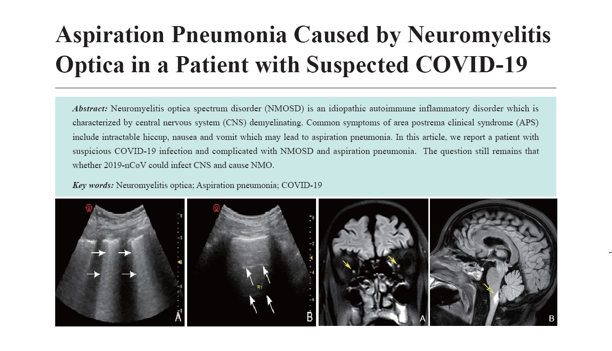
Neuromyelitis optica spectrum disorder (NMOSD) is an idiopathic autoimmune inflammatory disorder which is characterized by central nervous system (CNS) demyelinating. Common symptoms of area postrema clinical syndrome (APS) include intractable hiccup, nausea and vomit which may lead to aspiration pneumonia. In this article, we report a patient with suspicious COVID-19 infection and complicated with NMOSD and aspiration pneumonia. The question still remains that whether 2019-nCoV could infect CNS and cause NMO.
Key words: Neuromyelitis optica; Aspiration pneumonia; COVID-19
Zhao, MD Yuanyuan , Liu, MD Jie , Wu, MD Shengzheng , Li, MD Weihua , Li, MD Keyan , Chen, MD Zhiye , Wu, MD Dudu , Zhang, MD Ming , Zhang, MD Ningbo , Zhou Xuan , Shan Xuexia , Gao, MD Shunji , Lv, MD Faqin . Aspiration Pneumonia Caused by Neuromyelitis Optica in a Patient with Suspected COVID-19[J]. ADVANCED ULTRASOUND IN DIAGNOSIS AND THERAPY, 2020 , 4(2) : 138 -141 . DOI: 10.37015/AUDT.2020.200031
| [1] | World Health Organization. Coronavirus disease (COVID-19) outbreak. 12 January, 2012. https://www.who.int/ emergencies/diseases/novel-coronavirus-2019. Available from: |
| [2] | Soummer A, Perbet S, Brisson H, Arbelot C, Constantin JM, Lu Q, et al. Lung Ultrasound Study Group. Ultrasound assessment of lung aeration loss during a successful weaning trial predicts postextubation distress. Crit Care Med 2012; 40:2064-72. |
| [3] | General Office of National Health Commission, General Office of National Administration of Traditional Chinese Medicine. Diagnostic and treatment protocol for Novel Coronavirus Pneumonia (Trial version 7). Available from:http://www.nhc.gov.cn/yzygj/s7653p/202003/46c9294a7dfe4cef80dc7f5912eb1989.shtml. |
| [4] | Pan Y, Guan H, Zhou S, Wang Y, Li Q, Zhu T, et al. Initial CT findings and temporal changes in patients with the novel coronavirus pneumonia (2019-nCoV): a study of 63 patients in Wuhan, China. Eur Radiol 2020. DOI: 10.1007/s00330-020-06731-x. |
| [5] | JI GH, Huang MH, Zhang Q, Wang WB, Wang P, Qin XT, et al. CT manifestations and dynamic changes of corona virus disease 2019. Chinese Journal of Medical Imaging Technology 2020, 36:242-247. |
| [6] | Zhang PJ. Diagnosis and differential diagnosis of pulmonary groun-glass opacities with multi-layered spiral CT. China Medical Herald 2012, 4:95-97. |
| [7] | National Health Commission Capacity Building and Continuing Education Center; War Trauma and Critical Care Ultrasound Committee of Ultrasonic Equipment Technical Committee of China Association of Medical Equipment; Remote and Mobile Ultrasound Professional Committee of Ultrasonic Equipment Technical Committee of China Association of Medical Equipment. Expert consensus on the application of COVID-19 severe ultrasound (the draft during the war). Chin J Crit Care 2012, 40:185-195. [In Chinese]. Available from: http://111.40.160.75:802/CN/10.3969/j.issn.1002-1949.2020.03.001 |
| [8] | Zhong HJ, Zhao ZZ, Song XB, Lu XJ, Zhou Y, Song JJ, et al. Clinical points and experience in nucleic acid testing of SARS-CoV-2. Int J Lab Med 2020; 41:523-526. |
| [9] | General Office of National Health Commission. Technical guidelines on prevention and control of novel coronavirus infection in medical institutions (First edition). [In Chinese]. Available from:http://www.nhc.gov.cn/yzygj/s7659/202001/b91fdab7c304431eb082d67847d27e14.shtml. |
| [10] | Wingerchuk DM, Hogancamp WF, O'Brien PC, Weinshenker BG. The clinical course of neuromyelitis optica (Devic's syndrome). Neurology 1999; 53:1107-14. |
| [11] | Jarius S, Wildemann B. The history of neuromyelitis optica. J Neuroinflammation 2013; 10:8. |
| [12] | Wingerchuk DM, Lennon VA, Pittock SJ, Lucchinetti CF, Weinshenker BG. Revised diagnostic criteria for neuromyelitis optica. Neurology 2006; 66:1485-9. |
| [13] | Lennon VA, Wingerchuk DM, Kryzer TJ, Pittock SJ, Lucchinetti CF, Fujihara K, et al. A serum autoantibody marker of neuromyelitis optica: distinction from multiple sclerosis. Lancet 2004; 364:2106-12. |
| [14] | Lennon VA, Kryzer TJ, Pittock SJ, Verkman AS, Hinson SR. IgG marker of optic-spinal multiple sclerosis binds to the aquaporin-4 water channel. J Exp Med 2005; 202:473-7. |
| [15] | Wingerchuk DM, Banwell B, Bennett JL, Cabre P, Carroll W, Chitnis T, et al. International Panel for NMO Diagnosis. International consensus diagnostic criteria for neuromyelitis optica spectrum disorders. Neurology 2015; 85:177-89. |
| [16] | Wang QP, Guan JL, Pan W, Kastin AJ, Shioda S. A diffusion barrier between the area postrema and nucleus tractus solitarius. Neurochem Res 2008; 33:2035-43. |
| [17] | Longatti P, Porzionato A, Basaldella L, Fiorindi A, De Caro P, Feletti A. The human area postrema: clear-cut silhouette and variations shown in vivo. J Neurosurg 2015; 122:989-95. |
| [18] | Hou D, Yang GS, Guo T, Zhou F, Yu D. Infection and neuromyelitis optica spectrum disorders. J Cent South Univ (Med Sci) 2020; 5:181-186. |
/
| 〈 |
|
〉 |
Share: WeChat
Copyright ©2018 Advanced Ultrasound in Diagnosis and Therapy
|
 Advanced Ultrasound in Diagnosis and Therapy (AUDT)
is licensed under a Creative Commons Attribution 4.0 International License.
Advanced Ultrasound in Diagnosis and Therapy (AUDT)
is licensed under a Creative Commons Attribution 4.0 International License.