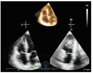
| [1] |
Khush KK, Cherikh WS, Chambers DC, Harhay MO, Hayes D Jr, Hsich E, et al. The international thoracic organ transplant registry of the international society for heart and lung transplantation: thirty-sixth adult heart transplantation report-2019; focus theme: donor and recipient size match. J Heart Lung Transplant 2019;38:1056-1066.
|
| [2] |
Badano LP, Miglioranza MH, Edvardsen T, Colafranceschi AS, Muraru D, Bacal F, et al. European association of cardiovascular imaging/cardiovascular imaging department of the brazilian society of cardiology recommendations for the use of cardiac imaging to assess and follow patients after heart transplantation. Eur Heart J Cardiovasc Imaging 2015;16:919-948.
|
| [3] |
Khush KK, Potena L, Cherikh WS, Chambers DC, Harhay MO, Hayes D Jr. The international thoracic organ transplant registry of the international society for heart and lung transplantation: 37th adult heart transplantation report-2020; focus on deceased donor characteristics. J Heart Lung Transplant 2020;39:1016-1027.
|
| [4] |
Stewart S, Winters GL, Fishbein MC, Tazelaar HD, Kobashigawa J, Abrams J, et al. Revision of the 1990 working formulation for the standardization of nomenclature in the diagnosis of heart rejection. J Heart Lung Transplant 2005;24:1710-1720.
|
| [5] |
Mehra MR, Crespo-Leiro MG, Dipchand A, Ensminger SM, Hiemann NE, Kobashigawa JA, et al. International society for heart and lung transplantation working formulation of a standardized nomenclature for cardiac allograft vasculopathy-2010. J Heart Lung Transplant 2010;29:717-727.
|
| [6] |
Miller CA, Fildes JE, Ray SG, Doran H, Yonan N, Williams SG, et al. Non-invasive approaches for the diagnosis of acute cardiac allograft rejection. Heart 2013;99:445-453.
|
| [7] |
Dandel M and Hetzer R. Post-transplant surveillance for acute rejection and allograft vasculopathy by echocardiography: usefulness of myocardial velocity and deformation imaging. J Heart Lung Transplant 2017;36:117-131.
|
| [8] |
Zhu S, Li M, Tian F, Wang S, Li Y, Yin P, et al. Diagnostic value of myocardial strain using two-dimensional speckle-tracking echocardiography in acute cardiac allograft rejection: a systematic review and meta-analysis. Echocardiography 2020;37:561-569.
|
| [9] |
Sade LE, Hazirolan T, Kozan H, Ozdemir H, Hayran M, Eroglu S, et al. T1 Mapping by cardiac magnetic resonance and multidimensional speckle-tracking strain by echocardiography for the detection of acute cellular rejection in cardiac allograft recipients. JACC Cardiovasc Imaging 2019;12:1601-1614.
|
| [10] |
Clemmensen TS, Logstrup BB, Eiskjaer H, Poulsen SH. Evaluation of longitudinal myocardial deformation by 2-dimensional speckle-tracking echocardiography in heart transplant recipients: relation to coronary allograft vasculopathy. J Heart Lung Transplant 2015;34:195-203.
|
| [11] |
Clemmensen TS, Eiskjaer H, Logstrup BB, Tolbod LP, Harms HJ, Bouchelouche K, et al. Noninvasive detection of cardiac allograft vasculopathy by stress exercise echocardiographic assessment of myocardial deformation. J Am Soc Echocardiogr 2016;29:480-490.
|
| [12] |
Mitchell C, Rahko PS, Blauwet LA, Canaday B, Finstuen JA, Foster MC, et al. Guidelines for performing a comprehensive transthoracic echocardiographic examination in adults: recommendations from the American Society of Echocardiography. J Am Soc Echocardiogr 2019;32:1-64.
|
| [13] |
Rudski LG, Lai WW, Afilalo J, Hua L, Handschumacher MD, Chandrasekaran K, et al. Guidelines for the echocardiographic assessment of the right heart in adults:a report from the american society of echocardiography endorsed by the European association of echocardiography, a registered branch of the European society of cardiology, and the Canadian society of echocardiography. J Am Soc Echocardiogr 2010;23:685-713.
|
| [14] |
Lang RM, Badano LP, Mor-Avi V, Afilalo J, Armstrong A, Ernande L, et al. Recommendations for cardiac chamber quantification by echocardiography in adults: an update from the American Society of Echocardiography and the European Association of Cardiovascular Imaging. J Am Soc Echocardiogr 2015;28:1-39.
|
| [15] |
Lv Q, Sun W, Wang J, Wu C, Li H, Shen X, et al. Evaluation of biventricular functions in transplanted hearts using 3-dimensional speckle-tracking echocardiography. J Am Heart Assoc 2020;18: e015742.
|
| [16] |
Dorosz JL, Lezotte DC, Weitzenkamp DA, Allen LA, Salcedo EE. Performance of 3-dimensional echocardiography in measuring left ventricular volumes and ejection fraction: a systematic review and meta-analysis. J Am Coll Cardiol 2012;15:1799-1808.
|
| [17] |
Seo Y, Ishizu T, Atsumi A, Kawamura R, Aonuma K. Three-dimensional speckle tracking echocardiography. Circ J 2014;78:1290-1301.
|
| [18] |
Zhu S, Sun W, Qiao W, Li M, Li Y, Liang B, et al. Real time three-dimensional echocardiographic quantification of left atrial volume in orthotopic heart transplant recipients: Comparisons with cardiac magnetic resonance imaging. Echocardiography 2020;37:1243-1250.
|
| [19] |
Muraru D, Spadotto V, Cecchetto A, Romeo G, Aruta P, Ermacora D, et al. New speckle-tracking algorithm for right ventricular volume analysis from three-dimensional echocardiographic data sets: validation with cardiac magnetic resonance and comparison with the previous analysis tool. Eur Heart J Cardiovasc Imaging 2016;17:1279-1289.
|
| [20] |
Zhang Y, Wu C, Sun W, Zhu S, Zhang Y, Xie Y, et al. Left heart chamber volumetric assessment by automated three-dimensional echocardiography in heart transplant recipients. Frontiers in cardiovascular medicine 2022;9:877051.
|
| [21] |
Nagata Y, Wu VC, Kado Y, Otani K, Lin FC, Otsuji Y, et al. Prognostic value of right ventricular ejection fraction assessed by transthoracic 3D echocardiography. Circ Cardiovasc Imaging 2017;10:e005384.
|
| [22] |
Sun W, Lv Q, Yang Y, et al. Evaluation of right ventricular function by fully automated three-dimensional echocardiography right ventricular quantification software in patients after heart transplantation. Chin J Ultrasonogr 2021;30:584-591.
|
| [23] |
Wilhelmi M, Pethig K, Wilhelmi M, Nguyen H, Strüber M, Haverich A. Heart transplantation: echocardiographic assessment of morphology and function after more than 10 years of follow-up. Ann Thorac Surg 2002;74:1075-1079.
|
| [24] |
Goland S, Siegel RJ, Burton K, De Robertis MA, Rafique A, Schwarz E, et al. Changes in left and right ventricular function of donor hearts during the first year after heart transplantation. Heart 2011;97:1681-1686.
|
| [25] |
Ingvarsson A, Werther Evaldsson A, Waktare J, Braun O, Smith GJ, et al. Echocardiographic assessment of chamber size and ventricular function during the first year after heart transplantation. Clin Physiol Funct Imaging 2021;41:355-365.
|
| [26] |
Ingvarsson A, Werther Evaldsson A, Waktare J, Nilsson J, Smith GJ, Stagmo M, et al. Normal reference ranges for transthoracic echocardiography following heart transplantation. J Am Soc Echocardiogr 2018;31:349-360.
|
| [27] |
Ran H, Zhang PY, Wan LL, Ma XW, Dong J. Heart transplantation ten-year follow-ups: Deformation differentiation comparison of myocardial performance in left ventricle and right ventricle. Clin Physiol Funct Imaging 2020;40:415-422.
|
| [28] |
Senior R, Becher H, Monaghan M, et al. Clinical practice of contrast echocardiography: recommendation by the European Association of Cardiovascular Imaging (EACVI) 2017. Eur Heart J Cardiovasc Imaging 2017;18:1205-1205.
|
| [29] |
Mingo-Santos S, Monivas-Palomero V, Garcia-Lunar I, Agati L, Zamorano J, Vanoverschelde JL, et al. Usefulness of two-dimensional strain parameters to diagnose acute rejection after heart transplantation. J Am Soc Echocardiogr 2015;28:1149-1156.
|
| [30] |
Goirigolzarri Artaza J, Mingo Santos S, Larrañaga JM, Osa A, Sutil-Vega M, Ruiz Ortiz M, et al. Validation of the usefulness of 2-dimensional strain parameters to exclude acute rejection after heart transplantation: a multicenter study. Rev Esp Cardiol (Engl Ed) 2021;74:337-344.
|
| [31] |
Ciarka A, Cordeiro F, Droogne W, Van Cleemput J, Voigt JU. Speckle-tracking-based global longitudinal and circumferential strain detect early signs of antibody-mediated rejection in heart transplant patients. Eur Heart J Cardiovasc Imaging 2022;23:1520-1529.
|
| [32] |
Clemmensen TS, Logstrup BB, Eiskjaer H, Poulsen SH. Changes in longitudinal myocardial deformation during acute cardiac rejection: the clinical role of two-dimensional speckle-tracking echocardiography. J Am Soc Echocardiogr 2015;28:330-339.
|
| [33] |
Narang A, Blair JE, Patel MB, Mor-Avi V, Fedson SE, Uriel N, et al. Myocardial perfusion reserve and global longitudinal strain as potential markers of coronary allograft vasculopathy in late-stage orthotopic heart transplantation. Int J Cardiovasc Imaging 2018;34:1607-1617.
|
| [34] |
Bhatia SJ, Kirshenbaum JM, Shemin RJ, Cohn LH, Collins JJ, Di Sesa VJ, et al. Time course of resolution of pulmonary hypertension and right ventricular remodeling after orthotopic cardiac transplantation. Circulation 1987;76:819-826.
|
| [35] |
Clemmensen TS, Eiskjaer H, Løgstrup BB, Andersen MJ, Mellemkjaer S, Poulsen SH. Echocardiographic assessment of right heart function in heart transplant recipients and the relation to exercise hemodynamics. Transpl Int 2016;29:909-920.
|
| [36] |
Lakatos BK, Tokodi M, Assabiny A, Tősér Z, Kosztin A, Doronina A, et al. Dominance of free wall radial motion in global right ventricular function of heart transplant recipients. Clin Transplant 2018;32:e13192.
|
| [37] |
Monivas Palomero V, Mingo Santos S, Goirigolzarri Artaza J, Rodriguez Gonzalez E, Restrepo Córdoba MA, Jiménez Sanchez D, et al. Two-dimensional speckle tracking echocardiography in heart transplant patients: two-year follow-up of right and left ventricular function. Echocardiography 2016;33:703-713.
|
| [38] |
Harrington JK, Richmond ME, Woldu KL, Pasumarti N, Kobsa S, Freud LR. Serial changes in right ventricular systolic function among rejection-free children and young adults after heart transplantation. J Am Soc Echocardiogr 2019;32:1027-1035.
|
| [39] |
Davies RR, Russo MJ, Morgan JA, Sorabella RA, Naka Y, Chen JM. Standard versus bicaval techniques for orthotopic heart transplantation: an analysis of the United Network for Organ Sharing database. J Thorac Cardiovasc Surg 2010;140:700-708.
|
| [40] |
Schnoor M, Schäfer T, Lühmann D, Sievers HH. Bicaval versus standard technique in orthotopic heart transplantation: a systematic review and meta-analysis. J Thorac Cardiovasc Surg 2007;134:1322-1331.
|
| [41] |
Urbano-Moral JA, Arias-Godinez JA, Ahmad R, Malik R, Kiernan MS, DeNofrio D, et al. Evaluation of myocardial mechanics with three-dimensional speckle tracking echocardiography in heart transplant recipients: comparison with two-dimensional speckle tracking and relationship with clinical variables. Eur Heart J Cardiovasc Imaging 2013;14:1167-1173.
|
| [42] |
Bishawi M, Zanotti G, Shaw L, MacKenzie M, Castleberry A, Bartels K, et al. Tricuspid valve regurgitation immediately after heart transplant and long-term outcomes. Ann Thorac Surg 2019;107:1348-1355.
|
| [43] |
Kim HR, Kim HJ, Lee SE, Jung SH, Yun TJ, Kim JJ, et al. Prevalence and risk factors of post-heart transplant tricuspid regurgitation. Transplantation 2022;106:e297-e303.
|
| [44] |
Kwon MH, Shemin RJ. Tricuspid valve regurgitation after heart transplantation. Annals of cardiothoracic surgery 2017;6:270-274.
|
| [45] |
Aziz TM, Burgess MI, Rahman AN, Campbell CS, Deiraniya AK, Yonan NA. Risk factors for tricuspid valve regurgitation after orthotopic heart transplantation. Ann Thorac Surg 1999; 68:1247-1251.
pmid: 10543487
|
| [46] |
Herrmann G, Simon R, Haverich A, Cremer J, Dammenhayn L, Schäfers HJ, et al. Left ventricular function, tricuspid incompetence, and incidence of coronary artery disease late after orthotopic heart transplantation. Eur J Cardiothorac Surg 1989;3:111-117
|
| [47] |
Wartig M, Tesan S, Gäbel J, Jeppsson A, Selimovic N, Holmberg E, et al. Tricuspid regurgitation influences outcome after heart transplantation. J Heart Lung Transplant 2014;33:829-835.
|
| [48] |
Guo A, Alnsasra H, Kitahara H, Rodrigo M, Medvedofsky D. Tricuspid valve injury after heart transplantation: how to monitor for rejection? Eur Heart J Cardiovasc Imaging 2021;22:e91.
|
| [49] |
Sherman-Weber S, Axelrod P, Suh B, Rubin S, Beltramo D, Manacchio J, et al. Infective endocarditis following orthotopic heart transplantation: 10 cases and a review of the literature. Transpl Infect Dis 2004;6:165-170.
|
| [50] |
Aziz TM, Saad RA, Burgess MI, Campbell CS, Yonan NA. Clinical significance of tricuspid valve dysfunction after orthotopic heart transplantation. J Heart Lung Transplant 2002; 21:1101-1108.
|
| [51] |
Sze DY, Robbins RC, Semba CP, Razavi MK, Dake MD. Superior vena cava syndrome after heart transplantation: percutaneous treatment of a complication of bicaval anastomoses. J Thorac Cardiovasc Surg 1998;116:253-261.
|
| [52] |
Strecker T, Zimmermann I, Heinz M, Rösch J, Agaimy A, Weyand M. Successful venous angioplasty of superior vena cava syndrome after heart transplantation. Case reports in cardiology 2014;2014:490276.
|
| [53] |
Tadros HJ, Whelihan JT, Lopez-Colon D, Fudge JC, Vyas HV, Fricker FJ. Risk factors associated with post-transplant superior caval vein stenosis in paediatric heart transplantation. Cardiol Young 2021;31:1589-1594.
|
| [54] |
Portran P, Lavigne F, Jacquet-Lagreze M, Fellahi JL. Inferior vena cava stenosis after heart transplant: a rare cause of venoarterial extracorporeal membrane oxygenation weaning failure. Perfusion 2019;34:254-256.
|
| [55] |
Al-Dadah AS, Guthrie TJ, Pasque MK, Moon MR, Ewald GA, Moazami N. Clinical course and predictors of pericardial effusion following cardiac transplantation. Transplant Proc 2007;39:1589-1592.
|
| [56] |
Stämpfli SF, Özkartal T, Hagenbuch N, Bernhart S, Flammer AJ, Vecchiati A, et al. Pericardial effusion unrelated to surgery is a predictor of mortality in heart transplant patients. Cardiol J 2018;25:714-721.
|
| [57] |
Kindel SJ, Hsu HH, Hussain T, Johnson JN, McMahon CJ, Kutty S. Multimodality noninvasive imaging in the monitoring of pediatric heart transplantation. J Am Soc Echocardiogr 2017;30:859-870.
|
| [58] |
Stehlik J, Kobashigawa J, Hunt SA, Reichenspurner H, Kirklin JK. Honoring 50 years of clinical heart transplantation in circulation: In-depth state-of-the-art review. Circulation 2018;137:71-87.
|
| [59] |
Wu C, Zhang L, Li Y, Xie M. A rare case of pericardial lymphoma in a heart transplantation recipient. Med Ultrason 2020;22:255-256.
|
| [60] |
Kobashigawa J, Zuckermann A, Macdonald P, Leprince P, Esmailian F, Luu M. Report from a consensus conference on primary graft dysfunction after cardiac transplantation. J Heart Lung Transplant 2014;33:327-340.
|
 Advanced Ultrasound in Diagnosis and Therapy (AUDT)
is licensed under a Creative Commons Attribution 4.0 International License.
Advanced Ultrasound in Diagnosis and Therapy (AUDT)
is licensed under a Creative Commons Attribution 4.0 International License.