Original Research
- Image Features-based Learning Effectively Improves Inter-Observer Agreement for Beginners in Evaluating Thyroid Nodule with Ultrasound
- Ying Wang, MD, Luying Gao, MD, Yuxin Jiang, MD, Hui Pan, MD, Jun Zhao, MA, Xin Zhou, MA, Qiong Wu, MM, Ruyu Liu, MM, Bo Zhang, MD
- 2019, 3 (1): 1-5. DOI:10.37015/AUDT.2019.190801
- Abstract ( 875 ) HTML ( 30 ) PDF ( 2102KB ) ( 819 )
-
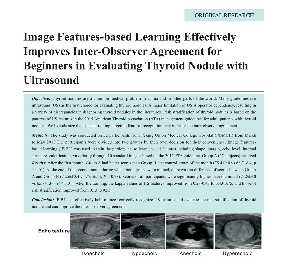
Objective: Thyroid nodules are a common medical problem in China and in other parts of the world. Many guidelines use ultrasound (US) as the first choice for evaluating thyroid nodules. A major limitation of US is operator dependency, resulting in a variety of discrepancies in diagnosing thyroid nodules in the literatures. Risk stratification of thyroid nodules is based on the patterns of US features in the 2015 American Thyroid Association (ATA) management guidelines for adult patients with thyroid nodules. We hypothesize that special training targeting features recognition may increase the inter-observer agreement.
Methods: The study was conducted on 52 participants from Peking Union Medical College Hospital (PUMCH) from March to May 2018.The participants were divided into two groups by their own decision for their convenience. Image featuresbased learning (IF-BL) was used to train the participants to learn special features including shape, margin, echo level, internal structure, calcification, vascularity through 10 standard images based on the 2015 ATA guideline. Group A (27 subjects) received IF-BL during the first month, and Group B (25 subjects) received IF-BL during the second month. All participants evaluated US features and risk stratification in 60 US images of 20 thyroid nodules before and after the training. The test results were graded by a teaching assistant according to the rule of 0.5 points assigned to every feature and 2 points assigned to risk stratification, with a total of 100 points. Inter-observer agreements of US features and risk stratification were assessed and compared before and after the training.
Results: After the first month, Group A had better scores than Group B, the control group of the month (75.4±9.4 vs 68.7±8.4, p = 0.01). At the end of the second month during which both groups were trained, there was no difference of scores between Group A and Group B (74.5±10.4 vs 75.1±7.4, P = 0.78). Scores of all participants were significantly higher than the initial (74.8±9.0 vs 65.8±13.6, P < 0.01). After the training, the kappa values of US features improved from 0.28-0.43 to 0.43-0.75, and those of risk stratification improved from 0.13 to 0.55.
Conclusion: IF-BL can effectively help trainees correctly recognize US features and evaluate the risk stratification of thyroid nodule and can improve the inter-observer agreement. -
Graphic Paper
- Potential Threat of Tracheal Diverticulum to Thermal Ablation Treatment of Thyroid Nodule
- Jianquan Zhang, MD, Lei Yan, MD, Zongping Diao, MD, Hongqiong Chen, MD, Jie Cheng, MD
- 2019, 3 (1): 6-11. DOI:10.37015/AUDT.2019.190802
- Abstract ( 529 ) HTML ( 5 ) PDF ( 1181KB ) ( 515 )
-
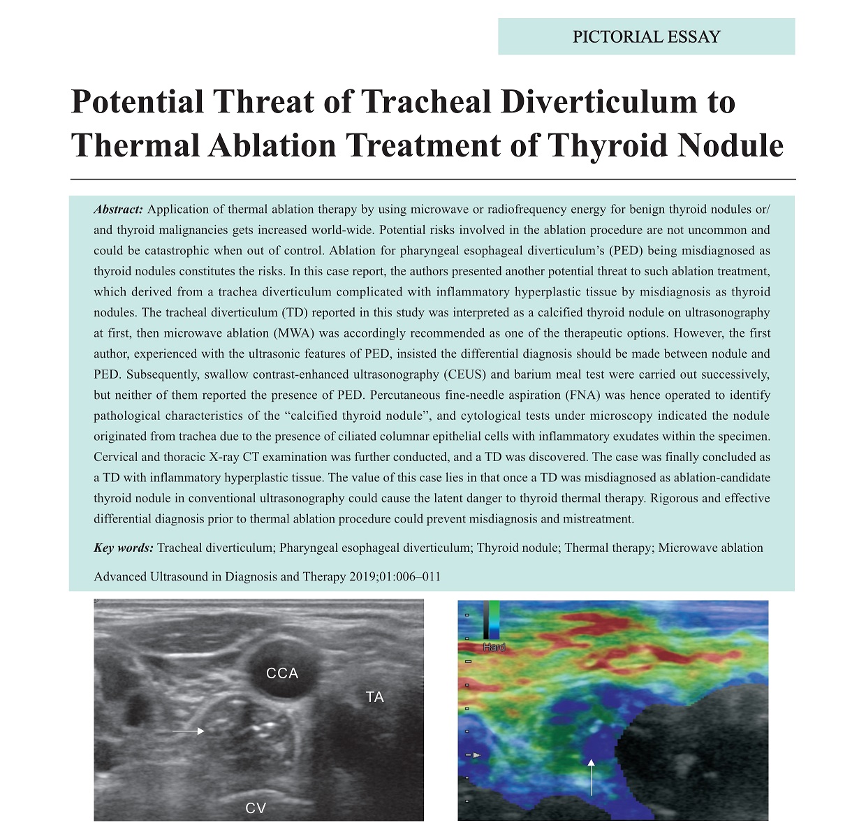
Application of thermal ablation therapy by using microwave or radiofrequency energy for benign thyroid nodules or/ and thyroid malignancies gets increased world-wide. Potential risks involved in the ablation procedure are not uncommon and could be catastrophic when out of control. Ablation for pharyngeal esophageal diverticulum’s (PED) being misdiagnosed as thyroid nodules constitutes the risks. In this case report, the authors presented another potential threat to such ablation treatment, which derived from a trachea diverticulum complicated with inflammatory hyperplastic tissue by misdiagnosis as thyroid nodules. The tracheal diverticulum (TD) reported in this study was interpreted as a calcified thyroid nodule on ultrasonography at first, then microwave ablation (MWA) was accordingly recommended as one of the therapeutic options. However, the first author, experienced with the ultrasonic features of PED, insisted the differential diagnosis should be made between nodule and PED. Subsequently, swallow contrast-enhanced ultrasonography (CEUS) and barium meal test were carried out successively, but neither of them reported the presence of PED. Percutaneous fine-needle aspiration (FNA) was hence operated to identify pathological characteristics of the “calcified thyroid nodule”, and cytological tests under microscopy indicated the nodule originated from trachea due to the presence of ciliated columnar epithelial cells with inflammatory exudates within the specimen. Cervical and thoracic X-ray CT examination was further conducted, and a TD was discovered. The case was finally concluded as a TD with inflammatory hyperplastic tissue. The value of this case lies in that once a TD was misdiagnosed as ablation-candidate thyroid nodule in conventional ultrasonography could cause the latent danger to thyroid thermal therapy. Rigorous and effective differential diagnosis prior to thermal ablation procedure could prevent misdiagnosis and mistreatment.
-
Case Report
- Ultrasonic Imaging of Clear Cell Sarcoma of Kidney: A Case Report
- Muyi Mao, MD, Bei Xia, MD, Weiling Chen, MD, Jianming Song, MD
- 2019, 3 (1): 12-17. DOI:10.37015/AUDT.2019.190803
- Abstract ( 454 ) HTML ( 14 ) PDF ( 2597KB ) ( 451 )
-
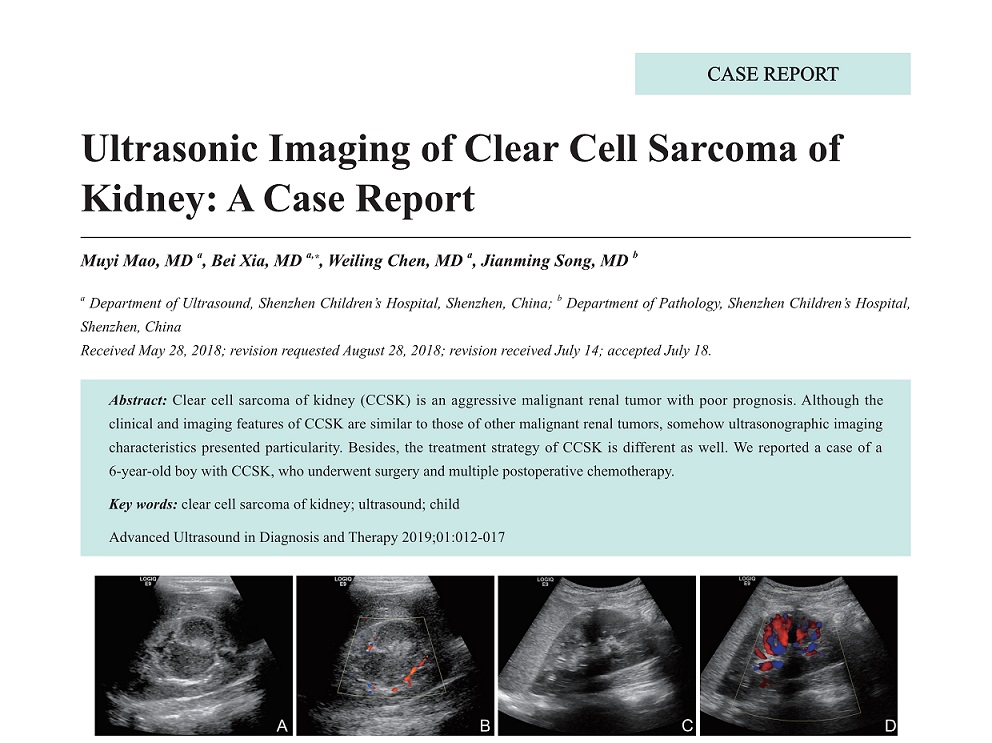
Clear cell sarcoma of kidney (CCSK) is an aggressive malignant renal tumor with poor prognosis. Although the clinical and imaging features of CCSK are similar to those of other malignant renal tumors, somehow ultrasonographic imaging characteristics presented particularity. Besides, the treatment strategy of CCSK is different as well. We reported a case of a 6-year-old boy with CCSK, who underwent surgery and multiple postoperative chemotherapy.
-
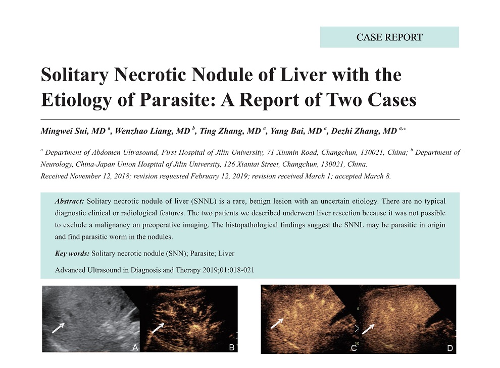
- Solitary Necrotic Nodule of Liver with the Etiology of Parasite: A Report of Two Cases
- Mingwei Sui, MD, Wenzhao Liang, MD, Ting Zhang, MD, Yang Bai, MD, Dezhi Zhang, MD
- 2019, 3 (1): 18-21. DOI:10.37015/AUDT.2019.190804
- Abstract ( 399 ) HTML ( 15 ) PDF ( 690KB ) ( 816 )
-
Solitary necrotic nodule of liver (SNNL) is a rare, benign lesion with an uncertain etiology. There are no typical diagnostic clinical or radiological features. The two patients we described underwent liver resection because it was not possible to exclude a malignancy on preoperative imaging. The histopathological findings suggest the SNNL may be parasitic in origin and find parasitic worm in the nodules.
-
- Horner Syndrome as a Complication Following Microwave Ablation of Secondary Hyperparathyroidism (sHPT): A Case Report
- Ying Wei, MD, Lili Peng, MD, Zhenlong Zhao, MD, Mingan Yu, PhD, MD
- 2019, 3 (1): 22-25. DOI:10.37015/AUDT.2019.190805
- Abstract ( 388 ) HTML ( 3 ) PDF ( 1058KB ) ( 389 )
-
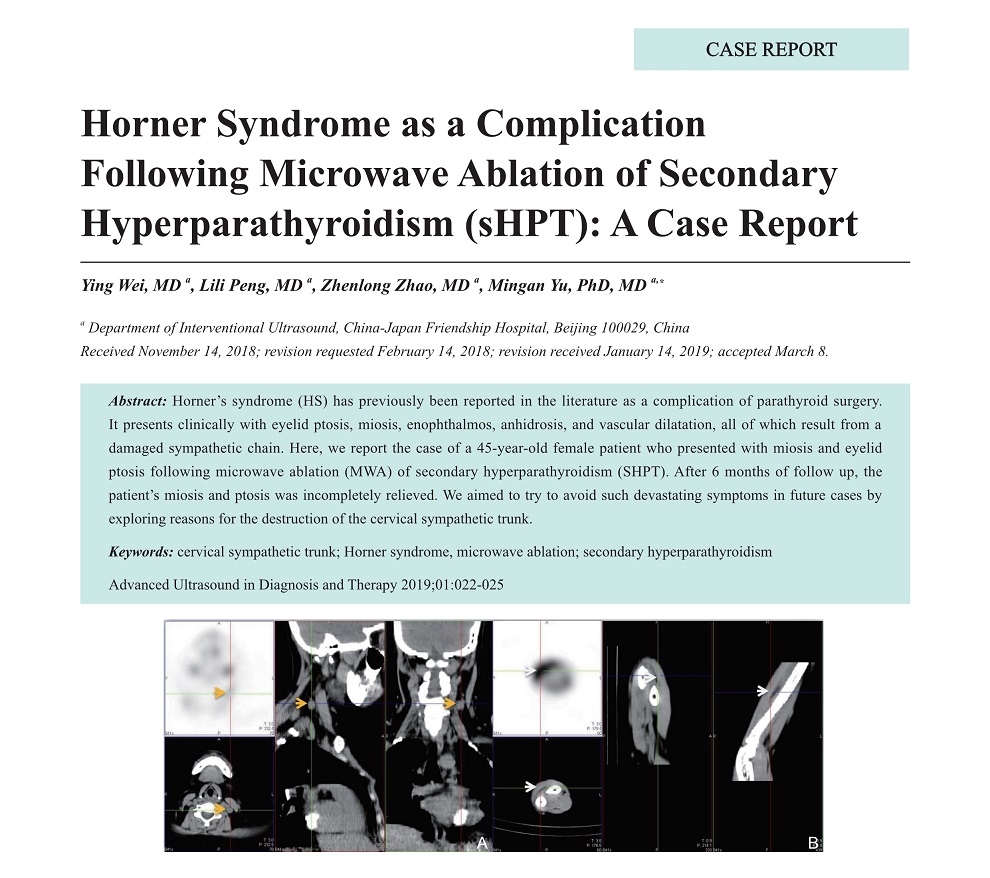
Horner’s syndrome (HS) has previously been reported in the literature as a complication of parathyroid surgery. It presents clinically with eyelid ptosis, miosis, enophthalmos, anhidrosis, and vascular dilatation, all of which result from a damaged sympathetic chain. Here, we report the case of a 45-year-old female patient who presented with miosis and eyelid ptosis following microwave ablation (MWA) of secondary hyperparathyroidism (SHPT). After 6 months of follow up, the patient’s miosis and ptosis was incompletely relieved. We aimed to try to avoid such devastating symptoms in future cases by exploring reasons for the destruction of the cervical sympathetic trunk.
-