[an error occurred while processing this directive]

Advanced Ultrasound in Diagnosis and Therapy ›› 2025, Vol. 9 ›› Issue (3): 277-282.doi: 10.26599/AUDT.2025.240008

• • 上一篇    下一篇


  • 收稿日期:2024-12-15 修回日期:2025-02-14 接受日期:2025-03-04 出版日期:2025-09-30 发布日期:2025-10-13

Sonographic Assessment of Uterine and Ovarian Development in Normal Girls aged 2-18 Years in Pakistan

Irshad Ayeshaa, Muhammad Yousaf Farooq Syedb,c,*(), Fatima Mehreena, Gilani Syed Amirc,d, Yousaf Muhammada   

  1. aUniversity Institute of Radiological Sciences & Medical Imaging Technology, Faculty of Allied Health Sciences, The University of Lahore
    bRadiography and Imaging Technology, Green International University, Lahore, Pakistan
    cOffice of Research, Innovation and Commercialization, Green International University, Lahore. Pakistan
    dDepartment of Medical Diagnostic imaging, College of Health Sciences, University of Sharjah, United Arab Emirates
  • Received:2024-12-15 Revised:2025-02-14 Accepted:2025-03-04 Online:2025-09-30 Published:2025-10-13
  • Contact: Radiography and Imaging Technology, Green International University, Lahore, Pakistan. Office of Research, Innovation and Commercialization, Green International University, Lahore, Pakistan. e-mail:yousafgelani@gmail.com(SMY F)

Abstract:

Objective Uterine and ovarian development is influenced by both age and hormonal milieu. Sonographic assessment of normal pubertal and pre-puberty girls provides critical insights into the physiological trajectory of female gonadal maturation and its potential pathological deviations.
Methods This was a cross-sectional study conducted at Gilani Ultrasound Center, Lahore, Pakistan. The duration of the study was 9 months. Uterus length, width, height, volume, right/left ovary volume and Fundo/Cervical ratio were measured.
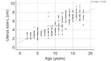
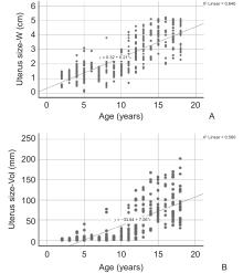
Results A total of 384 subjects were included in our study, categorizing them into pre-puberty (age 2-6 years), early puberty (age 7-11 years), and late puberty (age 12-18 years) groups. In the pre-puberty group (n = 111, mean age 4.29 ± 1.20 years), uterine measurements revealed a mean length of 2.95 cm, height of 0.97 cm, width of 1.41 cm, and volume of 4.82 cm3. The mean volumes of the right and left ovaries were 1.24 cm3 and 1.10 cm3, respectively, with a mean F/C ratio of 1.33. For the early puberty group (n = 99, mean age 9.15 ± 1.45 years), uterine measurements included a mean length of 4.03 cm, height of 1.22 cm, width of 1.80 cm, and volume of 12.37 cm3. In the late puberty group (n = 31, mean age 12.63 ± 1.21 years), uterine measurements showed a mean length of 5.29 cm, height of 1.82 cm, width of 2.65 cm, and volume of 31.11 cm3. The mean volumes of the right and left ovaries were 4.70 cm3 and 5.26 cm3, respectively, with a mean F/C ratio of 1.26.
Conclusion Uterine and ovarian dimensions, including volumes, correlate directly with age and pubertal status, except for the (Fundus/Cervical) ratio, which shows individual variability. This normative data could serve as a basis for the evaluation of Uterine and ovarian dimensions and volume in the local population.

Key words: Normotensive, Uterus volume, Ovarian volume, Puberty

"

VariablesPre-pubertyPuberty
2-6 years7-11 years12-18 years7-11 years12-18 years
n11199318135
Age (years)4.29 ± 1.209.15 ± 1.4512.63 ± 1.219.50 ± 1.3115.21 ± 2.01
Uterus length (cm)2.95 ± 0.664.03 ± 1.095.29 ± 1.224.96 ± 1.657.18 ± 0.92
Uterus height (cm)0.97 ± 0.501.22 ± 0.621.82 ± 0.681.75 ± 0.862.99 ± 0.56
Uterus width (cm)1.41 ± 0.531.80 ± 0.702.65 ± 0.742.58 ± 1.043.81 ± 0.79
Uterus volume (cm3)4.82 ± 5.2012.37 ± 15.5431.11 ± 28.0231.52 ± 31.8286.16 ± 38.75
Right ovary volume (cm3)1.24 ± 0.833.13 ± 2.064.70 ± 2.623.33 ± 1.506.64 ± 2.67
Left ovary volume (cm3)1.10 ± 0.682.68 ± 1.745.26 ± 3.593.10 ± 1.426.83 ± 2.68
F/C ratio1.33 ± 0.421.26 ± 0.291.26 ± 0.291.34 ± 0.411.47 ± 0.34

"

ItemsTotal (n)Pre-puberty (n)Puberty (n)
Uterus volume (cm3)0-5026523332
50-10075867
101-15033033
151-20010010
201-250101
Uterus height (cm)0.1-1.01321302
1.1-2.099936
2.1-3.0971681
3.1-4.051249
4.1-5.0505
Uterus width (cm)0.1-1.037370
1.1-2.01431385
2.1-3.0825527
3.1-4.0621052
4.1-5.053152
5.1-6.0707
Uterus length (cm)0.1-2.012120
2.1-4.01601582
4.1-6.0735914
6.1-8.011412102
8.1-10.025025
Right ovary volume
(cm3)
0-5.027522154
5.1-10.0912071
10.1-15.018018
Left ovary volume
(cm3)
0-5.026921950
5.1-10.0931974
10.1-15.022319
F/C ratio0.1-1.045396
1.1-2.0328198130
2.1-3.01147
Total384241143

"

Model Summary
ModelRR SquareAdjusted R SquareP value
10.849 a0.7200.719< 0.001

"

"

[1] Asăvoaie C, Fufezan O, Coşarca M. Ovarian and uterine ultrasonography in pediatric patients. Pictorial essay. Medical Ultrasonography 2014; 16: 160-167.
[2] Motlagh ME, Rabbani A, Kelishadi R, Mirmoghtadaee P, Shahryari S, Ardalan G, et al. Timing of puberty in Iranian girls according to their living area: a national study. J Res Med Sci 2011; 16: 276.
[3] Soriano-Guillén L, Tena-Sempere M, Seraphim CE, Latronico AC, Argente J. Precocious sexual maturation: Unravelling the mechanisms of pubertal onset through clinical observations. J Neuroendocrinol 2022; 34: e12979.
[4] Viner RM, Allen NB, Patton GC. Puberty, developmental processes, and health interventions. In: Bundy DAP, Silva ND, Horton S, Jamison DT, Patton GC, editors. Child and Adolescent Health and Development. Washington (DC): The World Bank 2017.
[5] Razzaghy-Azar M, Ghasemi F, Hallaji F, Ghasemi A, Ghasemi M. Sonographic measurement of uterus and ovaries in premenarcheal healthy girls between 6 and 13 years old: correlation with age and pubertal status. J Clin Ultrasound 2011; 39: 64-73.
[6] Talarico V, Rodio MB, Viscomi A, Galea E, Galati MC, Raiola G. The role of pelvic ultrasound for the diagnosis and management of central precocious puberty: An update. Acta Biomed 2021; 92: e2021480.
[7] Takhreem M, Akram Q. Essentials of Ultrasound for Practical Scanning. InUltrasound in Rheumatology: A Practical Guide for Diagnosis 2021 May 16 (pp. 1-15). Cham: Springer International Publishing.
[8] Bhargava SK. Principles and practice of ultrasonography. Jaypee Brothers Medical Publishers 2002; 30.
[9] Herter LD, Golendziner E, Flores JA, Moretto M, Di Domenico K, Becker Jr E, et al. Ovarian and uterine findings in pelvic sonography: comparison between prepubertal girls, girls with isolated thelarche, and girls with central precocious puberty. J Ultrasound Med 2002; 21: 1237-1246.
[10] Emmanuel M, Bokor BR. Tanner stages. 2017.
[11] Assens M, Dyre L, Henriksen LS, Brocks V, Sundberg K, Jensen LN, et al. Menstrual pattern, reproductive hormones, and transabdominal 3D ultrasound in 317 adolescent girls. J Clin Endocrinol Metab 2020; 105: e3257-e3266.
[12] Sample WF, Lippe BM, Gyepes MT. Gray-scale ultrasonography of the normal female pelvis. Radiology 1977; 125: 477-483.
[13] Giorlandino C, Gleicher N, Taramanni C, Vizzone A, Gentili P, Mancuso S, et al. Ovarian development of the female child and adolescent: I. Morphology. Int J Gynaecol Obstet 1989; 29: 57-63.
[14] Badouraki M, Christoforidis A, Economou I, Dimitriadis AS, Katzos G. Sonographic assessment of uterine and ovarian development in normal girls aged 1 to 12 years. J Clin Ultrasound 2008; 36: 539-544.
[15] de Vries L, Horev G, Schwartz M, Phillip M. Ultrasonographic and clinical parameters for early differentiation between precocious puberty and premature thelarche. Eur J Endocrinol 2006; 154: 891-898.
[16] Ivarsson SA, Nilsson KO, Persson PH. Ultrasonography of the pelvic organs in prepubertal and postpubertal girls. Arch Dis Child 1983; 58: 352-354.
[17] Stanhope R, Adams J, Jacobs HS, Brook CG. Ovarian ultrasound assessment in normal children, idiopathic precocious puberty, and during low dose pulsatile gonadotrophin releasing hormone treatment of hypogonadotrophic hypogonadism. Arch Dis Child 1985; 60: 116-119.
[18] Orsini LF, Salardi S, Pilu G, Bovicelli L, Cacciari E. Pelvic organs in premenarcheal girls: real-time ultrasonography. Radiology 1984; 153: 113-116.
No related articles found!
Viewed
Full text


Abstract

Cited

  Shared   
  Discussed   
No Suggested Reading articles found!
[an error occurred while processing this directive]