[an error occurred while processing this directive]

Advanced Ultrasound in Diagnosis and Therapy ›› 2025, Vol. 9 ›› Issue (3): 283-289.doi: 10.26599/AUDT.2025.240033

• • 上一篇    下一篇


  • 收稿日期:2024-07-13 修回日期:2024-12-05 接受日期:2024-12-27 出版日期:2025-09-30 发布日期:2025-10-13

Inter and Intra-Rater Reliability of Musculoskeletal Ultrasound to Measure Plantar Fascia Thickness in Patients with Established Plantar Fasciitis

Farooq Syed Muhammad Yousafa,b,*(), Rasool Kinzac, Gilani Syed Amird, Fatima Mehreenc, Malik Sajid Shaheenc   

  1. aRadiography and Imaging Technology, Green International University, Lahore, Pakistan
    bOffice of Research, Innovation and Commercialization, Green International University, Lahore, Pakistan
    cUniversity Institute of Radiological Sciences & Medical Imaging Technology, Faculty of Allied Health Sciences, The University of Lahore
    dDepartment of Medical Diagnostic Imaging, College of Health Sciences, University of Sharjah, Sharjah, UAE
  • Received:2024-07-13 Revised:2024-12-05 Accepted:2024-12-27 Online:2025-09-30 Published:2025-10-13
  • Contact: Radiography and Imaging Technology, Green International University, Lahore, Pakistan. Office of Research, Innovation and Commercialization, Green International University, Lahore, Pakistan. e-mail:yousafgelani@gmail.com(SMY F)

Abstract:

Objective Plantar fascia (PF) is a thick connective tissue on the plantar surface of the foot that plays a crucial role in maintaining the longitudinal arch. Plantar fasciitis, characterized by inflammation of the medial tuberosity of the calcaneus, is one of the most common causes of heel pain. Imaging is essential for accurate diagnosis, with ultrasonography widely applied to evaluate PF thickness, monitor therapeutic outcomes, and guide interventions. However, its application is limited by operator dependency. This study aimed to investigate the inter- and intra-rater reliability of musculoskeletal ultrasound in measuring PF thickness in patients with plantar fasciitis.
Methods In this cross-sectional analytical study, 40 participants were enrolled, including 26 females and 14 males. The reliability of PF measurements on different sides was assessed using Cronbach’s alpha and intraclass correlation coefficients (ICC).
Results A total of 40 participants (age range: 20-60 years) were included in the study. PF thickness in patients with plantar fasciitis measured by Observer 1 ranged from 3.8-6.9 mm (left) and 3.2-6.0 mm (right), whereas measurements by Observer 2 ranged from 2.9-7.1 mm (left) and 3.2-6.0 mm (right). Intra-rater reliability showed ICCs ranging from 0.618-0.857 for Observer 1 and 0.76-0.92 for Observer 2, indicating moderate (> 0.60) to excellent reliability.
Conclusion PF is a deep structure, and its visualization may be influenced by operator technique. Ultrasound measurement of tendon thickness shows good reliability in patients with established plantar fasciitis.

Key words: Plantar fasciitis, Musculoskeletal ultrasound, Thickness, Intra-rater, Inter-rater, Reliability, Heel Pain

"

"

"

VariablesNMeanS.D.Cronbach’s AlphaICC95% CIP-value
Medial supine Right OB 1404.4600.940.9230.8570.746-0.9220.000
Medial supine Right OB 2404.2100.770.9230.8570.855-0.9590.000
Medial supine Left OB 1404.4130.960.8070.6820.472-0.8180.000
Medial supine Left OB 2404.3730.650.8070.6820.641-0.9000.000
Central supine Right OB 1404.4030.8570.9160.7860.448-0.9050.000
Central supine Right OB 2404.0930.6960.9160.7860.618-0.9500.000
Central supine Left OB1404.3901.01950.8590.7530.579-0.8610.000
Central supine Left OB 2404.4330.65770.8590.7530.733-0.9250.000
Lateral supine Right OB 1404.3450.78610.8740.7760.615-0.8750.000
Lateral supine Right OB 2404.0880.61110.8740.7760.762-0.9330.000
Lateral supine Left OB 1404.4731.02610.8780.7820.627-0.8780.000
Lateral supine Left OB 2404.3850.70770.8780.7820.770-0.9350.000
Medial prone Right OB 1404.8251.32460.8640.7440.558-0.8580.000
Medial prone Right OB 2405.1301.22090.8640.7440.716-0.9230.000
Medial prone Left OB 1404.5981.57180.8650.7610.592-0.8660.000
Medial prone Left OB 2405.0881.60280.8650.7610.744-0.9280.000
Central prone Right OB 1404.7251.12200.8960.7900.615-0.8870.000
Central prone Right OB 2405.0181.20190.8960.7900.761-0.9400.000
Central prone Left OB 1404.6081.34400.8710.7500.557-0.8630.000
Central prone Left OB 2404.9781.46190.8710.7500.716-0.9260.000
Lateral prone Right OB1404.5501.12980.8700.7300.488-0.8590.000
Lateral prone Right OB 2404.9481.18080.8700.7300.656-0.9240.000
Lateral Prone Left OB 1404.4951.26980.7950.6180.347-0.7860.000
Lateral Prone Left OB 2405.0601.58050.7950.6180.515-0.8800.000

"

"

"

"

"

[1] Ieong E, Afolayan J, Carne A, Solan M. Ultrasound scanning for recalcitrant plantar fasciopathy. Basis of a new classification. Skeletal radiol 2013; 42: 393-398.
[2] Gordon R, Wong C, Crawford EJ. Ultrasonographic evaluation of low energy extracorporeal pulse activated therapy (EPAT) for chronic plantar fasciitis. Foot Ankle Int 2012; 33: 202-207.
[3] Akfirat M, Sen C, Günes T. Ultrasonographic appearance of the plantar fasciitis. Clin Imaging 2003; 27: 353-357.
[4] Ehrmann C, Maier M, Mengiardi B, Pfirrmann CW, Sutter R. Calcaneal attachment of the plantar fascia: MR findings in asymptomatic volunteers. Radiology 2014; 272: 807-814.
[5] Cornwall MW, McPoil TG. Plantar fasciitis: etiology and treatment. J Orthop Sports Phys Ther 1999; 29: 756-760.
[6] Khammas ASA, Mahmud R, Hassan HA, Ibrahim I, Mohammed SS. An assessment of plantar fascia with ultrasound findings in patients with plantar fasciitis: a systematic review. J Ultrasound 2023; 26: 13-38.
[7] Kane D, Grassi W, Sturrock R, Balint PV. Musculoskeletal ultrasound--a state of the art review in rheumatology. Part 2: Clinical indications for musculoskeletal ultrasound in rheumatology. Rheumatology (Oxford) 2004; 43: 829-838.
[8] Park YH, Kim HJ, Kim W, Choi JW. Reliability of ultrasound measurement of plantar fascia thickness: a systematic review. J Am Podiatr Med Assoc 2023; 113.
[9] McNally M, Mousavi ME, Mohseni-Bandpei MA, Shakourirad A, Safari MR, Kashani RV, et al. Intra-and inter-rater reliability of ultrasound in plantar fascia thickness measurement. Iranian Journal of Radiology 2018; 15.
[10] McNally EG, Shetty S. Plantar fascia: imaging diagnosis and guided treatment. Semin Musculoskelet Radiol 2010; 14: 334-343.
[11] Wang X, Xu L, Hu X, Zhao H, Yin J. Musculoskeletal ultrasound for the diagnosis of plantar fasciitis: An accuracy and diagnostic yield study. Int J Gen Med 2023; 16: 4765-4771.
[12] Vohra PK, Japour CJ. Ultrasound-guided plantar fascia release technique: a retrospective study of 46 feet. J Am Podiatr Med Assoc 2009; 99: 183-190.
[13] 
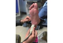
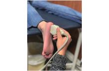
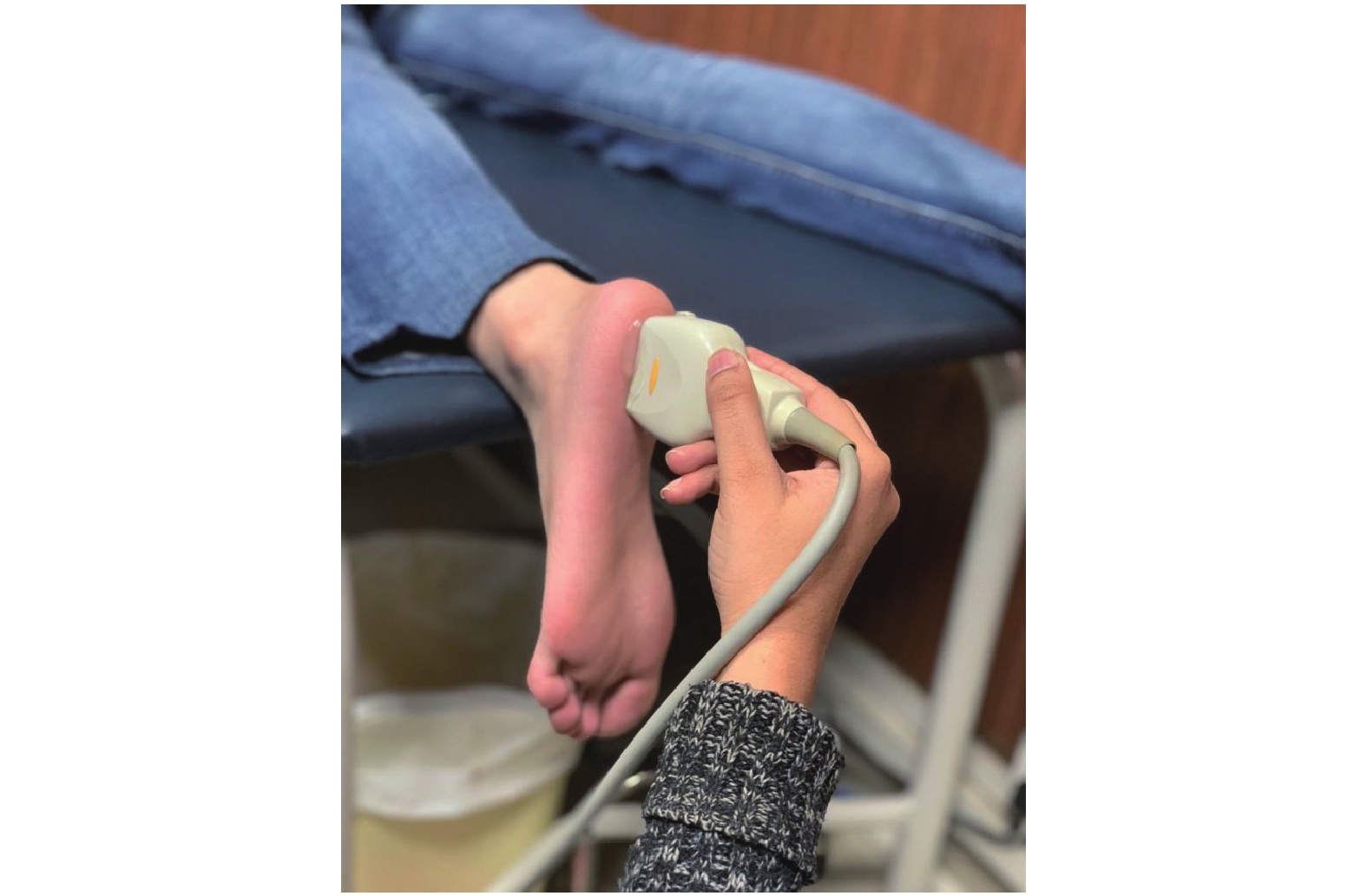
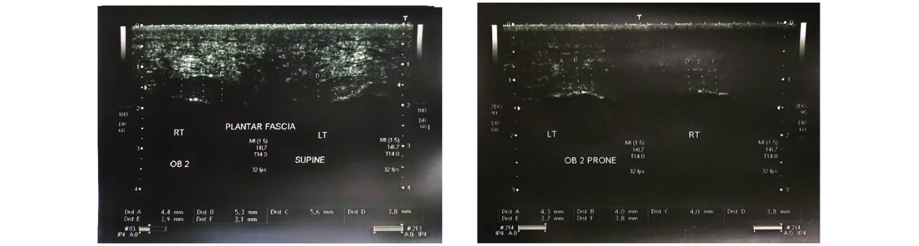
Ahn JH, Lee CW, Park C, Kim YC. Ultrasonographic examination of plantar fasciitis: a comparison of patient positions during examination. J Foot Ankle Res 2016; 9: 38.
[14] Kock N, Hadaya P. Minimum sample size estimation in PLS‐SEM: The inverse square root and gamma‐exponential methods. Info Systems J 2018; 28: 227-261.
[15] Cheng JW, Tsai WC, Yu TY, Huang KY. Reproducibility of sonographic measurement of thickness and echogenicity of the plantar fascia. J Clin Ultrasound 2012; 40: 14-19.
[16] Rathleff MS, Moelgaard C, Lykkegaard Olesen J. Intra- and interobserver reliability of quantitative ultrasound measurement of the plantar fascia. J Clin Ultrasound 2011; 39: 128-134.
[17] Mohseni-Bandpei MA, Nakhaee M, Mousavi ME, Shakourirad A, Safari MR, Vahab Kashani R. Application of ultrasound in the assessment of plantar fascia in patients with plantar fasciitis: a systematic review. Ultrasound Med Biol 2014; 40: 1737-1754.
[18] Bisi-Balogun A, Rector M. Clinical utility of ultrasound measurements of plantar fascia width and cross-sectional area: a novel technique. J Am Podiatr Med Assoc 2017; 107: 375-381.
[19] Cardinal E, Chhem RK, Beauregard CG, Aubin B, Pelletier M. Plantar fasciitis: sonograpic evaluation. Radiology 1996; 201: 257-259.
[20] Gibbon WW, Long G. Ultrasound of the plantar aponeurosis (fascia). Skeletal Radiol 1999; 28: 21-26.
[21] Ozdemir H, Yilmaz E, Murat A, Karakurt L, Poyraz AK, Ogur E. Sonographic evaluation of plantar fasciitis and relation to body mass index. Eur Radiol 2005; 54: 443-447.
[22] Wall JR, Harkness MA, Crawford A. Ultrasound dignosis of plantar fasciitis. Foot & Ankle 1993; 148: 465-470.
No related articles found!
Viewed
Full text


Abstract

Cited

  Shared   
  Discussed   
No Suggested Reading articles found!
[an error occurred while processing this directive]