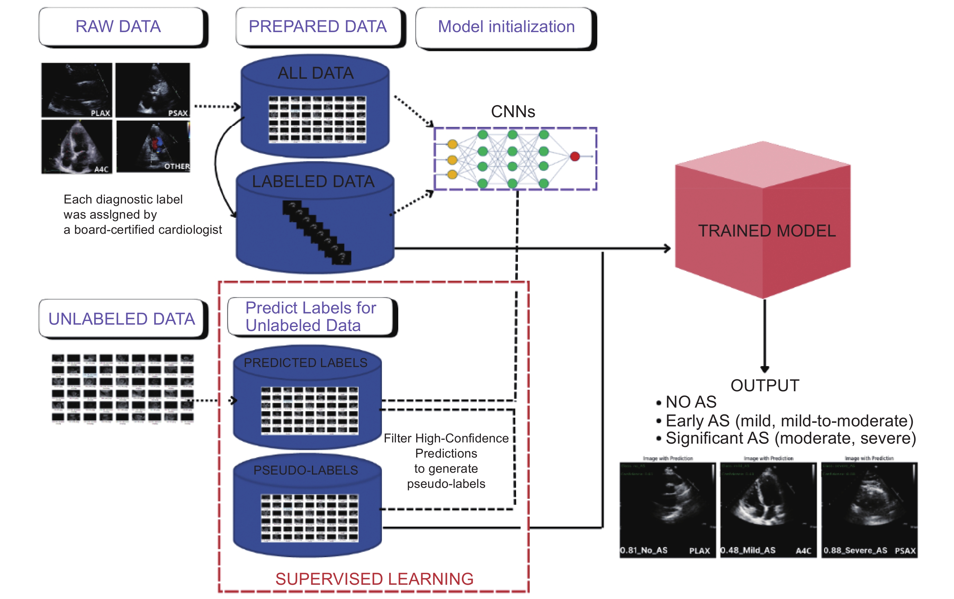
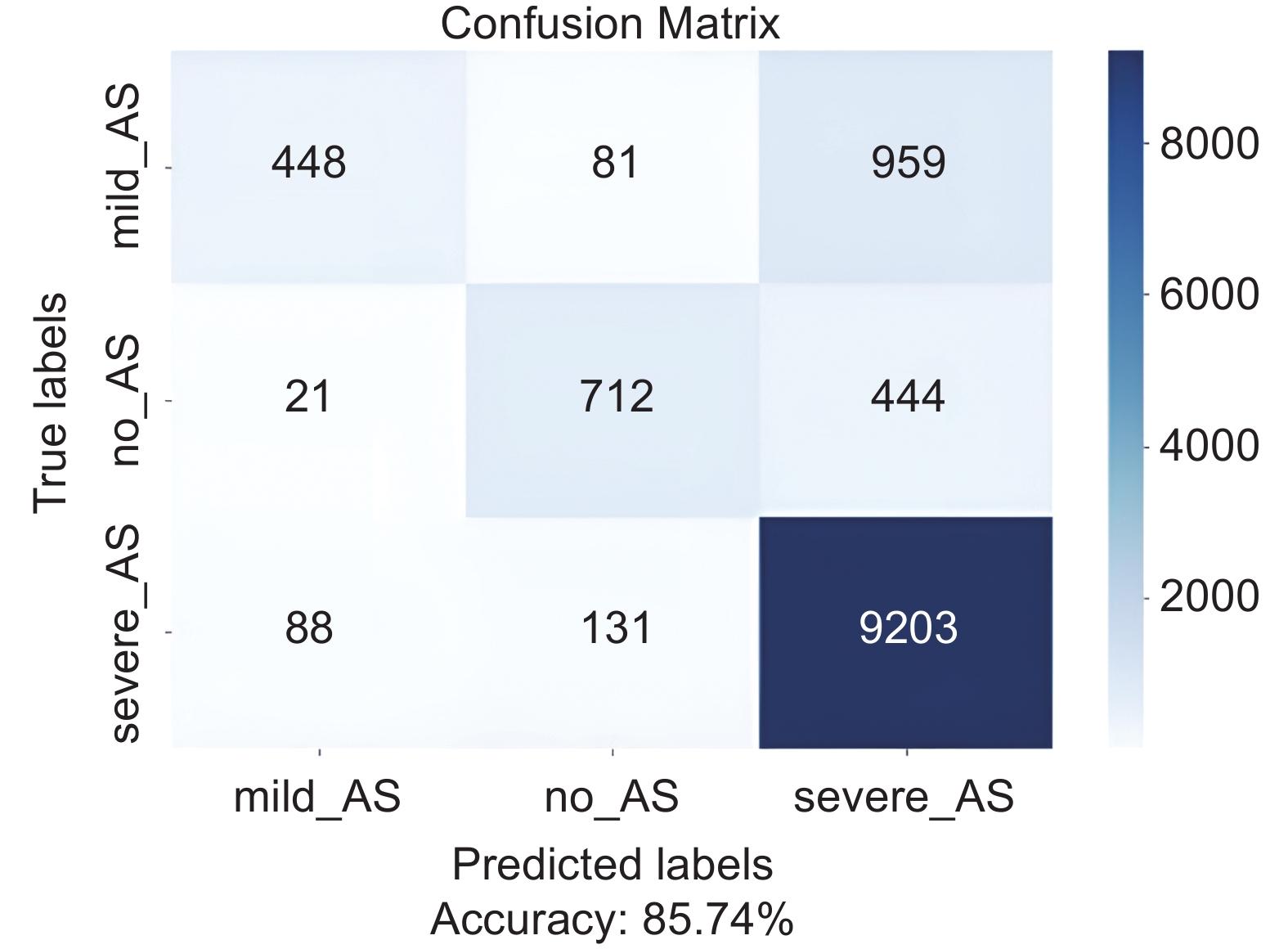
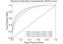
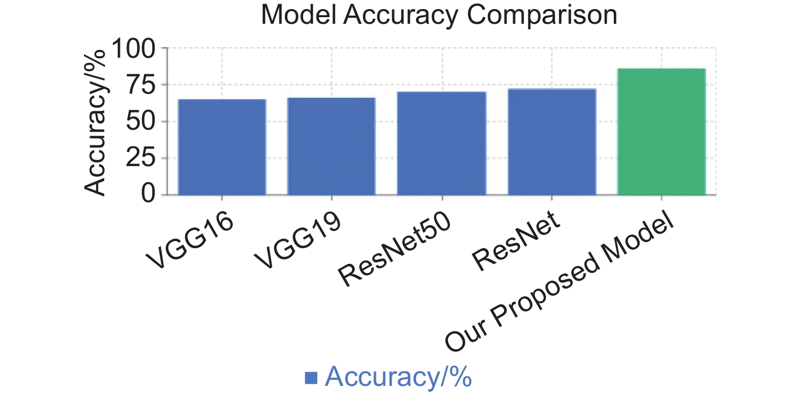
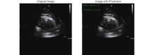
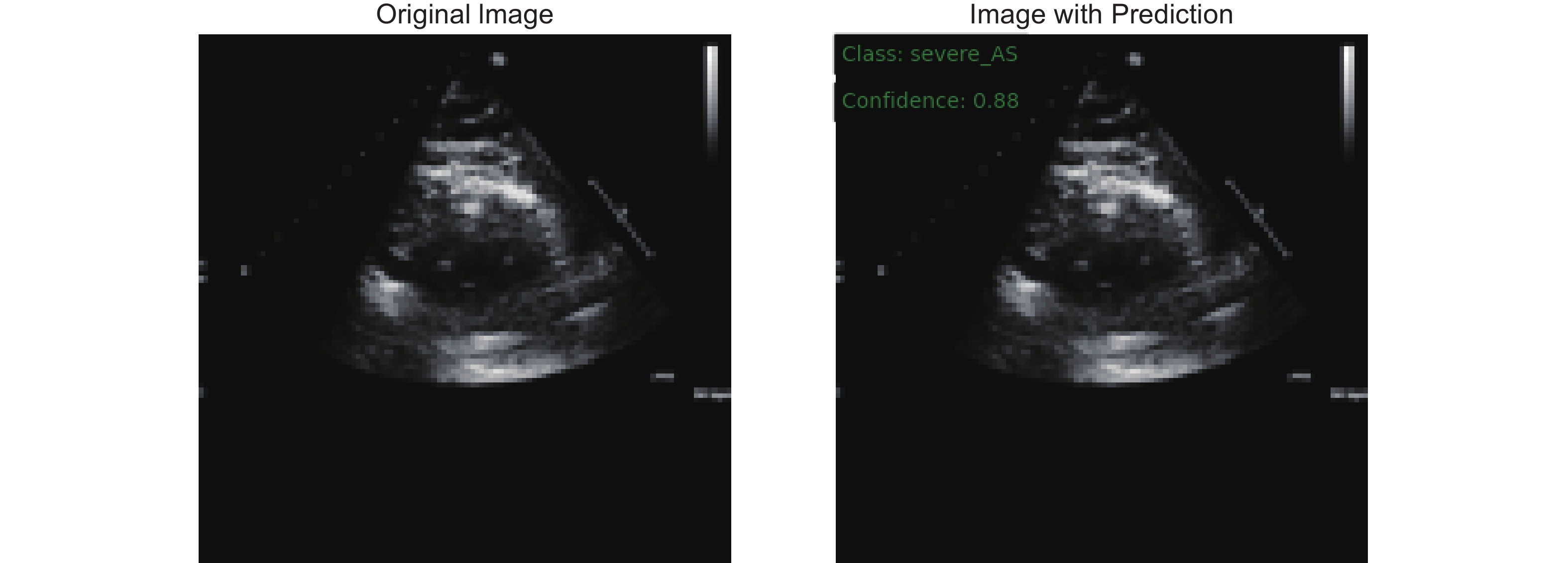
| [1] |
Zhenyi L, Ya C, Xinqi W, Lan Y, Anni C, Zhaojun L, et al. Left and right ventricular interaction: insight from echocardiography imaging. Advanced Ultrasound in Diagnosis and Therapy 2024; 8: 195-204.
|
| [2] |
Vahanian A, Beyersdorf F, Praz F, Milojevic M, Baldus S, Bauersachs J, et al. 2021 ESC/EACTS Guidelines for the management of valvular heart disease. Eur Heart J 2022; 43: 561-632.
|
| [3] |
Aluru JS, Barsouk A, Saginala K, Rawla P, Barsouk A. Valvular heart disease epidemiology. Med Sci (Basel) 2022; 10: 32
|
| [4] |
Guedeney P, Collet JP. Aortic stenosis: an update. Rev Med Interne 2022; 43: 145-151.
|
| [5] |
Chen W, Xie Y, Zhang Z, Zhu Y, Zhang Y, Zhu S, et al. Artificial intelligence-assisted medical imaging in interventional management of valvular heart disease. Advanced Ultrasound in Diagnosis & Therapy 2023; 7: 217-227.
|
| [6] |
Pachulski RT, Chan KL. Progression of aortic valve dysfunction in 51 adult patients with congenital bicuspid aortic valve: assessment and follow up by Doppler echocardiography. Br Heart J 1993; 69: 237-240.
|
| [7] |
Muhammad Y, Tahir M, Hayat M, Chong KT. Early and accurate detection and diagnosis of heart disease using intelligent computational model. Sci Rep 2020; 10: 19747.
|
| [8] |
Tribouilloy C, Rusinaru D, Maréchaux S, Castel AL, Debry N, Maizel J, et al. Low-gradient, low-flow severe aortic stenosis with preserved left ventricular ejection fraction: characteristics, outcome, and implications for surgery. J Am Coll Cardiol 2015; 65: 55-66.
|
| [9] |
Zoghbi WA, Galan A, Quinones MA. Accurate assessment of aortic stenosis severity by Doppler echocardiography independent of aortic jet velocity. Am Heart J 1988; 116: 855-863.
|
| [10] |
Bang W C, Seong Y K, Lee J. The impact of deep learning on ultrasound in diagnosis and therapy: enhancing clinical decision support, workflow efficiency, quantification, image registration, and real-time assistance. Advanced Ultrasound in Diagnosis & Therapy (AUDT) 2023; 7: 204-216.
|
| [11] |
Hagendorff A, Knebel F, Helfen A, Knierim J, Sinning C, Stöbe S, et al. Expert consensus document on the assessment of the severity of aortic valve stenosis by echocardiography to provide diagnostic conclusiveness by standardized verifiable documentation. Clin Res Cardiol 2020; 109: 271-288.
|
| [12] |
Chernyshov A, Grue JF, Nyberg J, Grenne B, Dalen H, Aase SA, et al. Automated segmentation and quantification of the right ventricle in 2-D echocardiography. Ultrasound Med Biol 2024; 50: 540-548.
|
| [13] |
Gill H, Fernandes J, Chehab O, Prendergast B, Redwood S, Chiribiri A, et al. Evaluation of aortic stenosis: from Bernoulli and Doppler to Navier-Stokes. Trends Cardiovasc Med 2023; 33: 32-43.
|
| [14] |
Guzzetti E, Capoulade R, Tastet L, Garcia J, Le Ven F, Arsenault M, et al. Estimation of stroke volume and aortic valve area in patients with aortic stenosis: a comparison of echocardiography versus cardiovascular magnetic resonance. J Am Soc Echocardiogr 2020; 33: 953-963.e5.
|
| [15] |
Saikrishnan N, Kumar G, Sawaya FJ, Lerakis S, Yoganathan AP. Accurate assessment of aortic stenosis: a review of diagnostic modalities and hemodynamics. Circulation 2014; 129: 244-253.
|
| [16] |
Wessler BS, Huang Z, Long GM Jr, Pacifici S, Prashar N, Karmiy S, et al. Automated detection of aortic stenosis using machine learning. J Am Soc Echocardiogr 2023; 36: 411-420.
|
| [17] |
Sun X, Yin Y, Yang Q, Huo T. Artificial intelligence in cardiovascular diseases: diagnostic and therapeutic perspectives. Eur J Med Res 2023; 28: 242.
|
| [18] |
Milosevic M, Jin Q, Singh A, Amal S. Applications of artificial intelligence in multi-modal imaging for cardiovascular disease. Front Radiol 2024; 3: 1294068.
|
| [19] |
Chen X, Yang F, Zhang P, Lin X, Wang W, Pu H, et al. Artificial intelligence–assisted left ventricular diastolic function assessment and grading: multiview versus single view. J Am Soc Echocardiogr 2023; 36: 1064-1078.
|
| [20] |
Holste G, Oikonomou EK, Mortazavi BJ, Coppi A, Faridi KF, Miller EJ, et al. Severe aortic stenosis detection by deep learning applied to echocardiography. Eur Heart J 2023; 44: 4592-4604.
|
| [21] |
Krishna H, Desai K, Slostad B, Bhayani S, Arnold JH, Ouwerkerk W, et al. Fully automated artificial intelligence assessment of aortic stenosis by echocardiography. J Am Soc Echocardiogr 2023; 36: 769-777.
|
| [22] |
Huang Z, Long G, Wessler BS, Hughes MC. Tmed 2: a dataset for semi-supervised classification of echocardiograms. In DataPerf: Benchmarking Data for Data-Centric AI Workshop 2022.
|
| [23] |
Huang Z, Sidhom MJ, Wessler BS, Hughes MC. Fix-A-Step: semi-supervised learning from uncurated unlabeled data. arXiv preprint arXiv:2208.11870 2023.
|
| [24] |
Huang Z, Long G, Wessler B, Hughes MC. A new semi-supervised learning benchmark for classifying view and diagnosing aortic stenosis from echocardiograms. Machine Learning for Healthcare Conference 2021; 149: 614-647.
|