

| [1] |
Ravine D, Gibson RN, Donlan J, Sheffield LJ. An ultrasound renal cyst prevalence survey: specificity data for inherited renal cystic disease. Am J Kidney Dis 1993; 22:803-807.
doi: 10.1016/S0272-6386(12)70338-4 |
| [2] |
Kissane JM. The morphology of renal cystic diseas. Perspect Nephrol Hypertens 1976; 4:31-63.
pmid: 1264568 |
| [3] |
Quaia E, Bussani R, Cova M, Mucelli RP. Radiologic-pathologic correlations of intratumoral tissue components in the most common solid and cystic renal tumors. Pictorial revie. Eur Radiol 2005; 15:1734-1744.
doi: 10.1007/s00330-005-2698-9 |
| [4] |
Hayakawa M, Hatano T, Tsuji A, Nakajima F, Ogawa Y. Patients with renal cysts associated with renal cell carcinoma and the clinical implications of cyst puncture: a study of 223 case. Urology 1996; 47:643-646.
pmid: 8650859 |
| [5] |
Bosniak MA. The current radiological approach to renal cyst. Radiology 1986; 158:1-10.
pmid: 3510019 |
| [6] |
Israel GM, Hindman N, Bosniak MA. Evaluation of cystic renal masses: comparison of CT and MR imaging by using the Bosniak classification syste. Radiology 2004; 231:365-371.
pmid: 15128983 |
| [7] |
Silverman SG, Pedrosa I, Ellis JH, Hindman NM, Schieda N, Smith AD, et al. Bosniak classification of cystic renal masses, version 2019: an update proposal and needs assessmen. Radiology 2019; 292:475-488.
doi: 10.1148/radiol.2019182646 pmid: 31210616 |
| [8] |
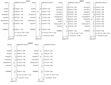
Zhou L, Tang L, Yang T, Chen W. Comparison of contrast-enhanced ultrasound with MRI in the diagnosis of complex cystic renal masses: a meta-analysi. Acta Radiol 2018; 59:1254-1263.
doi: 10.1177/0284185118755575 |
| [9] | Lan D, Qu HC, Li N, Zhu XW, Liu YL, Liu CL. The value of contrast-enhanced ultrasonography and contrast-enhanced CT in the diagnosis of malignant renal cystic lesions: a meta-analysi. PLoS One 2016; 11:e0155857. |
| [10] |
Sanz E, Hevia V, Gomez V, Alvarez S, Fabuel JJ, Martinez L, et al. Renal complex cystic masses: usefulness of contrast-enhanced ultrasound (CEUS) in Their assessment and its agreement with computed tomograph. Curr Urol Rep 2016; 17 :89.
doi: 10.1007/s11934-016-0646-7 |
| [11] |
Chen Y, Wu N, Xue T, Hao Y, Dai J. Comparison of contrast-enhanced sonography with MRI in the diagnosis of complex cystic renal masse. J Clin Ultrasound 2015; 43:203-209.
doi: 10.1002/jcu.22232 pmid: 25179487 |
| [12] |
Ferreira AM, Reis RB, Kajiwara PP, Silva GE, Elias J, Jr., Muglia VF. MRI evaluation of complex renal cysts using the Bosniak classification: a comparison to CT. Abdom Radiol (NY) 2016; 41:2011-2019.
doi: 10.1007/s00261-016-0797-5 pmid: 27271286 |
| [13] |
Zhong J, Cao F, Guan X, Chen J, Ding Z, Zhang M. Renal cyst masses (Bosniak category II-III) may be over evaluated by the Bosniak criteria based on MR finding. Medicine (Baltimore) 2017; 96:e9361.
doi: 10.1097/MD.0000000000009361 |
| [14] |
Defortescu G, Cornu JN, Bejar S, Giwerc A, Gobet F, Werquin C, et al. Diagnostic performance of contrast-enhanced ultrasonography and magnetic resonance imaging for the assessment of complex renal cysts: a prospective stud. Int J Urol 2017; 24:184-189.
doi: 10.1111/iju.13289 pmid: 28147450 |
| [15] |
Edenberg J, Gloersen K, Osman HA, Dimmen M, Berg GV. The role of contrast-enhanced ultrasound in the classification of CT-indeterminate renal lesion. Scand J Urol 2016; 50:445-451.
pmid: 27609413 |
| [16] | Nicolau C, Bunesch L, Pano B, Salvador R, Ribal MJ, Mallofre C, et al. Prospective evaluation of CT indeterminate renal masses using US and contrast-enhanced ultrasoun. Abdom Imaging 2014; 40:542-551. |
| [17] |
Xu Y, Zhang S, Wei X, Pan Y, Hao J. Contrast enhanced ultrasonography prediction of cystic renal mass in comparison to histopatholog. Clin Hemorheol Microcirc 2014; 58:429-438.
doi: 10.3233/CH-131799 |
| [18] |
Qiu X, Zhao Q, Ye Z, Meng L, Yan C, Jiang TA. How does contrast-enhanced ultrasonography influence Bosniak classification for complex cystic renal mass compared with conventional ultrasonograph. Medicine (Baltimore) 2020; 99:e19190.
doi: 10.1097/MD.0000000000019190 |
| [19] | Oh TH, Seo IY. The role of Bosniak classification in malignant tumor diagnosis: a single institution experienc. Investig Clin Urol 2016; 57:100-105. |
| [20] |
Reese AC, Johnson PT, Gorin MA, Pierorazio PM, Allaf ME, Fishman EK, et al. Pathological characteristics and radiographic correlates of complex renal cyst. Urol Oncol 2014; 32:1010-1016.
doi: 10.1016/j.urolonc.2014.02.022 pmid: 25022857 |
| [21] | Keseroglu B, Ozgur BC, Tastemur S, Irkilata L, Doluoglu OG, Yuceturk CN. Bosniak classification and other variables in the prediction of renal cystic masse. J Coll Physicians Surg Pak 2019; 29:456-458. |
| [22] |
Kim DY, Kim JK, Min GE, Ahn HJ, Cho KS. Malignant renal cysts: diagnostic performance and strong predictors at MDC. Acta Radiol 2010; 51:590-598.
doi: 10.3109/02841851003641826 |
| [23] |
Kim MH, Yi R, Cho KS, Choi HJ. Three-phase, contrast-enhanced, multidetector CT in the evaluation of complicated renal cysts: comparison of the postcontrast phase combinatio. Acta Radiol 2014; 55:372-377.
doi: 10.1177/0284185113495837 |
| [24] |
Tse JR, Shen J, Yoon L, Kamaya A. Bosniak classification version 2019 of cystic renal masses assessed with MRI. AJR Am J Roentgenol 2020; 215:413-419.
doi: 10.2214/AJR.19.22740 |
| [25] |
Bai X, Sun SM, Xu W, Kang HH, Li L, Jin YQ, et al. MRI-based Bosniak Classification of cystic renal masses, version 2019: interobserver agreement, impact of readers' experience, and diagnostic performanc. Radiology 2020; 297:597-605.
doi: 10.1148/radiol.2020200478 |
| [26] | Ascenti G, Mazziotti S, Zimbaro G, Settineri N, Magno C, Melloni D, et al. Complex cystic renal masses: characterization with contrast-enhanced US. Radiology 2007; 243:158-165. |
| [27] | Sanz E, Hevia V, Gómez V, Álvarez S, Fabuel JJ, Martínez L, et al. Renal complex cystic masses: usefulness of contrast-enhanced ultrasound (CEUS) in their assessment and its agreement with computed tomography. Curr Urol Rep 2016; 17:89. |
| [28] |
Defortescu G, Cornu JN, Béjar S, Giwerc A, Gobet F, Werquin C, et al. Diagnostic performance of contrast-enhanced ultrasonography and magnetic resonance imaging for the assessment of complex renal cysts: a prospective stud. Int J Urol 2017; 24:184-189.
doi: 10.1111/iju.13289 pmid: 28147450 |
| [29] | Pacheco EO, Torres US, Alves AMA, Bekhor D, D'Ippolito G.Bosniak classification of cystic renal masses version 2019 does not increase the interobserver agreement or the proportion of masses categorized into lower Bosniak classes for non-subspecialized readers on CT or MR. Eur J Radiol 2020; 131:109270. |
| No related articles found! |
|
||