Advanced Ultrasound in Diagnosis and Therapy ›› 2025, Vol. 9 ›› Issue (4): 326-346.doi: 10.26599/AUDT.2025.250098
Previous Articles Next Articles
Yu Xiao jiea, Song Zheng laia, Chang Xue yonga, Yu Jiea,*( ), Liang Pinga,*()
Received:2025-10-05
Revised:2025-10-25
Accepted:2025-11-15
Online:2025-12-30
Published:2025-11-06
Contact:
Department of Interventional Ultrasound, Fifth Medical center of Chinese PLA General Hospital, 28# 4th Middle West Road, Beijing, China (Jie Yu, Ping Liang), e-mail: jiemi301@163.com (J Y);liangping301@hotmail.com (P L).,
About author:1Ping Liang and Jie Yu contributed equally to this work.
Yu Xiao jie, Song Zheng lai, Chang Xue yong, Yu Jie, Liang Ping. Artificial Intelligence in Ultrasound Diagnosis of Liver Nodules: A Comprehensive Review of B-Mode and Contrast-enhanced Applications. Advanced Ultrasound in Diagnosis and Therapy, 2025, 9(4): 326-346.
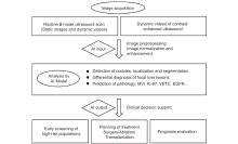
Figure 1
Flowchart showing the role of AI throughout the diagnostic workflow. MVI, microvascular invasion; VETC, encapsulating tumor clusters; EGFR, Epidermal growth factor receptor."
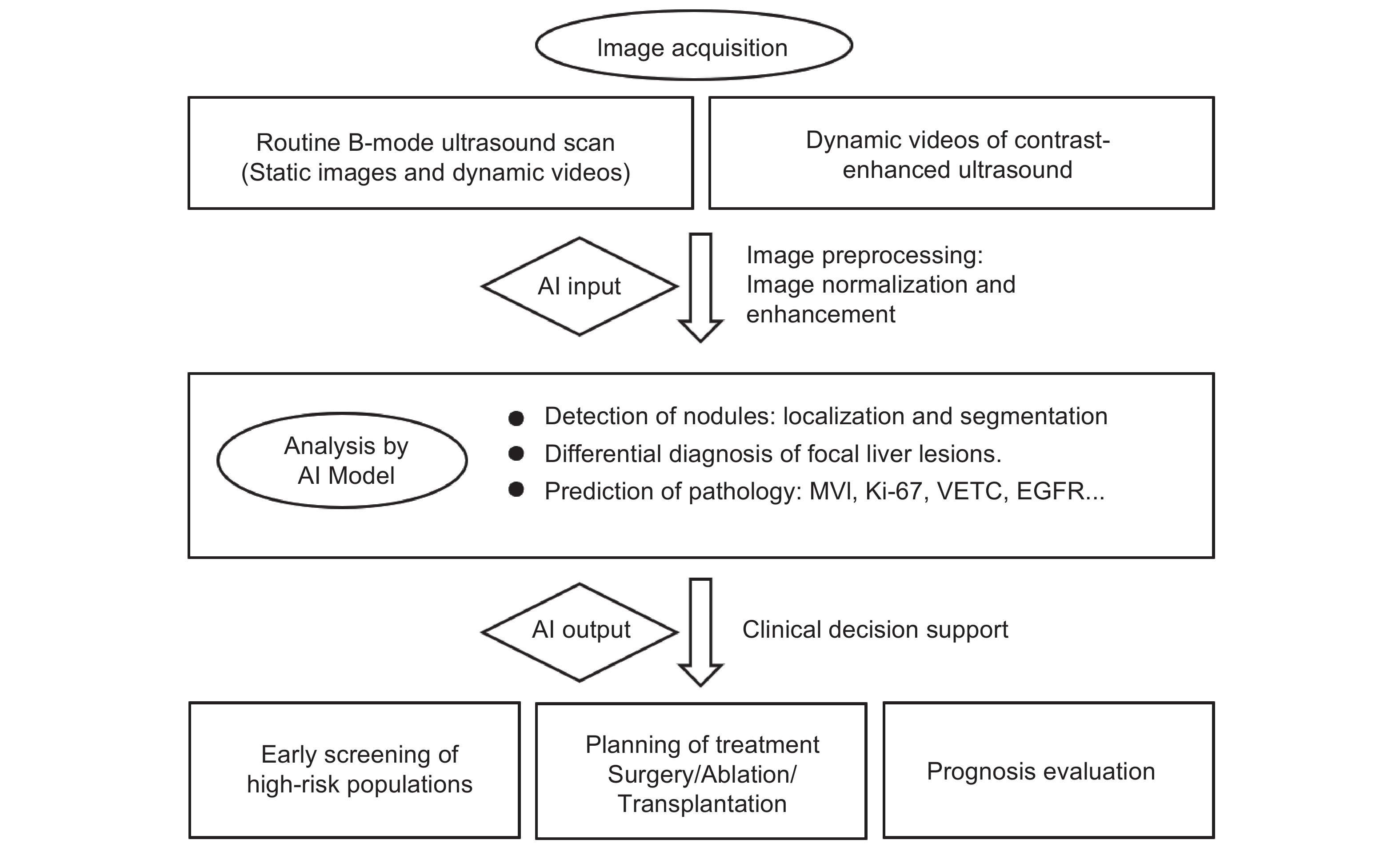
Table 1
Application of AI models in ultrasound detection of liver nodules"
| Author (year) | Objective | Mode | AI | Validation method | Multicenter study | Cases | Performance |
| Lu et al. 2025 [ | Detection | B-mode | DL | Train/test split, external validation | Yes | 11960 patients, 21934 images | Recall: 0.94 |
| Wu et al. 2025 [ | Detection | B-mode | DL | Train/validation/test split | No | 1576 patients | AP: 0.51 (MAL), 0.56 (BEN) |
| Rhyou et al. 2025 [ | Detection | B-mode | DL | Train/test split | No | 1134 images | Recall: 0.89 |
| Tian et al. 2023 [ | Detection | CEUS | DL | / | No | 33 patients | IOU: 0.864 |
| Dadoun et al. 2022 [ | Detection | B-mode | DL | Train/test split, external validation | Yes | 1074 patients, 2706 images | Recall: 0.97 |
| Tiyarattanachai et al. 2022 [ | Detection | B-mode | DL | Train/test split | No | 334 patients, (446 videos) | Recall: 0.90 |
| Tiyarattanachai et al. 2022 [ | Detection | B-mode | DL | Train/test split | No | 504 patients | The detection rate of non-expert group lesions was increased by 15.5% |
| Karako et al. 2022 [ | Detection | B-mode | DL | / | No | 1 patient, (91 images) | AP: 0.549 |
| Hao et al. 2022 [ | Segmentation | Doppler, CEUS, elastic US Multi-mode | DL | / | No | 100 patients | AP: 0.719 |
| Mishra et al. 2019 [ | Segmentation | CEUS | DL | Cross-validation | No | 152 images | Dice Index: 0.94 |
| Gatos et al. 2015 [ | Segmentation | CEUS | ML | Leave-one-out | No | 52 patients | LOU: 0.89 |
Table 2
Application of AI Models in the Diagnosis of Benign and Malignant Liver Nodules via Ultrasound"
| Author (year) | Objective | Mode | AI | Validation method | Multicenter study | Cases | AUC | Accuracy (%) | Sensitivity (%) | Specificity (%) |
| Lu et al. 2025 [ | BEN, MAL | B-mode | DL | Train/test split, external validation | Yes | 11960 patients, 21934 images | 0.87 | / | 95.6 | 78.7 |
| Du et al. 2025 [ | BEN, MAL | B-mode | ML | Train/test split, external validation | Yes | 1052 patients | 0.9 | 85.9 | 92.8 | 77.9 |
| Rhyou et al. 2025 [ | BEN, MAL | B-mode | DL | Train/test split | No | 1134 images | / | 96.2 | 97.6 | 98.1 |
| Mitrea et al. 2023 [ | BEN, MAL | B-mode | ML, DL | Train/test/validation split | Yes | 296 patients | 0.99 | 98.9 | 99.2 | 98.6 |
| Han et al. 2023 [ | BEN, MAL | B-mode | DL | Cross validation | Yes | 153 patients | / | 88.9 | 88.3 | 89.5 |
| Xu et al. 2023 [ | BEN, MAL | B-mode | DL | Train/test split, external validation | Yes | 11468 patients, 50063 images | 0.90-0.93 | / | 82.7-86.0 | 84.3-92.7 |
| Dadoun et al. 2022 [ | BEN, MAL | B-mode | DL | Train/test split, external validation | Yes | 1074 patients, 2706 images | / | 81 | 82 | 82 |
| Yamakawa et al. 2021 [ | BEN, MAL | B-mode | DL | Cross validation | Yes | 23756 patients, 70950 images | / | 94.3 | 82.9 | 96.7 |
| Hassan et al. 2021 [ | BEN, MAL | B-mode | DL | Cross validation | No | 352 patients | / | 92 | / | / |
| Ryu et al. 2021 [ | BEN, MAL | B-mode | DL | Train/test split | No | 3873 patients, 4309 images | 0.97 | 90.4 | 95 | 86 |
| Sato et al. 2021 [ | BEN, MAL | B-mode | DL | Train/test split | No | 1080 patients | 0.72 | 68.5 | 67.3 | 69.8 |
| Xi et al. 2021 [ | BEN, MAL | B-mode | DL | Train/validation/test split | No | 596 patients, 911 images | 0.83 | 84 | / | / |
| Yang et al. 2020 [ | BEN, MAL | B-mode | DL | Train/test split, external validation | Yes | 2143 patients, 24343 images | 0.92 | 84.7 | 86.5 | 85.5 |
| Tiyarattanachai et al. 2019 [ | BEN, MAL | B-mode | DL | External testing | No | 683 patients, 2698 images | 0.89 | 81 | 76 | 85 |
| Yamakawa et al. 2019 [ | BEN, MAL | B-mode | DL | Cross validation | No | 324 lesions, 980 images | / | 94.8 | 93.8 | 95.2 |
| Acharya et al. 2018 [ | BEN, MAL | B-mode | DL | Cross validation | No | 101 patients, 463 images | / | 93 | 90.8 | 97.4 |
| Kamiyama et al. 2024 [ | BEN, MAL | CEUS | DL | Cross validation | No | 181 patients | / | 85.2 | 93.2 | 63.3 |
| Urhut et al. 2023 [ | BEN, MAL | CEUS | DL | Train/test split | No | 49 patients, 59 lesions | 0.74 | 83 | 74 | 100 |
| Liu et al. 2022 [ | BEN, MAL | CEUS | DL | External test | Yes | 303 patients | 0.96 | 94 | 96.6 | 90.5 |
| Turco et al. 2022 [ | BEN, MAL | CEUS | ML | Cross validation | No | 72 patients | 0.84 | 82 | 76 | 92 |
| Zhang et al. 2021 [ | BEN, MAL | CEUS | ML | Cross validation | No | 153 patients | 0.9 | 88.2 | 86.9 | 89.4 |
| Hu et al. 2021 [ | BEN, MAL | CEUS | DL | Train/test split | No | 574 patients | 0.93 | 91 | 92.7 | 85.1 |
| Ta et al. 2018 [ | BEN, MAL | CEUS | DL, ML | Cross validation | Yes | 105 patients | 0.88 | 81.1 | 90 | 71.1 |
| Guo et al. 2018 [ | BEN, MAL | CEUS | DL | Cross validation | No | 93 patients | / | 90.3 | 93.6 | 86.9 |
| Kondo et al. 2017 [ | BEN, MAL | CEUS | ML | Leave-one-out Cross validation | No | 98 patients | / | 91.8 | 94 | 87.1 |
| Guo et al. 2017 [ | BEN, MAL | CEUS | DL | Cross validation | No | 93 patients | 0.95 | 90.4 | 93.6 | 86.9 |
| Dandan et al. 2017 [ | BEN, MAL | CEUS | ML | Cross validation | No | 5 patients | / | 98.9 | 98.9 | 98.9 |
| Qian et al. 2017 [ | BEN, MAL | CEUS | ML | Cross validation | No | 93 patients | 0.63 | 89.4 | 89.7 | 89.8 |
| Liang et al. 2016 [ | BEN, MAL | CEUS | ML | Cross validation | No | 353 patients | / | 92.7 | 97.3 | / |
| Gatos et al. 2015 [ | BEN, MAL | CEUS | ML | Leave-one-out Cross validation | No | 52 patients | 0.89 | 89 | 93.1 | 86.9 |
Table 3
Application of AI Models in the Multiclass Diagnosis of Ultrasound Liver Nodules"
| Author (year) | Objective | Mode | AI | Validation method | Multicenter study | Cases | AUC | Accuracy (%) | Sensitivity (%) | Specificity (%) |
| Chen et al. 2024 [ | HCC, ICC, cHCC-ICC | B-mode | DL | Train/test/validation split | No | 465 patients | 0.92 | 86 | 84.59 | 92.65 |
| Chaiteerakij et al. 2024 [ | HCC, ICC, FFI, FFS, HC, HA, and RN | B-mode | DL | Train/test split | No | 5444 patients | / | 94.8-99.9 | 74.3-98.8 | 97-100 |
| Nakata et al. 2023 [ | BLT, HC, HM, HCC | B-mode | ML, DL | Cross-validation | Yes | 6056 patients, 26440 images | 0.94 | 78.3 | 78.3 | 92.8 |
| Yang et al. 2023 [ | HE | B-mode | DL | Train/test split, external validation | Yes | 6784 patients, 9631 images | 0.95 | 90.7-94.6 | 77.1-97.1 | 92.4-94.2 |
| Dadoun et al. 2022 [ | FLLs (HC, HE, FNH, HA, MM, HCC) | B-mode | DL | Train/test split, external validation | Yes | 1074 patients, 2706 images | 76 | 65 | 94 | |
| Naoshi et al. 2022 [ | HC, HH, HCC, HM | B-mode | DL | Train/test split, external validation | Yes | 29264 patients, 74427 images | / | 93.4-99.0 | 62.8-99.0 | 96.0-98.8 |
| Cheng et al. 2022 [ | HE | B-mode | DL | Cross-validation | Yes | 5028 patients, 9112 images | / | 86.2-94.2 | 87.1-95.4 | / |
| Peng et al. 2022 [ | alpha-Fetoprotein-Negative FLL, MAL | B-mode | ML | Train/validation split | No | 589 patients | 0.75 | 79.1 | 86.3 | 45.2 |
| Zhang et al. 2022 [ | HCC, AFNH | B-mode | DL | Train/test split | No | 407 patients | 0.94 | 86.4 | 96.1 | 76.9 |
| Wu et al. 2022 [ | HE | B-mode | DL | Cross-validation | No | 967 images | 0.98-0.99 | / | / | / |
| Yamakawa et al. 2021 [ | HC, HCC, HH, HM | B-mode | DL | 10-fold cross-validation | Yes | 23756 patients, 70950 images | / | 91.1 | / | / |
| Tiyarattanachai et al. 2021 [ | HC, HCC, HH, FFC, FFS | B-mode | DL | Train/test split, external validation | Yes | 3872 patients, 40397 images | / | 95.4 | 83.9 | 97.1 |
| Ren et al. 2021 [ | ICC, HCC | B-mode | ML | Train/test split, external validation | Yes | 226 patients | 0.87 | 86.2 | 88.9 | 86.7 |
| Mao et al. 2021 [ | HCC, HM, ICC | B-mode | ML | 5-fold cross-validation | No | 114 patients | 0.82 | 84 | 77 | 88 |
| Zhou et al. 2021 [ | HCC, other MAL | B-mode | DL | Train/test split | No | 172 patients | 0.74 | 78.4 | 57.1 | 91.3 |
| Peng et al. 2020 [ | HCC, ICC, cHCC-ICC | B-mode | ML | Train/test split | No | 668 patients | 0.73 0.78 | / | / | / |
| Lin et al. 2020 [ | HCC, ICC | B-mode | ML | Train/test/validation split | No | 124 patients | 0.94 | / | 100 | 77.8 |
| Sritunyarat et al. 2020 [ | HC, HCC, HH, FFI, FFS | B-mode | DL | / | No | 157 patients | / | 95 | 87 | 97 |
| Xu et al. 2020 [ | HA, HCC | B-mode | DL | 10-fold cross-validation | No | 79 patients | / | 83.8 | / | / |
| Yamakawa et al. 2019 [ | HC, HCC, HH, HM | B-mode | DL | 10-fold cross-validation | No | 324 lesions, 911 images | / | 88 | 80.4 | 96 |
| Schmauch et al. 2019 [ | HC, FNH, HCC, HH, HM | B-mode | DL | Train/test split | No | 544 patients | 0.89 | / | / | / |
| Mitrea et al. 2019 [ | HCC, HH | B-mode | DL | 5-fold cross-validation | / | 300 patients | 0.81 | 85.4 | 78 | 82.9 |
| Tiyarattanachai et al. 2019 [ | HC, HCC, HH, FFC, FFS | B-mode | DL | external testing | / | 683 patients | / | 69 | / | / |
| Hassan et al. 2017 [ | HC, HCC, HH | B-mode | DL | 10-fold cross-validation | Yes | 110 patients | / | 97.2 | 98 | 95.7 |
| Hwang et al. 2015 [ | HC, HH, MAL | B-mode | DL | / | No | 115 patients | / | 98.1 | / | / |
| Hassan et al. 2015 [ | HC, HCC, HH | B-mode | ML | 10-fold cross-validation | No | 110 patients | / | 96.5 | 97.6 | 92.5 |
| Ding et al. 2025 [ | FLLs (HCC, HM, ICC, HH, HA and others) | CEUS | MLP | Train/test split, external validation | Yes | 3725 lesions | 0.89 | 79-92 | / | 97 |
| Li et al. 2024 [ | HCC, others (CCA, cHCC-CCA) | CEUS | ML | Train/test split | No | 159 patients | 0.91 | 85.4 | 95.2 | 77.8 |
| Zhou et al. 2024 [ | HA, HH, FNH, RN, HCC, IHCC, HM | CEUS | DL | Train/test split | No | 420 patients | 0.91 | / | 83 | 82 |
| Mamuleanu et al. 2023 [ | FLL (HCC, HM, HH, other MAL and BEN) | CEUS | DL | Train/test split | No | 49 patients, 59 lesions | / | 58.6 | / | / |
| Urhut et al. 2023 [ | FLL (HCC, HM, CCA, MA, HH, HC, FNH, HA) | CEUS | DL | Train/test split | No | 49 patients, 59 lesions | / | 69.9 | 86.9 | 56.2 |
| Li et al. 2023 [ | HCC, HM | CEUS | ML | Cross-validation | No | 3210 patients | / | 83 | 77.3 | 88.6 |
| Kim et al. 2023 [ | HCC, FNH | CEUS | DL | Cross-validation | No | 145 patients | / | 98 | / | / |
| Wan et al. 2023 [ | FLL (HCC, ICC) | CEUS | DL, ML | Train/test split | No | 175 patients | 0.88 | 88.4 | 86.2 | 90.1 |
| Zhou et al. 2022 [ | FLL (HCC, FNH) | CEUS | ML | Internal | No | 186 patients | / | 96.7 | 98.7 | 94.7 |
| Wang and Xia 2022 [ | HBV-associated HCC | CEUS | DL | Internal | No | 80 patients | 0.93 | 85 | 85 | 85 |
| Schmiedt et al. 2022 [ | FLL (HCC, HH, FNH) | CEUS | DL | Cross-validation | No | 358 patients | / | 86.7 | / | / |
| Sirbu et al. 2022 [ | FLL (FNH, HCC, HH, hypo-and hyper-vascular metastasis) | CEUS | DL | Leave-one-out Cross validation | No | 444 patients | / | 90 | / | / |
| Cǎleanu et al. 2021 [ | FLL (FNH, HH, HCC, HM) | CEUS | DL | Leave-one-out Cross validation | No | 91 patients | / | 88 | / | / |
| Li et al. 2021 [ | FNH, HCC | CEUS | ML | Cross-validation | No | 226 patients | 0.93 | / | 93.5 | 84.9 |
| Mitrea et al. 2021 [ | HCC | CEUS | ML | Internal | No | 48 patients | 0.98 | 98 | 96.9 | 98.9 |
| Denis et al. 2020 [ | FNH, inflammatory-HA | CEUS | ML | Cross-validation | No | 46 patients | 0.97 | 95.9 | 93.4 | 97.6 |
| Huang et al. 2020 [ | FNH, HCC | CEUS | ML | Cross-validation | No | 342 patients | / | 94.4 | 94.8 | 93.6 |
| Sirbu et al. 2020 [ | FLL (FNH, HH, HCC, HM) | CEUS | DL | Internal | No | 95 patients | / | 95.7 | / | / |
| Pan et al. 2019 [ | HCC, FNH | CEUS | DL | Cross-validation | No | 242 lesions | 0.97 | 93.1 | 94.5 | 93.6 |
| Kondo et al. 2017 [ | FLL (HCC, HM, HH, FNH) | CEUS | ML | Leave-one-out Cross validation | No | 98 patients | / | 84.4-87.7 | / | / |
| Liang et al. 2016 [ | FLL (HCC, FNH, HH) | CEUS | ML | Cross-validation | No | 353 patients | / | 84.8 | 63.6-88.5 | / |
| Su et al. 2024 [ | HCC, ICC | B-mode, CEUS | ML | Train/test split | No | 280 lesions | 0.97 | 91 | 89.2 | 92.8 |
| Li et al. 2023 [ | HCC, others | B-mode, CEUS | DL | Train/validation/test split | No | 116 patients | 0.85 | 85 | 80 | 90 |
| Li et al. 2024 [ | HCC, ICC | B-mode, CEUS, SWE | DL | Train/test split | No | 86 patients, 852 images, 62 videos | 0.85 | 87.5 | 87.5 | 87.5 |
Table 4
Application of AI Models in Predicting Pathological Biomarkers for Liver Cancer"
| Author (year) | Objective | Mode | AI | Validation method | Multicenter study | Cases | AUC | Accuracy (%) | Sensitivity (%) | Specificity (%) |
| Zhang et al. 2024 [ | Grading | CEUS | ML | Cross-validation | No | 90 patients | 0.95 | / | 92.6 | 81.6 |
| Qin et al. 2023 [ | Grading | CEUS | ML, DL, combined | Train/test split | No | 272 patients, 1088 images | 0.93 | 91.5 | 93.8 | 90 |
| Ren et al. 2021 [ | Grading | B-mode | ML | Train/test split, external validation | Yes | 193 patients | 0.85 | 81.8 | 74 | 85.71 |
| Wang et al. 2021 [ | Grading | CEUS | ML | Cross-validation, Train/test split | No | 235 patients | 0.79 | 75.7 | 74.5 | 78.9 |
| Zhou et al. 2020 [ | Grading | B-mode | ML | Leave-one-out Cross-validation | No | 43 patients | 0.76 | / | / | / |
| Wang et al. 2025 [ | MVI | CEUS | DL | Train/validation split | No | 318 patients | 0.93 | 89.2 | 85.3 | 91.7 |
| Zhang et al. 2024 [ | MVI | CEUS | DL, ML | Train/validation split | Yes | 576 patients, 2304 images | 0.667-0.818 | 70.3-75.2 | 68.5-72.4 | 72.1-78 |
| Zhang et al. 2024 [ | MVI | CEUS | ML | Cross-validation | No | 90 patients | 0.88 | / | 81 | 83 |
| Qin et al. 2023 [ | MVI | CEUS | DL | Train/test split | No | 252 patients, 756 images | 0.86 | 77.8 | 52.4 | 93.9 |
| Zhang et al. 2022 [ | MVI | CEUS | DL | Train/test split | No | 436 images | 0.87 | 78.8 | 83.3 | 81 |
| Dong et al. 2022 [ | MVI | CEUS | ML | Leave-one-out Cross- validation | No | 100 patients | 0.8 | 77 | 84.4 | 73.5 |
| Dong et al. 2020 [ | MVI | B-mode | ML | Train/validation split | No | 322 patients | 0.74 | 63.4 | 89.2 | 48.4 |
| Dong et al. 2019 [ | MVI | B-mode | ML | Leave-one-out Cross-validation | No | 42 patients | 0.95 | 93 | 86 | 100 |
| Hu et al. 2019 [ | MVI | CEUS | ML | Train/validation split | No | 482 patients | 0.73 | / | / | / |
| Zhang et al. 2024 [ | Ki-67 | CEUS | ML | Train/test split, external validation | Yes | 310 patients | 0.856 | 76.8 | 79.3 | 74.1 |
| Qian et al. 2024 [ | Ki-67 | B-mode | ML | Train/test split | No | 153 patients | 0.762 | 75 | 85.7 | 65.2 |
| Zhang et al. 2023 [ | Ki-67 | B-mode | ML | Train/test split, external validation | Yes | 244 patients | 0.742 | 78.8 | 71.4 | 80.8 |
| Qian et al. 2023 [ | Ki-67 | B-mode | ML | Train/validation split | No | 118 patients | 0.87 | 87.5 | 73.7 | 88.2 |
| Dong et al. 2022 [ | Ki-67 | CEUS | ML | Train/test split | No | 101 patients | 0.77 | 70 | 74.6 | 80 |
| Dai et al. 2018 [ | Ki-67 | B-mode | ML | Leave-one-out Cross-validation | No | 133 patients | 0.75 | / | / | / |
| Ma et al. 2024 [ | EGFR | B-mode | ML | Train/test split | No | 198 patients | 0.82 | 81.7 | 83.3 | 80.0 |
| Xu et al. 2025 [ | VETC | CEUS | DL | Train/test split | No | 242 patients | 0.9 | 81 | 81 | 81 |
| Wang et al. 2023 [ | T Cell-Inflamed GEP | CEUS | ML | 5-fold cross-validation | No | 268 patients | 0.889 | 81.3 | 79.9 | 82.8 |
| Yao et al. 2018 [ | PD-1, Ki-67, MVI | B-mode, SWE, SWV | ML | Leave-one-out Cross-validation | No | 111 patients | PD-1:0.97 Ki67:0.94 MVI:0.98 | PD-1: 92 Ki67: 93 MVI: 95 | PD-1: 100 Ki67: 95 MVI: 91 | PD-1: 88 Ki67: 91 MVI: 100 |
| [1] | Siegel RL , Kratzer TB , Giaquinto AN , Sung H , Jemal A . Cancer statistics, 2025. CA Cancer J Clin 2025; 75: 10-45 |
| [2] |
Kaltenbach TEM , Engler P , Kratzer W , Oeztuerk S , Seufferlein T , Haenle MM , et al . Prevalence of benign focal liver lesions: ultrasound investigation of 45,319 hospital patients. Abdom Radiol 2016; 41: 25-32.
doi: 10.1007/s00261-015-0605-7 |
| [3] |
Qin YJ , Tang CL , Li JH , Gong JP . Liver cancer in China: the analysis of mortality and burden of disease trends from 2008 to 2021. Bmc Cancer 2024; 24: 594.
doi: 10.1186/s12885-024-12334-2 |
| [4] | Zhang BH , Yang BH , Tang ZY . Randomized controlled trial of screening for hepatocellular carcinoma. J Cancer Res Clin 2004; 130: 417-422 |
| [5] |
Vitale A , Trevisani F , Farinati F , Cillo U . Treatment of hepatocellular carcinoma in the precision medicine era: from treatment stage migration to therapeutic hierarchy. Hepatology 2020; 72: 2206-2218.
doi: 10.1002/hep.31187 |
| [6] |
Tzartzeva K , Obi J , Rich NE , Parikh ND , Marrero JA , Yopp A , et al . Surveillance imaging and alpha fetoprotein for early detection of hepatocellular carcinoma in patients with cirrhosis: a meta-analysis. Gastroenterology 2018; 154: 1706-1718.e1701.
doi: 10.1053/j.gastro.2018.01.064 |
| [7] |
Tiyarattanachai T , Apiparakoon T , Marukatat S , Sukcharoen S , Yimsawad S , Chaichuen O , et al . The feasibility to use artificial intelligence to aid detecting focal liver lesions in real-time ultrasound: a preliminary study based on videos. Sci Rep 2022; 12: 7749.
doi: 10.1038/s41598-022-11506-z |
| [8] |
Karako K , Mihara Y , Arita J , Ichida A , Bae SK , Kawaguchi Y , et al . Automated liver tumor detection in abdominal ultrasonography with a modified faster region-based convolutional neural networks (faster R-CNN) architecture. Hepatobiliary Surg Nutr 2022; 11: 675-683.
doi: 10.21037/hbsn-21-43 |
| [9] |
Tian H , Cai W , Ding W , Liang P , Yu J , Huang Q . Long-term liver lesion tracking in contrast-enhanced ultrasound videos via a siamese network with temporal motion attention. Front Physiol 2023; 14: 1180713.
doi: 10.3389/fphys.2023.1180713 |
| [10] |
Lu R-F , She C-Y , He D-N , Cheng M-Q , Wang Y , Huang H , et al . AI enhanced diagnostic accuracy and workload reduction in hepatocellular carcinoma screening. Npj Digit Med 2025; 8: 500.
doi: 10.1038/s41746-025-01892-9 |
| [11] |
Amarapurkar D , Han KH , Chan HLY , Ueno Y . Application of surveillance programs for hepatocellular carcinoma in the Asia–Pacific Region. J Gastroen Hepatol 2009; 24: 955-961.
doi: 10.1111/j.1440-1746.2009.05805.x |
| [12] |
Yang Q , Wei J , Hao X , Kong D , Yu X , Jiang T , et al . Improving B-mode ultrasound diagnostic performance for focal liver lesions using deep learning: a multicentre study. Ebiomedicine 2020; 56: 102777.
doi: 10.1016/j.ebiom.2020.102777 |
| [13] |
Rhyou S-Y , Yoo J-C . Automated ultrasonography of hepatocellular carcinoma using discrete wavelet transform based deep-learning neural network. Med Image Anal 2025; 101: 103453.
doi: 10.1016/j.media.2025.103453 |
| [14] | Xu Y , Zheng B , Liu X , Wu T , Ju J , Wang S , et al . Improving artificial intelligence pipeline for liver malignancy diagnosis using ultrasound images and video frames. Brief Bioinform 2023; 24: 1-13 |
| [15] |
Du Z , Fan F , Ma J , Liu J , Yan X , Chen X , et al . Development and validation of an ultrasound-based interpretable machine learning model for the classification of ≤ 3 cm hepatocellular carcinoma:a multicentre retrospective diagnostic study. Eclinicalmedicine 2025; 81: 103098.
doi: 10.1016/j.eclinm.2025.103098 |
| [16] |
Han X , Gong B , Guo L , Wang J , Ying S , Li S , et al . B-mode ultrasound based CAD for liver cancers via multi-view privileged information learning. Neural Networks 2023; 164: 369-381.
doi: 10.1016/j.neunet.2023.03.028 |
| [17] |
Hu HT , Wang W , Chen LD , Ruan SM , Chen SL , Li X , et al . Artificial intelligence assists identifying malignant versus benign liver lesions using contrast‐enhanced ultrasound. J Gastroen Hepatol 2021; 36: 2875-2883.
doi: 10.1111/jgh.15522 |
| [18] |
Liu L , Tang C , Li L , Chen P , Tan Y , Hu X , et al . Deep learning radiomics for focal liver lesions diagnosis on long-range contrast-enhanced ultrasound and clinical factors. Quant Imag Med Surg 2022; 12: 3213-3226.
doi: 10.21037/qims-21-1004 |
| [19] | Benson AB , D’Angelica MI , Abbott DE , Anaya DA , Anders R , Are C , et al. Hepatobiliary cancers, version 2.2021, NCCN Clinical Practice Guidelines in Oncology. J Natl Compr Canc Ne 2021; 19:541-565. |
| [20] |
Budke CM , Deplazes P , Torgerson PR . Global socioeconomic impact of cystic echinococcosis. Emerg Infect Dis 2006; 12: 296-303.
doi: 10.3201/eid1202.050499 |
| [21] |
Nishida N , Yamakawa M , Shiina T , Mekada Y , Nishida M , Sakamoto N , et al . Artificial intelligence (AI) models for the ultrasonographic diagnosis of liver tumors and comparison of diagnostic accuracies between AI and human experts. J Gastroenterol 2022; 57: 309-321.
doi: 10.1007/s00535-022-01849-9 |
| [22] |
Nakata N , Siina T . Ensemble learning of multiple models using deep learning for multiclass classification of ultrasound images of hepatic masses. Bioengineering 2023; 10: 69.
doi: 10.3390/bioengineering10010069 |
| [23] |
Chaiteerakij R , Ariyaskul D , Kulkraisri K , Apiparakoon T , Sukcharoen S , Chaichuen O , et al . Artificial intelligence for ultrasonographic detection and diagnosis of hepatocellular carcinoma and cholangiocarcinoma. Sci Rep 2024; 14: 20617.
doi: 10.1038/s41598-024-71657-z |
| [24] |
Cheng J , Wang HX , Li R , Li XM , Zhou X , Yang X , et al . A two-stage multiresolution neural network for automatic diagnosis of hepatic echinococcosis from ultrasound images: a multicenter study. Med Phys 2022; 49: 3199-3212.
doi: 10.1002/mp.15548 |
| [25] |
Yang Y , Cairang Y , Jiang Ta , Zhou J , Zhang L , Qi B , et al . Ultrasound identification of hepatic echinococcosis using a deep convolutional neural network model in China:a retrospective, large-scale, multicentre, diagnostic accuracy study. Lancet Digit Health 2023; 5: e503-e514.
doi: 10.1016/S2589-7500(23)00091-2 |
| [26] |
Li JM , Li HR , Xiao F , Liu RQ , Chen YX , Xue ML , et al . Comparison of machine learning models and CEUS LI-RADS in differentiation of hepatic carcinoma and liver metastases in patients at risk of both hepatitis and extrahepatic malignancy. Cancer Imaging 2023; 23: 63.
doi: 10.1186/s40644-023-00573-8 |
| [27] |
Zhou HY , Ding JM , Zhou Y , Wang YD , Zhao L , Shih CC , et al . Malignancy diagnosis of liver lesion in contrast enhanced ultrasound using an end-to-end method based on deep learning. Bmc Med Imaging 2024; 24: 68.
doi: 10.1186/s12880-024-01247-y |
| [28] |
Ding W , Meng Y , Ma J , Pang C , Wu J , Tian J , et al . Contrast-enhanced ultrasound-based AI model for multi-classification of focal liver lesions. J Hepatol 2025; 83: 426-439.
doi: 10.1016/j.jhep.2025.01.011 |
| [29] |
Guo YS , Li TX , Gong BX , Hu Y , Wang SC , Yang L , et al . From images to genes: radiogenomics based on artificial intelligence to achieve non-invasive precision medicine in cancer patients. Adv Sci 2025; 12: e2408069.
doi: 10.1002/advs.202408069 |
| [30] |
Erstad DJ , Tanabe KK . Prognostic and therapeutic implications of microvascular invasion in hepatocellular carcinoma. Ann Surg Oncol 2019; 26: 1474-1493.
doi: 10.1245/s10434-019-07227-9 |
| [31] |
Lim KC , Chow PKH , Allen JC , Chia GS , Lim M , Cheow PC , et al . Microvascular invasion is a better predictor of tumor recurrence and overall survival following surgical resection for hepatocellular carcinoma compared to the milan criteria. Ann Surg 2011; 254: 108-113.
doi: 10.1097/SLA.0b013e31821ad884 |
| [32] | Hu H-t , Wang Z , Huang X-w , Chen S-l , Zheng X , Ruan S-m , et al . Ultrasound-based radiomics score: a potential biomarker for the prediction of microvascular invasion in hepatocellular carcinoma. Eur Radiol 2018; 29: 2890-2901 |
| [33] |
Wang Y , Xie W , Li C , Xu Q , Du Z , Zhong Z , et al . Automated microvascular invasion prediction of hepatocellular carcinoma via deep relation reasoning from dynamic contrast-enhanced ultrasound. Comput Med Imag Grap 2025; 124: 102606.
doi: 10.1016/j.compmedimag.2025.102606 |
| [34] |
Zhou L , Rui JA , Zhou WX , Wang SB , Chen SG , Qu Q . Edmondson-Steiner grade: a crucial predictor of recurrence and survival in hepatocellular carcinoma without microvascular invasio. Pathol Res Pract 2017; 213: 824-830.
doi: 10.1016/j.prp.2017.03.002 |
| [35] |
Xu X , Zhang HL , Liu QP , Sun SW , Zhang J , Zhu FP , et al . Radiomic analysis of contrast-enhanced CT predicts microvascular invasion and outcome in hepatocellular carcinoma. J Hepatol 2019; 70: 1133-1144.
doi: 10.1016/j.jhep.2019.02.023 |
| [36] |
Jonas S , Bechstein WO , Steinmüller T , Herrmann M , Radke C , Berg T , et al . Vascular invasion and histopathologic grading determine outcome after liver transplantation for hepatocellular carcinoma in cirrhosis. Hepatology 2001; 33: 1080-1086.
doi: 10.1053/jhep.2001.23561 |
| [37] |
Court CM , Harlander-Locke MP , Markovic D , French SW , Naini BV , Lu DS , et al . Determination of hepatocellular carcinoma grade by needle biopsy is unreliable for liver transplant candidate selection. Liver Transplant 2017; 23: 1123-1132.
doi: 10.1002/lt.24811 |
| [38] | Wu M , Tan H , Gao F , Hai J , Ning P , Chen J , et al . Predicting the grade of hepatocellular carcinoma based on non-contrast-enhanced MRI radiomics signature. Eur Radiol 2018; 29: 2802-2811 |
| [39] |
Wang W , Wu S-S , Zhang J-C , Xian M-F , Huang H , Li W , et al . Preoperative pathological grading of hepatocellular carcinoma using ultrasomics of contrast-enhanced ultrasound. Acad Radiol 2021; 28: 1094-1101.
doi: 10.1016/j.acra.2020.05.033 |
| [40] |
Qin X , Hu X , Xiao W , Zhu C , Ma Q , Zhang C . Preoperative evaluation of hepatocellular carcinoma differentiation using contrast-enhanced ultrasound-based deep-learning radiomics model. J Hepatocell Carcino 2023; 10: 157-168.
doi: 10.2147/JHC.S400166 |
| [41] | Luo YH , Ren FH , Liu YR , Shi ZH , Tan Z , Xiong HJ , et al . Clinicopathological and prognostic significance of high Ki-67 labeling index in hepatocellular carcinoma patients:a meta-analysis. Int J Clin Exp Med 2015; 8: 10235-10247 |
| [42] |
Nielsen TO , Leung SCY , Rimm DL , Dodson A , Acs B , Badve S , et al . Assessment of Ki67 in breast cancer: updated recommendations from the international Ki67 in breast cancer working group. J Natl Cancer Inst 2021; 113: 808-819.
doi: 10.1093/jnci/djaa201 |
| [43] |
Qian HW , Shen ZH , Zhou DF , Huang YH . Intratumoral and peritumoral radiomics model based on abdominal ultrasound for predicting Ki-67 expression in patients with hepatocellular cancer. Front Oncol 2023; 13: 1209111.
doi: 10.3389/fonc.2023.1209111 |
| [44] | Zhang L , Duan S , Qi Q , Li Q , Ren S , Liu S , et al . Noninvasive prediction of Ki-67 expression in hepatocellular carcinoma using machine learning-based ultrasomics. J Ultras Med 2022; 42: 1113-1122 |
| [45] |
Zhang D , Zhang X-Y , Lu W-W , Liao J-T , Zhang C-X , Tang Q , et al . Predicting Ki-67 expression in hepatocellular carcinoma: nomogram based on clinical factors and contrast-enhanced ultrasound radiomics signatures. Abdom Radiol 2024; 49: 1419-1431.
doi: 10.1007/s00261-024-04191-1 |
| [46] |
Fang JH , Zhou HC , Zhang C , Shang LR , Zhang L , Xu J , et al . A novel vascular pattern promotes metastasis of hepatocellular carcinoma in an epithelial-mesenchymal transition-independent manner. Hepatology 2015; 62: 452-465.
doi: 10.1002/hep.27760 |
| [47] |
Xu W , Zhang H , Zhang R , Zhong X , Li X , Zhou W , et al . Deep learning model based on contrast-enhanced ultrasound for predicting vessels encapsulating tumor clusters in hepatocellular carcinoma. Eur Radiol 2024; 35: 989-1000.
doi: 10.1007/s00330-024-10985-0 |
| [48] |
Arteaga CL , Engelman JA . ERBB receptors: from oncogene discovery to basic science to mechanism-based cancer therapeutics. Cancer Cell 2014; 25: 282-303.
doi: 10.1016/j.ccr.2014.02.025 |
| [49] |
Jin HJ , Shi YP , Lv YY , Yuan SX , Ramirez CFA , Lieftink C , et al . EGFR activation limits the response of liver cancer to lenvatinib. Nature 2021; 595: 730-734.
doi: 10.1038/s41586-021-03741-7 |
| [50] |
Ma Y , Duan S , Ren S , Bu D , Li Y , Cai X , et al . Machine learning based ultrasomics noninvasive predicting EGFR expression status in hepatocellular carcinoma patients. Front Med 2024; 11: 1483291.
doi: 10.3389/fmed.2024.1483291 |
| [51] |
Ayers M , Lunceford J , Nebozhyn M , Murphy E , Loboda A , Kaufman DR , et al . IFN-γ-related mRNA profile predicts clinical response to PD-1 blockade. J Clin Invest 2017; 127: 2930-2940.
doi: 10.1172/JCI91190 |
| [52] |
Wang Y , Weng W , Liang R , Zhou Q , Hu H , Li M , et al . Predicting T cell-inflamed gene expression profile in hepatocellular carcinoma based on dynamic contrast-enhanced ultrasound radiomics. J Hepatocell Carcino 2023; 10: 2291-2303.
doi: 10.2147/JHC.S437415 |
| [53] |
Dong Y , Zuo D , Qiu Y-J , Cao J-Y , Wang H-Z , Wang W-P . Prediction of histological grades and Ki-67 expression of hepatocellular carcinoma based on sonazoid contrast enhanced ultrasound radiomics signatures. Diagnostics 2022; 12: 2175.
doi: 10.3390/diagnostics12092175 |
| [54] | Li R , Xu XC , Lin JY , Huang LH , Zeng YG , Zheng W , et al . Automatic classification of liver cancer in b-ultrasound and contrast-enhanced ultrasound based on convolution neural network. Prog Biochem Biophys 2023; 50: 668-675 |
| [55] | Zhou BY , Shi YL , Guo LH , Mou LC , Zhu XX , Zhao CK . Artificial intelligence technology enables ultrasonography in precision diagnosis and treatment of liver diseases. Zhongguo Xue Xi Chong Bing Fang Zhi Za Zhi. 2022; 34: 458-464 |
| [56] |
Zhang W , Guo Q , Zhu Y , Wang M , Zhang T , Cheng G , et al . Cross-institutional evaluation of deep learning and radiomics models in predicting microvascular invasion in hepatocellular carcinoma: validity, robustness, and ultrasound modality efficacy comparison. Cancer Imaging 2024; 24: 142.
doi: 10.1186/s40644-024-00790-9 |
| [57] |
Brooks JA , Kallenbach M , Radu I-P , Berzigotti A , Dietrich CF , Kather JN , et al . Artificial intelligence for contrast-enhanced ultrasound of the liver: a systematic review. Digestion 2024; 106: 227-244.
doi: 10.1159/000541540 |
| [58] |
Su LY , Xu M , Chen YL , Lin MX , Xie XY . Ultrasomics in liver cancer: developing a radiomics model for differentiating intrahepatic cholangiocarcinoma from hepatocellular carcinoma using contrast-enhanced ultrasound. World J Radiol 2024; 16: 247-255.
doi: 10.4329/wjr.v16.i7.247 |
| [59] |
Ren S , Qi Q , Liu S , Duan S , Mao B , Chang Z , et al . Preoperative prediction of pathological grading of hepatocellular carcinoma using machine learning-based ultrasomics: a multicenter study. Eur J Radiol 2021; 143: 109891.
doi: 10.1016/j.ejrad.2021.109891 |
| [60] |
Qian HW , Huang YH , Xu LH , Fu H , Lu BC . Role of peritumoral tissue analysis in predicting characteristics of hepatocellular carcinoma using ultrasound-based radiomics. Sci Rep 2024; 14: 11538.
doi: 10.1038/s41598-024-62457-6 |
| [61] |
Tiyarattanachai T , Apiparakoon T , Marukatat S , Sukcharoen S , Geratikornsupuk N , Anukulkarnkusol N , et al . Development and validation of artificial intelligence to detect and diagnose liver lesions from ultrasound images. Plos One 2021; 16: e0252882.
doi: 10.1371/journal.pone.0252882 |
| [62] |
Qin X , Zhu J , Tu Z , Ma Q , Tang J , Zhang C . Contrast-enhanced ultrasound with deep learning with attention mechanisms for predicting microvascular invasion in single hepatocellular carcinoma. Acad Radiol 2023; 30: S73-S80.
doi: 10.1016/j.acra.2022.12.005 |
| [63] |
Ren SS , Li Q , Liu SH , Qi QH , Duan SB , Mao B , et al . Clinical value of machine learning-based ultrasomics in preoperative differentiation between hepatocellular carcinoma and intrahepatic cholangiocarcinoma: a multicenter study. Front Oncol 2021; 11: 749137.
doi: 10.3389/fonc.2021.749137 |
| [64] |
Li MD , Li W , Lin MX , Lin XX , Hu HT , Wang YC , et al . Systematic comparison of deep-learning based fusion strategies for multi-modal ultrasound in diagnosis of liver cancer. Neurocomputing 2024; 603: 128257.
doi: 10.1016/j.neucom.2024.128257 |
| [65] |
Wan P , Xue HY , Liu CR , Chen F , Shao W , Qin J , et al . Transport-based anatomical-functional metric learning for liver tumor recognition using dual-view dynamic ceus imaging. Ieee T Bio-Med Eng 2023; 70: 1012-1023.
doi: 10.1109/TBME.2022.3207473 |
| [66] |
Wu CH , Sheu JC , Chou PL , Lee J , Nien HC . Automatic real-time detection and diagnosis of liver tumor with ultrasound. J Hepatocell Carcino 2025; 12: 1599-1611.
doi: 10.2147/JHC.S524311 |
| [67] |
Dadoun H , Rousseau AL , de Kerviler E , Correas JM , Tissier AM , Joujou F , et al . Deep learning for the detection, localization, and characterization of focal liver lesions on abdominal US images. Radiol-Artif Intell 2022; 4: e210110.
doi: 10.1148/ryai.210110 |
| [68] |
Tiyarattanachai T , Apiparakoon T , Chaichuen O , Sukcharoen S , Yimsawad S , Jangsirikul S , et al . Artificial intelligence assists operators in real-time detection of focal liver lesions during ultrasound: a randomized controlled study. Eur J Radiol 2023; 165: 110932.
doi: 10.1016/j.ejrad.2023.110932 |
| [69] | Zhang Y , Cui J , Wan W , Liu J , Uddin Z . Multimodal imaging under artificial intelligence algorithm for the diagnosis of liver cancer and its relationship with expressions of EZH2 and p57. Comput Intel Neurosc 2022; 2022: 1-11 |
| [70] |
Mishra D , Chaudhury S , Sarkar M , Soin AS . Ultrasound image segmentation: a deeply supervised network with attention to boundaries. Ieee T Bio-Med Eng 2019; 66: 1637-1648.
doi: 10.1109/TBME.2018.2877577 |
| [71] |
Gatos I , Tsantis S , Spiliopoulos S , Skouroliakou A , Theotokas I , Zoumpoulis P , et al . A new automated quantification algorithm for the detection and evaluation of focal liver lesions with contrast-enhanced ultrasound. Med Phys 2015; 42: 3948-3959.
doi: 10.1118/1.4921753 |
| [72] |
Mitrea D-A , Brehar R , Nedevschi S , Lupsor-Platon M , Socaciu M , Badea R . Hepatocellular carcinoma recognition from ultrasound images using combinations of conventional and deep learning techniques. Sensors 2023; 23: 2520.
doi: 10.3390/s23052520 |
| [73] | Yamakawa M , Shiina T , Tsugawa K , Nishida N , Kudo M . Deep-learning framework based on a large ultrasound image database to realize computer-aided diagnosis for liver and breast tumors. 2021 IEEE International Ultrasonics Symposium (IUS) 2021:1-4. |
| [74] | Hassan T , Alzoubi A , Du H , Jassim S , Agaian SS , Jassim SA , et al. Towards optimal cropping:breast and liver tumor classification using ultrasound images. Multimodal Image Exploitation and Learning 2021 2021; 11734:1-12. |
| [75] |
Ryu H , Shin SY , Lee JY , Lee KM , Kang H-j , Yi J . Joint segmentation and classification of hepatic lesions in ultrasound images using deep learning. Eur Radiol 2021; 31: 8733-8742.
doi: 10.1007/s00330-021-07850-9 |
| [76] |
Sato M , Kobayashi T , Soroida Y , Tanaka T , Nakatsuka T , Nakagawa H , et al . Development of novel deep multimodal representation learning‐based model for the differentiation of liver tumors on B‐mode ultrasound images. J Gastroen Hepatol 2022; 37: 678-684.
doi: 10.1111/jgh.15763 |
| [77] | Xi IL , Wu J , Guan J , Zhang PJ , Horii SC , Soulen MC , et al . Deep learning for differentiation of benign and malignant solid liver lesions on ultrasonography. Abdom Radiol 2020; 46: 534-543 |
| [78] | Tiyarattanachai T , Chaiteerakij R , Marukatat S , Rerknimitr R . Computer-assisted ultrasonographic image analysis for differentiation between hepatocellular carcinoma (Hcc) and benign focal liver lesions. Gastroenterology 2019; 156: S1211-S1211 |
| [79] | Yamakawa M , Shiina T , Nishida N , Kudo M . Computer aided diagnosis system developed for ultrasound diagnosis of liver lesions using deep learning. Ieee Int Ultra Sym 2019:2330-2333. |
| [80] |
Acharya UR , Koh JEW , Hagiwara Y , Tan JH , Gertych A , Vijayananthan A , et al . Automated diagnosis of focal liver lesions using bidirectional empirical mode decomposition features. Comput Biol Med 2018; 94: 11-18.
doi: 10.1016/j.compbiomed.2017.12.024 |
| [81] | Kamiyama N , Sugimoto K , Nakahara R , Kakegawa T , Itoi T . Deep learning approach for discrimination of liver lesions using nine time-phase images of contrast-enhanced ultrasound. J Med Ultrason 2023; 51: 83-93 |
| [82] |
Urhuț M-C , Săndulescu LD , Streba CT , Mămuleanu M , Ciocâlteu A , Cazacu SM , et al . Diagnostic performance of an artificial intelligence model based on contrast-enhanced ultrasound in patients with liver lesions: a comparative study with clinicians. Diagnostics 2023; 13: 3387.
doi: 10.3390/diagnostics13213387 |
| [83] |
Turco S , Tiyarattanachai T , Ebrahimkheil K , Eisenbrey J , Kamaya A , Mischi M , et al . Interpretable machine learning for characterization of focal liver lesions by contrast-enhanced ultrasound. IEEE Trans Ultrason Ferroelectr Freq Control 2022; 69: 1670-1681.
doi: 10.1109/TUFFC.2022.3161719 |
| [84] |
Zhang H , Guo L , Wang D , Wang J , Bao L , Ying S , et al . Multi-source transfer learning via multi-kernel support vector machine plus for B-mode ultrasound-based computer-aided diagnosis of liver cancers. IEEE J Biomed Health Inform 2021; 25: 3874-3885.
doi: 10.1109/JBHI.2021.3073812 |
| [85] |
Ta CN , Kono Y , Eghtedari M , Oh YT , Robbin ML , Barr RG , et al . Focal liver lesions: computer-aided diagnosis by using contrast-enhanced US cine recordings. Radiology 2018; 286: 1062-1071.
doi: 10.1148/radiol.2017170365 |
| [86] | Guo L-H , Wang D , Qian Y-Y , Zheng X , Zhao C-K , Li X-L , et al . A two-stage multi-view learning framework based computer-aided diagnosis of liver tumors with contrast enhanced ultrasound images. Clin Hemorheol Micro 2018; 69: 343-354 |
| [87] |
Kondo S , Takagi K , Nishida M , Iwai T , Kudo Y , Ogawa K , et al . Computer-aided diagnosis of focal liver lesions using contrast-enhanced ultrasonography with perflubutane microbubbles. Ieee T Med Imaging 2017; 36: 1427-1437.
doi: 10.1109/TMI.2017.2659734 |
| [88] | Guo LH , Wang D , Xu HX , Qian YY , Wang CF , Zheng X , et al. CEUS-based classification of liver tumors with deep canonical correlation analysis and multi-kernel learning. 2017 Ieee Engineering in Medicine and Biology Society (Embc) 2017:1748-1751. |
| [89] | Li DD , Zhang YK , Jin J . Kernel sparse representation based classification of focal liver lesions with contrast-enhanced ultrasound. Ieee Imtc P 2017:1870-1874. |
| [90] | Qian YY , Shi J , Zheng X , Zhang Q , Guo LH , Wang D , et al. Multimodal ultrasound imaging based diagnosis of liver cancers with a two-stage multi-view learning framework. 2017 Ieee Engineering in Medicine and Biology Society (Embc) 2017:3232-3235. |
| [91] |
Liang XD , Lin L , Cao QX , Huang R , Wang YT . Recognizing focal liver lesions in CEUS with dynamically trained latent structured models. Ieee T Med Imaging 2016; 35: 713-727.
doi: 10.1109/TMI.2015.2492618 |
| [92] |
Chen J , Zhang W , Bao J , Wang K , Zhao Q , Zhu Y , et al . Implications of ultrasound-based deep learning model for preoperatively differentiating combined hepatocellular-cholangiocarcinoma from hepatocellular carcinoma and intrahepatic cholangiocarcinoma. Abdom Radiol 2023; 49: 93-102.
doi: 10.1007/s00261-023-04089-4 |
| [93] |
Peng JB , Peng YT , Lin P , Wan D , Qin H , Li X , et al . Differentiating infected focal liver lesions from malignant mimickers: value of ultrasound-based radiomics. Clin Radiol 2022; 77: 104-113.
doi: 10.1016/j.crad.2021.10.009 |
| [94] |
Zhang WB , Hou SZ , Chen YL , Mao F , Dong Y , Chen JG , et al . Deep Learning for approaching hepatocellular carcinoma ultrasound screening dilemma: identification of α-fetoprotein-negative hepatocellular carcinoma from focal liver lesion found in high-risk patients. Front Oncol 2022; 12: 862297.
doi: 10.3389/fonc.2022.862297 |
| [95] |
Wu M , Yan CB , Wang XR , Liu Q , Liu ZH , Song T . Automatic classification of hepatic cystic echinococcosis using ultrasound images and deep learning. J Ultras Med 2022; 41: 163-174.
doi: 10.1002/jum.15691 |
| [96] |
Mao B , Ma J , Duan S , Xia Y , Tao Y , Zhang L . Preoperative classification of primary and metastatic liver cancer via machine learning-based ultrasound radiomics. Eur Radiol 2021; 31: 4576-4586.
doi: 10.1007/s00330-020-07562-6 |
| [97] |
Zhou H , Jiang T , Li QY , Zhang C , Zhang C , Liu YJ , et al . US-based deep learning model for differentiating hepatocellular carcinoma (HCC) from other malignancy in cirrhotic patients. Front Oncol 2021; 11: 672055.
doi: 10.3389/fonc.2021.672055 |
| [98] |
Peng Y , Lin P , Wu L , Wan D , Zhao Y , Liang L , et al . Ultrasound-based radiomics analysis for preoperatively predicting different histopathological subtypes of primary liver cancer. Front Oncol 2020; 10: 1646.
doi: 10.3389/fonc.2020.01646 |
| [99] | Ying L , Zhan F , Guoping J . Gray-Scale Ultrasound-based radiomics in distinguishing hepatocellular carcinoma from intrahepatic mass-forming cholangiocarcinoma. Chinese Journal of Medical Imaging 2020; 28: 269-272 |
| [100] | Sritunyarat Y , Chaiteerakij R , Tiyarattanachai T , Apiparakoon T , Marukatat S , Sukchareon S , et al . Performance of artificial intelligence in diagnosing focal liver lesions detected by various trans-abdominal ultrasonographic machines: a validation study. Gastroenterology 2020; 158: S1278-S1278 |
| [101] |
Xu SSD , Chang CC , Su CT , Phu PQ , Halim TI , Su SF . Classification of hepatocellular carcinoma and liver abscess by applying neural network to ultrasound images. Sensor Mater 2020; 32: 2745-2753.
doi: 10.18494/SAM.2020.2796 |
| [102] |
Schmauch B , Herent P , Jehanno P , Dehaene O , Saillard C , Aubé C , et al . Diagnosis of focal liver lesions from ultrasound using deep learning. Diagn Interv Imag 2019; 100: 227-233.
doi: 10.1016/j.diii.2019.02.009 |
| [103] | Mitrea D , Nedevschi S , Mitrea P , (Lupşor) MP , Badea R . HCC recognition within ultrasound images employing advanced textural features with deep learning techniques. 2019 12th International Congress on Image and Signal Processing, BioMedical Engineering and Informatics (CISP-BMEI) 2019:1-6. |
| [104] |
Hassan TM , Elmogy M , Sallam E . Diagnosis of focal liver diseases based on deep learning technique for ultrasound images. Arab J Sci Eng 2017; 42: 3127-3140.
doi: 10.1007/s13369-016-2387-9 |
| [105] | Hwang YN , Lee JH , Kim GY , Jiang YY , Kim SM . Classification of focal liver lesions on ultrasound images by extracting hybrid textural features and using an artificial neural network. Bio-Med Mater Eng 2015; 26: S1599-S1611 |
| [106] | Hassan TM , Elmogy M , Sallam E . A classification framework for diagnosis of focal liver diseases. 2015 Tenth International Conference on Computer Engineering & Systems (Icces) 2015:395-401. |
| [107] |
Li L , Liang X , Yu Y , Mao R , Han J , Peng C , et al . Radiomics-based machine learning classification strategy for characterization of hepatocellular carcinoma on contrast-enhanced ultrasound in high-risk patients with LI-RADS category M nodules. Indian J Radiol Imag 2024; 34: 405-415.
doi: 10.1055/s-0043-1777993 |
| [108] |
Mamuleanu M , Urhut CM , Sandulescu LD , Kamal C , Ana-Maria P , Ionescu AG , et al . An automated method for classifying liver lesions in contrast-enhanced ultrasound imaging based on deep learning algorithms. Diagnostics 2023; 13: 1062.
doi: 10.3390/diagnostics13061062 |
| [109] | Kim N , Lee WJ , Lee H-J . Deep learning classification of focal liver lesions with contrast-enhanced ultrasound from arterial phase recordings. 2023 International Conference on Electronics, Information, and Communication (ICEIC) 2023:1-3. |
| [110] |
Zhou JK , Pan FX , Li W , Hu HT , Wang W , Huang QH . Feature fusion for diagnosis of atypical hepatocellular carcinoma in contrast- enhanced ultrasound. IEEE Trans Ultrason Ferroelectr Freq Control 2022; 69: 114-123.
doi: 10.1109/TUFFC.2021.3110590 |
| [111] | Wang SM , Xia YH . Construction of a prediction model for chronic HBV-associated hepatocellular carcinoma based on ultrasound radiomics. J Radiat Res Appl Sc 2022; 15: 100487 |
| [112] | Schmiedt K , Simion G , Caleanu CD . Preliminary results on contrast enhanced ultrasound video stream diagnosis using deep neural architectures. 2022 International Symposium on Electronics and Telecommunications (ISETC) 2022:1-4. |
| [113] | Sîrbu CL , Simion G , Caleanu CD . Improving the diagnostic of contrast enhanced ultrasound imaging using optical flow for focal liver lesion detection. 2022 24th International Symposium on Symbolic and Numeric Algorithms for Scientific Computing, Synasc 2022:258-263. |
| [114] |
Caleanu CD , Sirbu CL , Simion G . Deep neural architectures for contrast enhanced ultrasound (CEUS) focal liver lesions automated diagnosis. Sensors 2021; 21: 4126.
doi: 10.3390/s21124126 |
| [115] |
Li W , Lv XZ , Zheng X , Ruan SM , Hu HT , Chen LD , et al . Machine learning-based ultrasomics improves the diagnostic performance in differentiating focal nodular hyperplasia and atypical hepatocellular carcinoma. Front Oncol 2021; 11: 544979.
doi: 10.3389/fonc.2021.544979 |
| [116] |
Mitrea D , Badea R , Mitrea P , Brad S , Nedevschi S . Hepatocellular carcinoma automatic diagnosis within ceus and B-mode ultrasound images using advanced machine learning methods. Sensors 2021; 21: 2202.
doi: 10.3390/s21062202 |
| [117] |
Denis de Senneville B , Frulio N , Laumonier H , Salut C , Lafitte L , Trillaud H . Liver contrast-enhanced sonography: computer-assisted differentiation between focal nodular hyperplasia and inflammatory hepatocellular adenoma by reference to microbubble transport patterns. Eur Radiol 2020; 30: 2995-3003.
doi: 10.1007/s00330-019-06566-1 |
| [118] |
Huang QH , Pan FX , Li W , Yuan FN , Hu HT , Huang JH , et al . Differential diagnosis of atypical hepatocellular carcinoma in contrast-enhanced ultrasound using spatio-temporal diagnostic semantics. Ieee J Biomed Health 2020; 24: 2860-2869.
doi: 10.1109/JBHI.2020.2977937 |
| [119] | Sîrbu CL , Simion G , Caleanu CD . Deep CNN for contrast-enhanced ultrasound focal liver lesions diagnosis. Int Symp Elec Teleco 2020:1-4. |
| [120] | Pan FX , Huang QH , Li XL . Classification of liver tumors with CEUS based on 3D-CNN. 2019 Ieee 4th International Conference on Advanced Robotics and Mechatronics (Icarm 2019) 2019:845-849. |
| [121] |
Zhang R , Li D , Chen Y , Xu W , Zhou W , Lin M , et al . Development and comparison of prediction models based on sonovue- and sonazoid-enhanced ultrasound for pathologic grade and microvascular invasion in hepatocellular carcinoma. Ultrasound Med Biol 2024; 50: 414-424.
doi: 10.1016/j.ultrasmedbio.2023.12.003 |
| [122] | Liu Z , Yi D , XIAWei Z , Xingyu Z , Qi W , et al . Prediction of grade of hepatocellular carcinoma by radiomics based on ultrasound. Chinese Journal of Medical Physics 2020; 37: 59-64 |
| [123] |
Zhang YF , Wei QY , Huang YN , Yao Z , Yan CJ , Zou XB , et al . Deep learning of liver contrast-enhanced ultrasound to predict microvascular invasion and prognosis in hepatocellular carcinoma. Front Oncol 2022; 12: 878061.
doi: 10.3389/fonc.2022.878061 |
| [124] | Dong Y , Zuo D , Qiu Y-J , Cao J-Y , Wang H-Z , Yu L-Y , et al . Preoperative prediction of microvascular invasion (MVI) in hepatocellular carcinoma based on kupffer phase radiomics features of sonazoid contrast-enhanced ultrasound (SCEUS):A prospective study. Clin Hemorheol Micro 2022; 81: 97-107 |
| [125] |
Dong Y , Zhou L , Xia W , Zhao XY , Zhang Q , Jian JM , et al . Preoperative prediction of microvascular invasion in hepatocellular carcinoma:initial application of a radiomic algorithm based on grayscale ultrasound images. Front Oncol 2020; 10: 353.
doi: 10.3389/fonc.2020.00353 |
| [126] |
Dong Y , Wang QM , Li Q , Li LY , Zhang Q , Yao Z , et al . Preoperative prediction of microvascular invasion of hepatocellular carcinoma: radiomics algorithm based on ultrasound original radio frequency signals. Front Oncol 2019; 9: 1203.
doi: 10.3389/fonc.2019.01203 |
| [127] | Meng D , Yi D , Hong H , Jiaying C , Jinhua Y , Wenping W . Predicting the expression of Ki-67 in hepatocellular carcinoma based on radiomics approach. Oncoradiology 2018; 27: 7-11 |
| [128] |
Yao Z , Dong Y , Wu GQ , Zhang Q , Yang DH , Yu JH , et al . Preoperative diagnosis and prediction of hepatocellular carcinoma: radiomics analysis based on multi-modal ultrasound images. Bmc Cancer 2018; 18: 1089.
doi: 10.1186/s12885-018-5003-4 |
| [1] | Hu Xuelin, Zhu Ye, Zhang Zisang, Quan Yuanting, Chen Wenwen, Chen Leichong, Xu Guangyu, Qin Luning, Xie Mingxing, Zhang Li. Explainable Artificial Intelligence in Echocardiography [J]. Advanced Ultrasound in Diagnosis and Therapy, 2025, 9(4): 409-425. |
| [2] | Guan Xin, Hu Xinyuan, Han Hong, Zhang Dezhi, Xu Huixiong. The Evolving Application of Ultrasound in the Precision Management of Small Hepatocellular Carcinoma [J]. Advanced Ultrasound in Diagnosis and Therapy, 2025, 9(4): 375-387. |
| [3] | Li Yanran, Cui Yuanjie, Wu Qingqing, Zhang Na. Current Applications of Artificial Intelligence in Obstetric Ultrasound [J]. Advanced Ultrasound in Diagnosis and Therapy, 2025, 9(4): 449-456. |
| [4] | Hou Wenfei, Chen Wanting, Liu Huazhen, Tang Jiajia, Yang Meng. Applications of Ultrasound Localization Microscopy in Abdominal Imaging [J]. Advanced Ultrasound in Diagnosis and Therapy, 2025, 9(4): 347-356. |
| [5] | Zhong Xian, Xie Xiaoyan. Multimodal Ultrasound Radiomics in Liver Disease: Current Status and Future Directions [J]. Advanced Ultrasound in Diagnosis and Therapy, 2025, 9(4): 388-408. |
| [6] | Xu Junmei, Tahmasebi Aylin, Mohammed Amr, Pour Bahareh Kian, Liu Ji-Bin, Eisenbrey John R.. Contrast-enhanced Ultrasound LI-RADS for Nonradiation Treatment Response Assessment in Liver Tumor: A Pictorial Review Based on LR-TR v2024 [J]. Advanced Ultrasound in Diagnosis and Therapy, 2025, 9(4): 357-374. |
| [7] | Zhang Xiaoqian, Zhang Jingwen, Dong Yijie, Zhou Jianqiao. Research Progress and Clinical Translation of Photoacoustic–ultrasound Fusion Imaging in Breast Cancer Diagnosis and Therapy [J]. Advanced Ultrasound in Diagnosis and Therapy, 2025, 9(4): 467-482. |
| [8] | Jin Tong, Yu Xiaohu, Ai Zheng, Guo Hongcheng. Artificial Intelligence in Ultrasound Imaging: A Review of Progress from Machine Learning to Large Language Model [J]. Advanced Ultrasound in Diagnosis and Therapy, 2025, 9(4): 483-496. |
| [9] | Xiang Xi, Yang Yujia, Wang Liyun, Qiu Li. Advances and Applications in Dermatological Ultrasound [J]. Advanced Ultrasound in Diagnosis and Therapy, 2025, 9(4): 457-466. |
| [10] | Zheng Hairong, Meng Long, Li Fei, Niu Lili, Qiu Weibao, Ma Teng, Liu Chengbo, Zhu Xuefeng, Wan Liwen, Cai Feiyan. Advance in Ultrasound Super-resolution Imaging, Cell Manipulation and Inter-brain Communication [J]. Advanced Ultrasound in Diagnosis and Therapy, 2025, 9(4): 307-325. |
| [11] | Yang Jie, Liu Xiaodi, Wang Wenhui, Liao Min, Wu Zhe, Galeano July, Perez-Buitrago Sandra, Lu Qiang. Therapeutic Focused Ultrasound: Advances in Antitumor Immunotherapy of Solid Tumors [J]. Advanced Ultrasound in Diagnosis and Therapy, 2025, 9(3): 245-253. |
| [12] | Shi Junni, Xu Jiatong, Chen Chuanjian, Xiang Guanghua, Zheng Wen, Chen Man. Ultrasound Viscoelasticity for Breast Tumor: High Diagnostic Performance at the Peritumoral Boundary [J]. Advanced Ultrasound in Diagnosis and Therapy, 2025, 9(3): 270-276. |
| [13] | Farooq Syed Muhammad Yousaf, Rasool Kinza, Gilani Syed Amir, Fatima Mehreen, Malik Sajid Shaheen. Inter and Intra-Rater Reliability of Musculoskeletal Ultrasound to Measure Plantar Fascia Thickness in Patients with Established Plantar Fasciitis [J]. Advanced Ultrasound in Diagnosis and Therapy, 2025, 9(3): 283-289. |
| [14] | Elkouahy Fatima Ezzahra, Bennis Ahmed, Merke Nicolas, Ouahid Hajar, Malali Hamid El, Taleb Lhoucine Ben, Mouhsen Azeddine. Advanced Diagnosis of Aortic Stenosis Disease Based on Ultrasound Images: A Novel Artificial Intelligence Approach [J]. Advanced Ultrasound in Diagnosis and Therapy, 2025, 9(3): 298-306. |
| [15] | Xia ShuJun, Zheng YuHang, Hua Qing, Wei MinYan, Wen Jing, Luo XiaoMao, Yan JiPing, Bai BaoYan, Liu Fang, Dong YiJie, Zhou JianQiao, behalf of The Chinese Artificial Intelligence Alliance for Thyroid on, Ultrasound Breast. Super-Resolution Ultrasound-Based Habitat Imaging: A Consensus Statement [J]. Advanced Ultrasound in Diagnosis and Therapy, 2025, 9(2): 97-102. |
| Viewed | ||||||
|
Full text |
|
|||||
|
Abstract |
|
|||||
Share: WeChat
Copyright ©2018 Advanced Ultrasound in Diagnosis and Therapy
|
 Advanced Ultrasound in Diagnosis and Therapy (AUDT)
is licensed under a Creative Commons Attribution 4.0 International License.
Advanced Ultrasound in Diagnosis and Therapy (AUDT)
is licensed under a Creative Commons Attribution 4.0 International License.