Advanced Ultrasound in Diagnosis and Therapy ›› 2025, Vol. 9 ›› Issue (4): 347-356.doi: 10.26599/AUDT.2025.250097
Previous Articles Next Articles
Hou Wenfeia, Chen Wantinga, Liu Huazhena, Tang Jiajiaa, Yang Menga,*( )
)
Received:2025-09-14
Revised:2025-10-21
Accepted:2025-10-21
Online:2025-12-30
Published:2025-11-06
Contact:
Department of Ultrasound, State Key Laboratory of Complex Severe and Rare Diseases, Chinese Academy of Medical Sciences and Peking Union Medical College Hospital, Chinese Academy of Medical Sciences and Peking Union Medical College, Shuaifuyuan No.1, Dongcheng District, Beijing 100730, China (Meng Yang), e-mail: yangmeng_pumch@126.com (M Y).,
Hou Wenfei, Chen Wanting, Liu Huazhen, Tang Jiajia, Yang Meng. Applications of Ultrasound Localization Microscopy in Abdominal Imaging. Advanced Ultrasound in Diagnosis and Therapy, 2025, 9(4): 347-356.
Table 1
Application of ultrasound localization microscopy in abdominal organs"
| Condition | Organ | Imaging model | Applications |
| Healthy | Kidney | Rat | Imaging the microvasculature of the kidney and obtaining vascular parameters [ Visualization of the glomerulus [ |
| Rabbit | Imaging the microvasculature of the kidney and obtaining vascular parameters [ | ||
| Pig | Imaging the microvasculature of the kidney and obtaining vascular parameters [ | ||
| Rhesus macaque | Imaging the microvasculature of the kidney and obtaining vascular parameters [ | ||
| Human | Visualization of the glomerulus [ Exploring the trade-off between microbubble concentration and localization quality [ | ||
| Liver | Rat | Imaging the portal vein and its downstream vessels [ | |
| Human | Imaging the microvasculature of the liver and obtaining vascular parameters [ | ||
| Pancreas | Rat | Imaging the microvasculature of the pancreas and obtaining vascular parameters [ | |
| ovary | Sheep | Imaging the microvasculature of the ovary and obtaining vascular parameters [ | |
| Tumors | Kidney | Chicken embryo | Evaluating microvascular structure, hemodynamics, and hypoxia in renal cell carcinoma [ |
| Human | Differential diagnosis of renal tumors and renal pseudotumors [ | ||
| Liver | Rabbit | Reconstructing the microvascular system of tumors and their surrounding tissues [ | |
| Rat | Evaluating the early treatment outcomes of transarterial chemotherapy embolization [ | ||
| Human | Differentiation between hepatocellular carcinoma, metastatic liver cancer, and focal nodular hyperplasia [ Investigating the correlation between ultrasound localization microscopy vascular parameters and histological features [ Spoke wheel sign imaging of focal nodular hyperplasia [ | ||
| Pancreas | Human | Imaging the microvasculature of pancreatic tumors and obtaining vascular parameters [ | |
| Prostate | Human | Imaging the microvasculature of prostate cancer and obtaining vascular parameters [ | |
| Chronic disease | Kidney | Rat | Evaluating renal vascular changes induced by obesity hypertension [ Evaluating renal vascular changes induced by obesity [ Evaluating renal vascular changes induced by diabetes [ |
| Mouse | Early diagnosis of renal fibrosis [ | ||
| Human | Imaging microvasculature in chronic kidney disease and obtaining vascular parameters [ | ||
| Liver | Mouse | Diagnosis of liver fibrosis [ | |
| Human | Microvascular imaging in liver failure [ | ||
| Minipig | Evaluating microvascular changes in fatty liver [ | ||
| Pancreas | Rat | Monitoring of pancreatic microvascular morphology and hemodynamics in diabetes [ | |
| Acute injury | Kidney | Rat | Monitoring microcirculation in acute kidney injury (AKI) [ |
| Mouse | Monitoring disease progression from AKI to chronic kidney disease [ Monitoring microcirculation and hypoxia status in AKI [ | ||
| Human | Monitoring microcirculation in AKI [ | ||
| Allograft | Kidney | Human | Imaging allografts and obtaining vascular parameters [ Evaluating microvascular changes in transplant renal artery stenosis [ Monitoring acute rejection and evaluating treatment efficacy [ |

Figure 1
Application of ultrasound localization microscopy to abdominal organs. The image of the human body on the left is adapted from Servier Medical Art (https://smart.servier.com/), licensed under CC BY 4.0 (https://creativecommons.org/licenses/by/4.0/). The image on the right is adapted from [43], licensed under CC BY 4.0 (https://creativecommons.org/licenses/by/4.0/), combined from figure 2A, figure 2G and figure 2J."
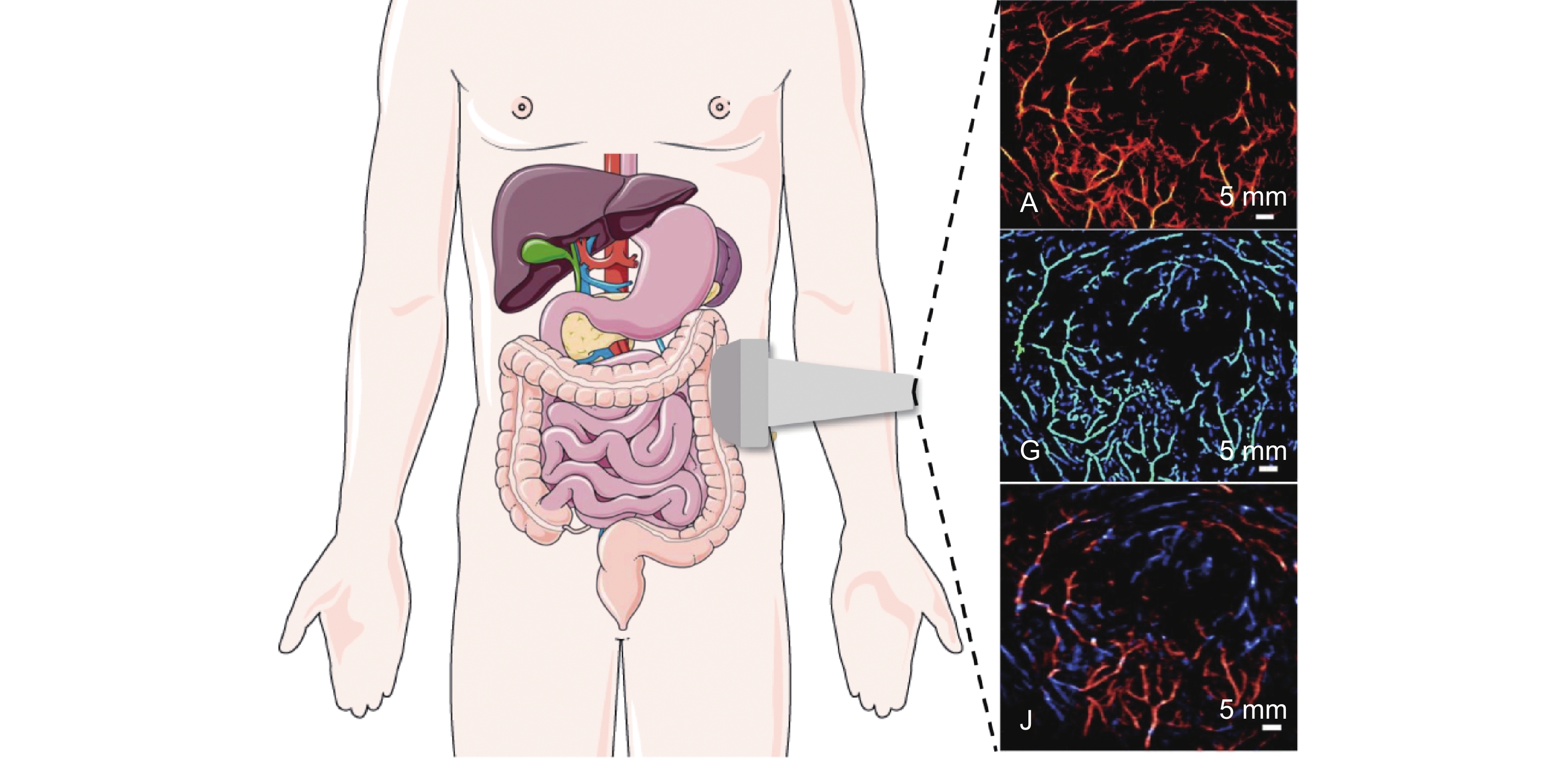
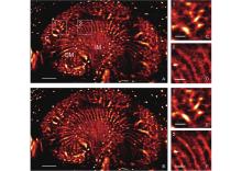
Figure 2
ULM imaging of rat renal microvasculature. (A) Vascular density map without motion correction; (B) Vascular density map with motion correction (C: cortex, OM: outer medulla, IM: inner medulla). (C-F) Enlarged views of white-boxed regions, (C-D) without motion correction; (E-F) after motion correction. Figure sourced from [21], creative commons license CC-BY-4.0 (https://creativecommons.org/licenses/by/4.0/). ULM, ultrasound localization microscopy"

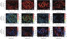
Figure 3
ULM parameter maps for different focal liver lesions. (A-D) Vascular density maps illustrate a peripherally distributed pattern (A), a well-distributed pattern (B), and an irregularly distributed pattern (C,D). (E-H) Velocity maps demonstrate high-speed feeding patterns (E,F) and low-speed supplying patterns (G,H). (I-L) Direction maps reveal several blood flow patterns: centrifugal (I), centripetal (J), eccentric (K), and mixed (L). The panels correspond to the following cases: (A,G,J) a 51-year-old female with liver metastasis; (B,E,I) a 35-year-old male with focal nodular hyperplasia; (C,F,K) a 71-year-old female with hepatocellular carcinoma; (D) a 65-year-old female with hepatocellular carcinoma; (H) a 45-year-old male with liver metastasis; and (L) another 65-year-old female with hepatocellular carcinoma. Figure sourced from [43], creative commons license CC-BY-4.0 (https://creativecommons.org/licenses/by/4.0/). ULM, ultrasound localization microscopy"
| [1] |
Hess ST , Girirajan TPK , Mason MD . Ultra-high resolution imaging by fluorescence photoactivation localization microscopy. Biophys J 2006; 91: 4258-4272.
doi: 10.1529/biophysj.106.091116 |
| [2] |
Betzig E , Patterson GH , Sougrat R , Lindwasser OW , Olenych S , Bonifacino JS , et al . Imaging intracellular fluorescent proteins at nanometer resolution. Science 2006; 313: 1642-1645.
doi: 10.1126/science.1127344 |
| [3] | Ruud J. G . van Sloun , Christensen-Jeffries K , Couture O , Dayton PA , Eldar YC , Hynynen K , et al. Super-resolution ultrasound imaging. Ultrasound Med Biol 2020; 46: 865-891 |
| [4] |
Errico C . Ultrafast ultrasound localization microscopy for deep super-resolution vascular imaging. Nature 2015; 527: 499-502.
doi: 10.1038/nature16066 |
| [5] | Kabir AU , Subramanian M , Kwon Y , Choi K . Linking tumour angiogenesis and tumour immunity. Nat Rev Immunol 2025;1-17. |
| [6] |
Fleig S , Magnuska ZA , Koczera P , Salewski J , Djudjaj S , Schmitz G , et al . Advanced ultrasound methods to improve chronic kidney disease diagnosis. Npj Imaging 2024; 2: 22.
doi: 10.1038/s44303-024-00023-5 |
| [7] |
Zhang W , Huang C , Yin T , Miao X , Deng H , Zheng R , et al . Ultrasensitive US microvessel imaging of hepatic microcirculation in the cirrhotic rat liver. Radiology 2023; 307: e220739.
doi: 10.1148/radiol.220739 |
| [8] |
Lin Y , Dong M , Liu Z , Xu M , Huang Z , Liu H , et al . A strategy of vascular-targeted therapy for liver fibrosis. Hepatol Baltim Md 2022; 76: 660-675.
doi: 10.1002/hep.32299 |
| [9] |
Molema G , Zijlstra JG , van Meurs M , Kamps JAAM . Renal microvascular endothelial cell responses in sepsis-induced acute kidney injury. Nat Rev Nephrol 2022; 18: 95-112.
doi: 10.1038/s41581-021-00489-1 |
| [10] |
Chen DC . Sepsis and intestinal microvascular endothelial dysfunction. Chin Med J (Engl) 2017; 130: 1137-1138.
doi: 10.4103/0366-6999.205865 |
| [11] |
George J , Lu Y , Tsuchishima M , Tsutsumi M . Cellular and molecular mechanisms of hepatic ischemia-reperfusion injury: The role of oxidative stress and therapeutic approaches. Redox Biol 2024; 75: 103258.
doi: 10.1016/j.redox.2024.103258 |
| [12] |
Sablik M , Sannier A , Raynaud M , Goutaudier V , Divard G , Astor BC , et al . Microvascular inflammation of kidney allografts and clinical outcomes. N Engl J Med 2025; 392: 763-776.
doi: 10.1056/NEJMoa2408835 |
| [13] |
Yan J , Huang B , Tonko J , Toulemonde M , Hansen-Shearer J , Tan Q , et al . Transthoracic ultrasound localization microscopy of myocardial vasculature in patients. Nat Biomed Eng 2024; 8: 689-700.
doi: 10.1038/s41551-024-01206-6 |
| [14] |
Opacic T , Dencks S , Theek B , Piepenbrock M , Ackermann D , Rix A , et al . Motion model ultrasound localization microscopy for preclinical and clinical multiparametric tumor characterization. Nat Commun 2018; 9: 1527.
doi: 10.1038/s41467-018-03973-8 |
| [15] |
Li M , Zhang X , Yan J , Shu H , Li Z , Ye C , et al . Non-invasive ultrasound localization microscopy (ULM) in azoospermia: connecting testicular microcirculation to spermatogenic functions. Theranostics 2024; 14: 4967-4982.
doi: 10.7150/thno.99668 |
| [16] |
Aziz MU , Eisenbrey JR , Deganello A , Zahid M , Sharbidre K , Sidhu P , et al . Microvascular flow imaging: A state-of-the-art review of clinical use and promise. Radiology 2022; 305: 250-264.
doi: 10.1148/radiol.213303 |
| [17] |
Tang Y , Dong Z , Wang N , Del Aguila A , Johnston N , Vu T , et al . Non-invasive deep-brain imaging with 3D integrated photoacoustic tomography and ultrasound localization microscopy (3D-PAULM). IEEE Trans Med Imaging 2025; 44: 994-1004.
doi: 10.1109/TMI.2024.3477317 |
| [18] |
Huang J , Wiacek A , Kempski KM , Palmer T , Izzi J , Beck S , et al . Empirical assessment of laser safety for photoacoustic-guided liver surgeries. Biomed Opt Express 2021; 12: 1205-1216.
doi: 10.1364/BOE.415054 |
| [19] |
Gitto S , Messina C , Chianca V , Tuscano B , Lazzara A , Corazza A , et al . Superb microvascular imaging (SMI) in the evaluation of musculoskeletal disorders: a systematic review. Radiol Med 2020; 125: 481-490.
doi: 10.1007/s11547-020-01141-x |
| [20] | Song P , Rubin JM , Lowerison MR . Super-resolution ultrasound microvascular imaging: Is it ready for clinical use? Z Med Phys 2023; 33:309-323. |
| [21] |
Foiret J , Zhang H , Ilovitsh T , Mahakian L , Tam S , Ferrara KW . Ultrasound localization microscopy to image and assess microvasculature in a rat kidney. Sci Rep 2017; 7: 13662.
doi: 10.1038/s41598-017-13676-7 |
| [22] |
Andersen SB , Taghavi I , Kjer HM , Søgaard SB , Gundlach C , Dahl VA , et al . Evaluation of 2D super-resolution ultrasound imaging of the rat renal vasculature using ex vivo micro-computed tomography. Sci Rep 2021; 11: 24335.
doi: 10.1038/s41598-021-03726-6 |
| [23] | McDermott A , Panduro NS , Taghavi I , Kjer HM , Søgaard SB , Nielsen MB , et al . Analysing the renal vasculature using super-resolution ultrasound imaging: Considerations for clinical and research applications. Diagn Basel Switz 2025; 15: 1515 |
| [24] | Andersen SB , Taghavi I , Søgaard SB , Hoyos CAV , Nielsen MB , Jensen JA , et al . Super-resolution ultrasound imaging can quantify alterations in microbubble velocities in the renal vasculature of rats. Diagn Basel Switz 2022; 12: 1111 |
| [25] |
Denis L , Bodard S , Hingot V , Chavignon A , Battaglia J , Renault G , et al . Sensing ultrasound localization microscopy for the visualization of glomeruli in living rats and humans. EBioMedicine 2023; 91: 104578.
doi: 10.1016/j.ebiom.2023.104578 |
| [26] | Chabouh G , Denis L , Bodard S , Lager F , Renault G , Chavignon A , et al . Whole organ volumetric sensing ultrasound localization microscopy for characterization of kidney structure. IEEE Trans Med Imaging 2024; 43: 40554063 |
| [27] | Huang C , Wai Lok U , Zhang J , Zhu XY , Krier JD , Stern A , et al. Optimizingin vivodata acquisition for robust clinical microvascular imaging using ultrasound localization microscopy. Phys Med Biol 2025;70. |
| [28] |
Song P , Trzasko JD , Manduca A , Huang R , Kadirvel R , Kallmes DF , et al . Improved super-resolution ultrasound microvessel imaging with spatiotemporal nonlocal means filtering and bipartite graph-based microbubble tracking. IEEE Trans Ultrason Ferroelectr Freq Control 2018; 65: 149-167.
doi: 10.1109/TUFFC.2017.2778941 |
| [29] |
Lei S , Zhang G , Zhu B , Long X , Jiang Z , Liu Y , et al . In vivo ultrasound localization microscopy imaging of the kidney’s microvasculature with block-matching 3-D denoising. IEEE Trans Ultrason Ferroelectr Freq Control 2022; 69: 523-533.
doi: 10.1109/TUFFC.2021.3125010 |
| [30] |
Yang J , Zhang J , An J , Dong F , Huang S , Guo W , et al . Hepatic portal venous perfusion imaging using vessel-labeling super-resolution ultrasound. Ultrasound Med Biol 2025; 51: 951-960.
doi: 10.1016/j.ultrasmedbio.2025.01.019 |
| [31] | Lok UW , Huang C , Gong P , Tang S , Yang L , Zhang W , et al. Fast super-resolution ultrasound microvessel imaging using spatiotemporal data with deep fully convolutional neural network. Phys Med Biol 2021;66. |
| [32] | Huang C , Zhang W , Gong P , Lok UW , Tang S , Yin T , et al. Super-resolution ultrasound localization microscopy based on a high frame-rate clinical ultrasound scanner: an in-human feasibility study. Phys Med Biol 2021;66. |
| [33] |
Zhang T , Yan J , Zhou X , Wu B , Zhang C , Tang M , et al . Application of ultrasound localization microscopy in evaluating the type 2 diabetes progression. Cyborg Bionic Syst 2025; 6: 0117.
doi: 10.34133/cbsystems.0117 |
| [34] |
Butler MB , Papageorgiou G , Kanoulas ED , Voulgaridou V , Wijkstra H , Mischi M , et al . Mapping of prostate cancer microvascular patterns using super-resolution ultrasound imaging. Eur Radiol Exp 2025; 9: 25.
doi: 10.1186/s41747-025-00561-6 |
| [35] |
Thuener JE . Urologic malignancies. Prim Care 2019; 46: 275-285.
doi: 10.1016/j.pop.2019.02.009 |
| [36] |
Roussel E , Capitanio U , Kutikov A , Oosterwijk E , Pedrosa I , Rowe SP , et al . Novel imaging methods for renal mass characterization: A collaborative review. Eur Urol 2022; 81: 476-488.
doi: 10.1016/j.eururo.2022.01.040 |
| [37] | Cantisani V , Bertolotto M , Clevert DA , Correas JM , Drudi FM , Fischer T , et al. EFSUMB 2020 proposal for a contrast-enhanced ultrasound-adapted bosniak cyst categorization - position statement. Ultraschall Med Stuttg Ger 1980. 2021; 42:154-166. |
| [38] |
Lowerison MR , Huang C , Lucien F , Chen S , Song P . Ultrasound localization microscopy of renal tumor xenografts in chicken embryo is correlated to hypoxia. Sci Rep 2020; 10: 2478.
doi: 10.1038/s41598-020-59338-z |
| [39] |
Bodard S , Denis L , Chabouh G , Anglicheau D , Hélénon O , Correas JM , et al . First clinical utility of sensing ultrasound localization microscopy (sULM): identifying renal pseudotumors. Theranostics 2025; 15: 233-244.
doi: 10.7150/thno.100897 |
| [40] |
Bergers G , Benjamin LE . Tumorigenesis and the angiogenic switch. Nat Rev Cancer 2003; 3: 401-410.
doi: 10.1038/nrc1093 |
| [41] |
Zhang W , Lowerison MR , Dong Z , Miller RJ , Keller KA , Song P . Super-resolution ultrasound localization microscopy on a rabbit liver VX2 tumor model: An initial feasibility study. Ultrasound Med Biol 2021; 47: 2416-2429.
doi: 10.1016/j.ultrasmedbio.2021.04.012 |
| [42] |
Brown KG , Li J , Margolis R , Trinh B , Eisenbrey JR , Hoyt K . Assessment of transarterial chemoembolization using super-resolution ultrasound imaging and a rat model of hepatocellular carcinoma. Ultrasound Med Biol 2023; 49: 1318-1326.
doi: 10.1016/j.ultrasmedbio.2023.01.021 |
| [43] |
Zeng QQ , An SZ , Chen CN , Wang Z , Liu JC , Wan MX , et al . Focal liver lesions: Multiparametric microvasculature characterization via super-resolution ultrasound imaging. Eur Radiol Exp 2024; 8: 138.
doi: 10.1186/s41747-024-00540-3 |
| [44] |
Wang F , Yu J , Lu X , Numata K , Ruan L , Zhang D , et al . Relationship between contrast-enhanced ultrasound combined with ultrasound resolution microscopy imaging and histological features of hepatocellular carcinoma. Abdom Radiol N Y 2025; 50: 3530-3542.
doi: 10.1007/s00261-025-04825-y |
| [45] |
Kang TW , Jeong WK , Kim YY , Min JH , Kim YK , Kim SH , et al . Comparison of super-resolution US and contrast material-enhanced US in detection of the spoke wheel sign in patients with focal nodular hyperplasia. Radiology 2021; 298: 82-90.
doi: 10.1148/radiol.2020200885 |
| [46] |
Qian-Qian Zeng , Ping Liang . Super-resolution US imaging of focal nodular hyperplasia. Radiology 2024; 311: e233130.
doi: 10.1148/radiol.233130 |
| [47] |
Litwin MS , Tan HJ . The diagnosis and treatment of prostate cancer: A review. JAMA 2017; 317: 2532-2542.
doi: 10.1001/jama.2017.7248 |
| [48] |
Huang X , Ye H , Hu Y , Lei Y , Tian Y , Huang X , et al . Ultrasound super-resolution imaging for non-invasive assessment of microvessel in prostate lesion. Cancer Imaging 2025; 25: 1.
doi: 10.1186/s40644-024-00819-z |
| [49] |
Kanoulas E , Butler M , Rowley C , Voulgaridou V , Diamantis K , Duncan WC , et al . Super-resolution contrast-enhanced ultrasound methodology for the identification of in vivo vascular dynamics in 2D. Invest Radiol 2019; 54: 500-516.
doi: 10.1097/RLI.0000000000000565 |
| [50] |
Romagnani P , Remuzzi G , Glassock R , Levin A , Jager KJ , Tonelli M , et al . Chronic kidney disease. Nat Rev Dis Primer 2017; 3: 17088.
doi: 10.1038/nrdp.2017.88 |
| [51] |
Kopitkó C , Gondos T , Fülöp T , Medve L . Reinterpreting renal hemodynamics: The importance of venous congestion and effective organ perfusion in acute kidney injury. Am J Med Sci 2020; 359: 193-205.
doi: 10.1016/j.amjms.2020.01.012 |
| [52] |
Qiu L , Zhang J , Yang Y , Zhang H , Lee FF , He Q , et al . In vivo assessment of hypertensive nephrosclerosis using ultrasound localization microscopy. Med Phys 2022; 49: 2295-2308.
doi: 10.1002/mp.15583 |
| [53] | Søgaard SB , Andersen SB , Taghavi I , Hoyos CAV , Christoffersen C , Hansen KL , et al . Super-resolution ultrasound imaging provides quantification of the renal cortical and medullary vasculature in obese zucker rats: A pilot study. Diagn Basel Switz 2022; 12: 1626 |
| [54] | Søgaard SB , Andersen SB , Taghavi I , Schou M , Christoffersen C , Jacobsen JCB , et al . Super-resolution ultrasound imaging of renal vascular alterations in zucker diabetic fatty rats during the development of diabetic kidney disease. Diagn Basel Switz 2023; 13: 3197 |
| [55] |
Zhang H , Huang L , Yang Y , Qiu L , He Q , Liu J , et al . Evaluation of early diabetic kidney disease using ultrasound localization microscopy: A feasibility study. J Ultrasound Med 2023; 42: 2277-2292.
doi: 10.1002/jum.16249 |
| [56] | Xu Y , Luo Y , Chen M , Peng Q , Niu C . Super-resolution ultrasound imaging of renal microcirculation in a murine model of renal fibrosis. J Ultrasound Med 2025. |
| [57] |
Chen Q , George MW , McMahon B , Rosenthal JA , Kim K , Tan RJ . Super-resolution ultrasound to assess kidney vascular changes in humans with kidney disease. Am J Kidney Dis 2025; 85: 393-395.
doi: 10.1053/j.ajkd.2024.06.021 |
| [58] |
Zhang R , Wang J , Liu T , Wang Y , Yang S , Yan F , et al . Proof of concept: Super-resolution ultrasound and viscoelastic imaging of hepatic microcirculation for early detection and staging of liver fibrosis in a murine model. J Ultrasound Med 2025; 44: 1481-1491.
doi: 10.1002/jum.16703 |
| [59] |
Chabouh G , Pradier B , Denis L , Baida R , Hingot V , Chavignon A , et al . Multiparametric ultrasound evaluation of metabolic dysfunction-associated fatty liver disease in minipigs. Ultrasound Med Biol 2025; 51: 1710-1719.
doi: 10.1016/j.ultrasmedbio.2025.06.011 |
| [60] |
Basile DP , Bonventre JV , Mehta R , Nangaku M , Unwin R , Rosner MH , et al . Progression after AKI: Understanding maladaptive repair processes to predict and identify therapeutic treatments. J Am Soc Nephrol 2016; 27: 687-697.
doi: 10.1681/ASN.2015030309 |
| [61] |
Basile DP , Yoder MC . Renal endothelial dysfunction in acute kidney ischemia reperfusion injury. Cardiovasc Hematol Disord Drug Targets 2014; 14: 3-14.
doi: 10.2174/1871529X1401140724093505 |
| [62] | Andersen SB , Taghavi I , Hoyos CAV , Søgaard SB , Gran F , Lönn L , et al . Super-resolution imaging with ultrasound for visualization of the renal microvasculature in rats before and after renal ischemia: A pilot study. Diagn Basel Switz 2020; 10: 862 |
| [63] |
Chen Q , Yu J , Rush BM , Stocker SD , Tan RJ , Kim K . Ultrasound super-resolution imaging provides a noninvasive assessment of renal microvasculature changes during mouse acute kidney injury. Kidney Int 2020; 98: 355-365.
doi: 10.1016/j.kint.2020.02.011 |
| [64] |
Zhao S , Zhang X , Bailey K , Pai S , Zhao Y , Chen Y . Label‐free dual‐modal photoacoustic/ultrasound localization imaging for studying acute kidney injury. Adv Sci 2025; 12: 2414306.
doi: 10.1002/advs.202414306 |
| [65] | Huang X , Zhang Y , Zhou Q , Deng Q . Value of ultrasound super-resolution imaging for the assessment of renal microcirculation in patients with acute kidney injury: A preliminary study. Diagn Basel Switz 2024; 14: 1192 |
| [66] |
Bodard S , Denis L , Hingot V , Chavignon A , Hélénon O , Anglicheau D , et al . Ultrasound localization microscopy of the human kidney allograft on a clinical ultrasound scanner. Kidney Int 2023; 103: 930-935.
doi: 10.1016/j.kint.2023.01.027 |
| [67] |
Li T , Sun H , Gai Y , Zhu L , Yang J , Ma C . Ultrasound localization microscopy for the assessment of microvascular abnormalities in transplant renal artery stenosis. Ultrasound Med Biol 2025; 51: 2120-2124.
doi: 10.1016/j.ultrasmedbio.2025.07.032 |
| [68] |
Bodard S , Denis L , Chabouh G , Battaglia J , Anglicheau D , Hélénon O , et al . Visualization of renal glomeruli in human native kidneys with sensing ultrasound localization microscopy. Invest Radiol 2024; 59: 561-568.
doi: 10.1097/RLI.0000000000001061 |
| [69] | Zhu L , Ma C , Li T . Ultrasound localization microscopy for early detection of acute kidney allograft rejection. Nephrol Dial Transplant 2025:gfaf106. |
| [70] |
Yin J , Dong F , An J , Guo T , Cheng H , Zhang J , et al . Pattern recognition of microcirculation with super-resolution ultrasound imaging provides markers for early tumor response to anti-angiogenic therapy. Theranostics 2024; 14: 1312-1324.
doi: 10.7150/thno.89306 |
| [1] | Hou Shilin, Bao Guocui, Sun Zhe, Li Guo, Zhang Bo, Dai Jiyan. Volumetric Imaging with 2D Array Ultrasound Transducers for Clinical Applications: A Review [J]. Advanced Ultrasound in Diagnosis and Therapy, 2025, 9(4): 437-448. |
| [2] | Guan Xin, Hu Xinyuan, Han Hong, Zhang Dezhi, Xu Huixiong. The Evolving Application of Ultrasound in the Precision Management of Small Hepatocellular Carcinoma [J]. Advanced Ultrasound in Diagnosis and Therapy, 2025, 9(4): 375-387. |
| [3] | Zhang Xiaoqian, Zhang Jingwen, Dong Yijie, Zhou Jianqiao. Research Progress and Clinical Translation of Photoacoustic–ultrasound Fusion Imaging in Breast Cancer Diagnosis and Therapy [J]. Advanced Ultrasound in Diagnosis and Therapy, 2025, 9(4): 467-482. |
| [4] | Jin Tong, Yu Xiaohu, Ai Zheng, Guo Hongcheng. Artificial Intelligence in Ultrasound Imaging: A Review of Progress from Machine Learning to Large Language Model [J]. Advanced Ultrasound in Diagnosis and Therapy, 2025, 9(4): 483-496. |
| [5] | Xiang Xi, Yang Yujia, Wang Liyun, Qiu Li. Advances and Applications in Dermatological Ultrasound [J]. Advanced Ultrasound in Diagnosis and Therapy, 2025, 9(4): 457-466. |
| [6] | Zheng Hairong, Meng Long, Li Fei, Niu Lili, Qiu Weibao, Ma Teng, Liu Chengbo, Zhu Xuefeng, Wan Liwen, Cai Feiyan. Advance in Ultrasound Super-resolution Imaging, Cell Manipulation and Inter-brain Communication [J]. Advanced Ultrasound in Diagnosis and Therapy, 2025, 9(4): 307-325. |
| [7] | Xia ShuJun, Zheng YuHang, Hua Qing, Wei MinYan, Wen Jing, Luo XiaoMao, Yan JiPing, Bai BaoYan, Liu Fang, Dong YiJie, Zhou JianQiao, behalf of The Chinese Artificial Intelligence Alliance for Thyroid on, Ultrasound Breast. Super-Resolution Ultrasound-Based Habitat Imaging: A Consensus Statement [J]. Advanced Ultrasound in Diagnosis and Therapy, 2025, 9(2): 97-102. |
| [8] | Cui Liuping, Liu Ran, Liu Yumei, Zhou Fubo, Tao Yunlu, Xing Yingqi. Reviews on Imaging-based Risk Prediction Models for Ischemic Stroke [J]. Advanced Ultrasound in Diagnosis and Therapy, 2025, 9(2): 117-126. |
| [9] | Wang Yan, Zhang Mingbo. The Development of Photothermal Imaging Technology in Clinical Applications [J]. Advanced Ultrasound in Diagnosis and Therapy, 2025, 9(2): 146-153. |
| [10] | Xiang Hongjin, Huang Lin, Zheng Zhu, Li Jiawu, Qiu Tingting, Wu Zhenru, Shi Yujun, Jiang Huabei, Ling Wenwu, Luo Yan. Evaluation of Hepatic Steatosis Grades with Thermoacoustic Imaging in a Rabbit Model [J]. Advanced Ultrasound in Diagnosis and Therapy, 2025, 9(2): 171-180. |
| [11] | Yu Huimin, Tang Ying, Niu Ningning, Wu Hongtao, Zhang Guoying, Wang Mingyang, Hao Xiaoye, Liu Jing. The Impact of B-type natriuretic peptide (BNP) on Transplanted Liver Hemodynamics and Short-Term Prognosis: A Single-Center Big Data Retrospective Study [J]. Advanced Ultrasound in Diagnosis and Therapy, 2025, 9(2): 189-196. |
| [12] | Chen Ya, Wang Xinqi, Chen Anni, Li Zhenyi, Yang Lan, Li Zhaojun, Jin Lin, Wang Xifu. The Application and Research Progress of Cardiac Magnetic Resonance in the Assessment of Right Ventricular-Pulmonary Arterial Coupling [J]. Advanced Ultrasound in Diagnosis and Therapy, 2024, 8(4): 183-194. |
| [13] | Li Zhenyi, Chen Ya, Wang Xinqi, Yang Lan, Chen Anni, Li Zhaojun, Jin Lin. Left and Right Ventricular Interaction: Insight from Echocardiography Imaging [J]. Advanced Ultrasound in Diagnosis and Therapy, 2024, 8(4): 195-204. |
| [14] | Yuhang Zheng, BS, Jianqiao Zhou, MD. Deep Learning in Ultrasound Localization Microscopy [J]. Advanced Ultrasound in Diagnosis and Therapy, 2024, 8(3): 86-92. |
| [15] | Guiwu Chen, MS, Zhizhong He, BS, Wenqin Liu, MS, Junjun Chen, BS, Xiaomin Liao, MS, Yuhuan Xie, BS. Multimodal Sonographic Findings of Embryonal Carcinoma in the Testis: A Case Report and Literature Review [J]. Advanced Ultrasound in Diagnosis and Therapy, 2024, 8(2): 74-77. |
| Viewed | ||||||
|
Full text |
|
|||||
|
Abstract |
|
|||||
Share: WeChat
Copyright ©2018 Advanced Ultrasound in Diagnosis and Therapy
|
 Advanced Ultrasound in Diagnosis and Therapy (AUDT)
is licensed under a Creative Commons Attribution 4.0 International License.
Advanced Ultrasound in Diagnosis and Therapy (AUDT)
is licensed under a Creative Commons Attribution 4.0 International License.