Advanced Ultrasound in Diagnosis and Therapy ›› 2025, Vol. 9 ›› Issue (4): 467-482.doi: 10.26599/AUDT.2025.250103
Previous Articles Next Articles
Zhang Xiaoqiana,b,1, Zhang Jingwena,b,1, Dong Yijiea,b, Zhou Jianqiaoa,b,*( )
)
Received:2025-10-02
Revised:2025-10-23
Accepted:2025-10-30
Online:2025-12-30
Published:2025-11-06
Contact:
Department of Ultrasound, Ruijin Hospital, Shanghai Jiao Tong University School of Medicine, 197 Ruijin Er Road, Shanghai, China (Yijie Dong, Jianqiao Zhou),e-mail: zhousu30@126.com (JQ Z).,
About author:1Xiaoqian Zhang and Jingwen Zhang contributed equally to this study.
Zhang Xiaoqian, Zhang Jingwen, Dong Yijie, Zhou Jianqiao. Research Progress and Clinical Translation of Photoacoustic–ultrasound Fusion Imaging in Breast Cancer Diagnosis and Therapy. Advanced Ultrasound in Diagnosis and Therapy, 2025, 9(4): 467-482.

Figure 1
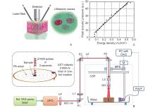
(A) Schematic illustration of the PA effect [11]; (B) Diagram of a representative PAI system [12]; (C) Relationship between the energy intensity of each laser pulse and the induced acoustic pressure, demonstrating a linear increase in acoustic pressure with input energy [13]. LDU, laser driving unit; CSP, circular scanning plate; S, sample; MPS, motor-pulley system; M, motor; DAQ, data acquisition card; R/A/F, receiver, amplifier, and filter for US signals; UST, ultrasound transducer."


Figure 3
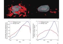
3D PA/US reconstructions of vascular networks in (A) an invasive breast cancer and (B) a breast fibroadenoma. Malignant tumors demonstrate dense peripheral vasculature and sparse central perfusion, whereas benign lesions exhibit more uniform vascular distribution. The probability density distribution of SO2 in the (C) intratumoral and (D) peritumoral regions further illustrates that malignant tumors have lower SO2 compared with benign counterparts [14]."


Figure 4
Representative PA/US fusion images demonstrating functional–structural contrasts between malignant and benign breast lesions. (A) Invasive ductal carcinoma (female, 40 years): marked peripheral vascular enrichment with limited or absent central PA signals, corresponding to angiogenic expansion at the margins and central hypoxia/necrosis; (B) Fibroadenoma (female, 36 years): relatively uniform PA signal distribution, reflecting preserved vascular architecture. This functional–structural contrast forms the mechanistic basis for PA/US-enhanced benign–malignant differentiation [5]."

| [1] | Sung H , Ferlay J , Siegel RL , Laversanne M , Soerjomataram I , Jemal A , et al . Global cancer statistics 2020: GLOBOCAN estimates of incidence and mortality worldwide for 36 cancers in 185 countries. CA: a cancer journal for clinicians 2021; 71: 209-249 |
| [2] |
Fan L , Strasser-Weippl K , Li J-J , St Louis J , Finkelstein DM , Yu K-D , et al . Breast cancer in China. Lancet Oncol 2014; 15: e279-e289.
doi: 10.1016/S1470-2045(13)70567-9 |
| [3] |
Oraevsky A , Clingman B , Zalev J , Stavros A , Yang W , Parikh J . Clinical optoacoustic imaging combined with ultrasound for coregistered functional and anatomical mapping of breast tumors. Photoacoustics 2018; 12: 30-45.
doi: 10.1016/j.pacs.2018.08.003 |
| [4] |
Lin L , Wang LV . The emerging role of photoacoustic imaging in clinical oncology. Nat Rev Clin Oncol 2022; 19: 365-384.
doi: 10.1038/s41571-022-00615-3 |
| [5] | Liu H , Wang M , Ji F , Jiang Y , Yang M . Mini review of photoacoustic clinical imaging: a noninvasive tool for disease diagnosis and treatment evaluation. J Biomed Opt 2024; 29: S11522 |
| [6] | Huang Z , Tian H , Luo H , Yang K , Chen J , Li G , et al. Assessment of oxygen saturation in breast lesions using photoacoustic imaging: correlation with benign and malignant disease. Clin Breast Cancer 2024; 24:e210-e218. e1. |
| [7] |
Chen J , Huang Z , Luo H , Li G , Ding Z , Tian H , et al . Development and validation of nomograms using photoacoustic imaging and 2D ultrasound to predict breast nodule benignity and malignancy. Postgrad Med J 2024; 100: 309-318.
doi: 10.1093/postmj/qgad146 |
| [8] |
Neuschler EI , Butler R , Young CA , Barke LD , Bertrand ML , Böhm-Vélez M , et al . A pivotal study of optoacoustic imaging to diagnose benign and malignant breast masses: a new evaluation tool for radiologists. Radiology 2018; 287: 398-412.
doi: 10.1148/radiol.2017172228 |
| [9] |
Liu H , Teng X , Yu S , Yang W , Kong T , Liu T . Recent advances in photoacoustic imaging: current status and future perspectives. Micromachines 2024; 15: 1007.
doi: 10.3390/mi15081007 |
| [10] | Doğan BE . Optoacoustic breast imaging: current status and future trends in clinical application. Society of Breast Imaging (SBI) Annual Meeting; United States: Society of Breast Imaging 2022. |
| [11] | Knieling F , Menezes JG , Claussen J , Schwarz M , Neufert C , Fahlbusch FB , et al. Raster-scanning optoacoustic mesoscopy for gastrointestinal imaging at high resolution. Gastroenterology 2018; 154:807-809. e3. |
| [12] |
Upputuri PK , Pramanik M . Performance characterization of low-cost, high-speed, portable pulsed laser diode photoacoustic tomography (PLD-PAT) system. Biomed Opt Express 2015; 6: 4118-4129.
doi: 10.1364/BOE.6.004118 |
| [13] |
Wang LV , Zhao X , Sun H , Ku G . Microwave-induced acoustic imaging of biological tissues. Review of scientific instruments 1999; 70: 3744-3748.
doi: 10.1063/1.1149986 |
| [14] |
Yang M , Zhao L , Yang F , Wang M , Su N , Zhao C , et al . Quantitative analysis of breast tumours aided by three-dimensional photoacoustic/ultrasound functional imaging. Sci Rep 2020; 10: 8047.
doi: 10.1038/s41598-020-64966-6 |
| [15] | Dantuma M , Lucka F , Kruitwagen S , Javaherian A , Alink L , van Meerdervoort RP , et al. Fully three-dimensional sound speed-corrected multi-wavelength photoacoustic breast tomography. arXiv preprint arXiv:230806754. 2023. |
| [16] | Suhonen M , Lucka F , Pulkkinen A , Arridge S , Cox B , Tarvainen T . Reconstructing initial pressure and speed of sound distributions simultaneously in photoacoustic tomography. arXiv preprint arXiv:250508482. 2025. |
| [17] | De Santi B , Kim L , Bulthuis RF , Lucka F , Manohar S . Automated three-dimensional image registration for longitudinal photoacoustic imaging. J Biomed Opt 2024; 29: S11515 |
| [18] |
Zhu X , Menozzi L , Cho S-W , Yao J . High speed innovations in photoacoustic microscopy. Npj Imaging 2024; 2: 46.
doi: 10.1038/s44303-024-00052-0 |
| [19] | A Study of Tumor Imaging With Multispectral Optoacoustic Tomography (MSOT) [NCT05488483] [Internet]. U.S. National Library of Medicine 2022-2025 [cited 2025-09-13]. Available from: https://clinicaltrials.gov/study/NCT05488483. |
| [20] |
Wang M , Zhao L , Wei Y , Li J , Qi Z , Su N , et al . Functional photoacoustic/ultrasound imaging for the assessment of breast intraductal lesions: preliminary clinical findings. Biomedical Optics Express 2021; 12: 1236-1246.
doi: 10.1364/BOE.411215 |
| [21] | Park J , Choi S , Knieling F , Clingman B , Bohndiek S , Wang LV , et al . Clinical translation of photoacoustic imaging. Nature Reviews Bioengineering 2025; 3: 193-212 |
| [22] |
Steinberg I , Huland DM , Vermesh O , Frostig HE , Tummers WS , Gambhir SS . Photoacoustic clinical imaging. Photoacoustics 2019; 14: 77-98.
doi: 10.1016/j.pacs.2019.05.001 |
| [23] |
Garcia-Uribe A , Erpelding TN , Krumholz A , Ke H , Maslov K , Appleton C , et al . Dual-modality photoacoustic and ultrasound imaging system for noninvasive sentinel lymph node detection in patients with breast cancer. Sci Rep 2015; 5: 15748.
doi: 10.1038/srep15748 |
| [24] |
Huang Z , Mo S , Wu H , Kong Y , Luo H , Li G , et al . Optimizing breast cancer diagnosis with photoacoustic imaging: an analysis of intratumoral and peritumoral radiomics. Photoacoustics 2024; 38: 100606.
doi: 10.1016/j.pacs.2024.100606 |
| [25] |
Huang Z , Wang M , Kong Y , Li G , Tian H , Wu H , et al . Photoacoustic-based intra-and peritumoral radiomics nomogram for the preoperative prediction of expression of Ki-67 in breast malignancy. Acad Radiol 2025; 32: 2422-2434.
doi: 10.1016/j.acra.2024.10.036 |
| [26] |
Abeyakoon O , Woitek R , Wallis M , Moyle P , Morscher S , Dahlhaus N , et al . An optoacoustic imaging feature set to characterise blood vessels surrounding benign and malignant breast lesions. Photoacoustics 2022; 27: 100383.
doi: 10.1016/j.pacs.2022.100383 |
| [27] |
Zhang R , Zhao L-y , Zhao C-y , Wang M , Liu S-r , Li J-c , et al . Exploring the diagnostic value of photoacoustic imaging for breast cancer: the identification of regional photoacoustic signal differences of breast tumors. Biomedical Optics Express 2021; 12: 1407-1421.
doi: 10.1364/BOE.417056 |
| [28] |
Lin L , Hu P , Tong X , Na S , Cao R , Yuan X , et al . High-speed three-dimensional photoacoustic computed tomography for preclinical research and clinical translation. Nat Commun 2021; 12: 882.
doi: 10.1038/s41467-021-21232-1 |
| [29] | Tong X , Liu CZ , Luo Y , Lin L , Dzubnar J , Invernizzi M , et al. Panoramic photoacoustic computed tomography with learning-based classification enhances breast lesion characterization. Nat Biomed Eng 2025:1-17. |
| [30] |
Wu Y , Huang K , Chen G , Lin L . Advances in photoacoustic imaging of breast cancer. Sensors 2025; 25: 4812.
doi: 10.3390/s25154812 |
| [31] |
Seiler SJ , Neuschler EI , Butler RS , Lavin PT , Dogan BE . Optoacoustic imaging with decision support for differentiation of benign and malignant breast masses: a 15-reader retrospective study. AJR Am J Roentgenol 2023; 220: 646-658.
doi: 10.2214/AJR.22.28470 |
| [32] |
Menezes GL , Pijnappel RM , Meeuwis C , Bisschops R , Veltman J , Lavin PT , et al . Downgrading of breast masses suspicious for cancer by using optoacoustic breast imaging. Radiology 2018; 288: 355-365.
doi: 10.1148/radiol.2018170500 |
| [33] | Ozcan BB , Wanniarachchi H , Mason RP , Dogan BE . Current status of optoacoustic breast imaging and future trends in clinical application: is it ready for prime time? Eur Radiol 2024; 34:6092-6107. |
| [34] | Food US , Drug A . Summary of Safety and Effectiveness Data (SSED): Imagio™ Breast Imaging System. FDA Premarket Approval Report. Silver Spring, MD: U.S. Food and Drug Administration, Center for Devices and Radiological Health; 2021. |
| [35] |
Brackstone M , Baldassarre FG , Perera FE , Cil T , Chavez Mac Gregor M , Dayes IS , et al . Management of the axilla in early-stage breast cancer: Ontario Health (Cancer Care Ontario) and ASCO guideline. J Clin Oncol 2021; 39: 3056-3082.
doi: 10.1200/JCO.21.00934 |
| [36] | Harrison B . Update on sentinel node pathology in breast cancer. Semin Diagn Pathol 2022 Sep;39:355-366. |
| [37] |
Li WB , Du ZC , Liu YJ , Gao JX , Wang JG , Dai Q , et al . Prediction of axillary lymph node metastasis in early breast cancer patients with ultrasonic videos based deep learning. Front Oncol 2023; 13: 1219838.
doi: 10.3389/fonc.2023.1219838 |
| [38] |
Huang Z , Wang M , Tian H , Li G , Wu H , Chen J , et al . Enhancing axillary lymph node diagnosis in breast cancer with a novel photoacoustic imaging-based radiomics nomogram: a comparative study of peritumoral regions. Acad Radiol 2025; 32: 1274-1286.
doi: 10.1016/j.acra.2024.10.018 |
| [39] | Schito L , Rey S , Tafani M , Zhang H , Wong CCL , Russo A , et al . Hypoxia-inducible factor 1-dependent expression of platelet-derived growth factor B promotes lymphatic metastasis of hypoxic breast cancer cells. Proc Natl Acad Sci U S A 2012; 109: E2707-E2716 |
| [40] | Nyayapathi N , Xia J . Photoacoustic imaging of breast cancer: a mini review of system design and image features. J Biomed Opt 2019; 24: 1-13 |
| [41] |
Stachs A , Thi AT-H , Dieterich M , Stubert J , Hartmann S , Glass Ä , et al . Assessment of ultrasound features predicting axillary nodal metastasis in breast cancer: the impact of cortical thickness. Ultrasound Int Open 2015; 1: E19-E24.
doi: 10.1055/s-0035-1555872 |
| [42] |
Huang Z , Mo S , Li G , Tian H , Wu H , Chen J , et al . Prognosticating axillary lymph node metastasis in breast cancer through integrated photoacoustic imaging, ultrasound, and clinical parameters. Breast Cancer Res 2025; 27: 123.
doi: 10.1186/s13058-025-02073-y |
| [43] | Zalev J , Richards LM , Clingman BA , Harris J , Cantu E , Menezes GLG , et al . Opto-acoustic imaging of relative blood oxygen saturation and total hemoglobin for breast cancer diagnosis. J Biomed Opt 2019; 24: 1-16 |
| [44] |
Dialani V , James D , Slanetz P . A practical approach to imaging the axilla. Insights Imaging 2015; 6: 217-229.
doi: 10.1007/s13244-014-0367-8 |
| [45] |
Chang JM , Shin HJ , Choi JS , Shin SU , Choi BH , Kim MJ , et al . Imaging protocol and criteria for evaluation of axillary lymph nodes in the NAUTILUS trial. J Breast Cancer 2021; 24: 554.
doi: 10.4048/jbc.2021.24.e47 |
| [46] |
Yang L , Gu Y , Wang B , Sun M , Zhang L , Shi L , et al . A multivariable model of ultrasound and clinicopathological features for predicting axillary nodal burden of breast cancer: potential to prevent unnecessary axillary lymph node dissection. BMC cancer 2023; 23: 1264.
doi: 10.1186/s12885-023-11751-z |
| [47] |
Riedel F , Schaefgen B , Sinn H-P , Feisst M , Hennigs A , Hug S , et al . Diagnostic accuracy of axillary staging by ultrasound in early breast cancer patients. Eur J Radiol 2021; 135: 109468.
doi: 10.1016/j.ejrad.2020.109468 |
| [48] |
Kim KJ , Park M , Joo B , Ahn SJ , Suh SH . Dynamic contrast-enhanced MRI and its applications in various central nervous system diseases. Investigative Magnetic Resonance Imaging 2022; 26: 256-264.
doi: 10.13104/imri.2022.26.4.256 |
| [49] |
Hadebe B , Harry L , Ebrahim T , Pillay V , Vorster M . The role of PET/CT in breast cancer. Diagnostics 2023; 13: 597.
doi: 10.3390/diagnostics13040597 |
| [50] |
Gentilini OD , Botteri E , Sangalli C , Galimberti V , Porpiglia M , Agresti R , et al . Sentinel lymph node biopsy vs no axillary surgery in patients with small breast cancer and negative results on ultrasonography of axillary lymph nodes: the SOUND randomized clinical trial. JAMA oncology 2023; 9: 1557-1564.
doi: 10.1001/jamaoncol.2023.3759 |
| [51] | Morrow M . Sentinel-lymph-node biopsy in early-stage breast cancer - is it obsolete? N Engl J Med 2025;392:1134-1136. |
| [52] | Abdulla AH , Althawadi R , Salman AZ , Altaei TH , Mahdi AM , Abdulla HA . Applying the SOUND trial for omitting axillary surgery in patients with early breast cancer in Bahrain. Eur J Breast Health 2024; 20: 270 |
| [53] |
Wong SL , Edwards MJ , Chao C , Tuttle TM , Noyes RD , Carlson DJ , et al . Sentinel lymph node biopsy for breast cancer: impact of the number of sentinel nodes removed on the false-negative rate. J Am Coll Surg 2001; 192: 684-689.
doi: 10.1016/S1072-7515(01)00858-4 |
| [54] | Rubio IT , Diaz-Botero S , Esgueva AJ , Espinosa-Bravo M . Increased detection of sentinel nodes in breast cancer patients with the use of superparamagnetic iron oxide tracer. American Society of Clinical Oncology 2014; 32: S100 |
| [55] |
Du Y , Wu R , Diao X . Photoacoustic imaging: an emerging tool for precision diagnosis and treatment of breast cancer. Acad Radiol 2025; 32: 2435-2437.
doi: 10.1016/j.acra.2025.03.006 |
| [56] |
Carriero A , Groenhoff L , Vologina E , Basile P , Albera M . Deep learning in breast cancer imaging: state of the art and recent advancements in early 2024. Diagnostics 2024; 14: 848.
doi: 10.3390/diagnostics14080848 |
| [57] |
Li G , Huang Z , Tian H , Wu H , Zheng J , Wang M , et al . Deep learning combined with attention mechanisms to assist radiologists in enhancing breast cancer diagnosis: a study on photoacoustic imaging. Biomedical Optics Express 2024; 15: 4689-4704.
doi: 10.1364/BOE.530249 |
| [58] |
Li G , Tang S , Huang Z , Wang M , Tian H , Wu H , et al . Photoacoustic imaging with attention-guided deep learning for predicting axillary lymph node status in breast cancer. Acad Radiol 2025; 32: 2453-2464.
doi: 10.1016/j.acra.2024.12.020 |
| [59] | Zubair M , Hussain M , Al-Bashrawi MA , Bendechache M , Owais M . A comprehensive review of techniques, algorithms, advancements, challenges, and clinical applications of multi-modal medical image fusion for improved diagnosis. Comput Methods Programs Biomed 2025:109014. |
| [60] |
Latha M , Kumar PS , Chandrika RR , Mahesh T , Kumar VV , Guluwadi S . Revolutionizing breast ultrasound diagnostics with EfficientNet-B7 and Explainable AI. BMC Med Imaging 2024; 24: 230.
doi: 10.1186/s12880-024-01404-3 |
| [61] | Yan L , Liang Z , Zhang H , Zhang G , Zheng W , Han C , et al . A domain knowledge-based interpretable deep learning system for improving clinical breast ultrasound diagnosis. Commun Med (Lond) 2024; 4: 90 |
| [1] | Hu Xuelin, Zhu Ye, Zhang Zisang, Quan Yuanting, Chen Wenwen, Chen Leichong, Xu Guangyu, Qin Luning, Xie Mingxing, Zhang Li. Explainable Artificial Intelligence in Echocardiography [J]. Advanced Ultrasound in Diagnosis and Therapy, 2025, 9(4): 409-425. |
| [2] | Guan Xin, Hu Xinyuan, Han Hong, Zhang Dezhi, Xu Huixiong. The Evolving Application of Ultrasound in the Precision Management of Small Hepatocellular Carcinoma [J]. Advanced Ultrasound in Diagnosis and Therapy, 2025, 9(4): 375-387. |
| [3] | Li Yanran, Cui Yuanjie, Wu Qingqing, Zhang Na. Current Applications of Artificial Intelligence in Obstetric Ultrasound [J]. Advanced Ultrasound in Diagnosis and Therapy, 2025, 9(4): 449-456. |
| [4] | Hou Wenfei, Chen Wanting, Liu Huazhen, Tang Jiajia, Yang Meng. Applications of Ultrasound Localization Microscopy in Abdominal Imaging [J]. Advanced Ultrasound in Diagnosis and Therapy, 2025, 9(4): 347-356. |
| [5] | Yu Xiao jie, Song Zheng lai, Chang Xue yong, Yu Jie, Liang Ping. Artificial Intelligence in Ultrasound Diagnosis of Liver Nodules: A Comprehensive Review of B-Mode and Contrast-enhanced Applications [J]. Advanced Ultrasound in Diagnosis and Therapy, 2025, 9(4): 326-346. |
| [6] | Zhong Xian, Xie Xiaoyan. Multimodal Ultrasound Radiomics in Liver Disease: Current Status and Future Directions [J]. Advanced Ultrasound in Diagnosis and Therapy, 2025, 9(4): 388-408. |
| [7] | Jin Tong, Yu Xiaohu, Ai Zheng, Guo Hongcheng. Artificial Intelligence in Ultrasound Imaging: A Review of Progress from Machine Learning to Large Language Model [J]. Advanced Ultrasound in Diagnosis and Therapy, 2025, 9(4): 483-496. |
| [8] | Xiang Xi, Yang Yujia, Wang Liyun, Qiu Li. Advances and Applications in Dermatological Ultrasound [J]. Advanced Ultrasound in Diagnosis and Therapy, 2025, 9(4): 457-466. |
| [9] | Zheng Hairong, Meng Long, Li Fei, Niu Lili, Qiu Weibao, Ma Teng, Liu Chengbo, Zhu Xuefeng, Wan Liwen, Cai Feiyan. Advance in Ultrasound Super-resolution Imaging, Cell Manipulation and Inter-brain Communication [J]. Advanced Ultrasound in Diagnosis and Therapy, 2025, 9(4): 307-325. |
| [10] | Yang Jie, Liu Xiaodi, Wang Wenhui, Liao Min, Wu Zhe, Galeano July, Perez-Buitrago Sandra, Lu Qiang. Therapeutic Focused Ultrasound: Advances in Antitumor Immunotherapy of Solid Tumors [J]. Advanced Ultrasound in Diagnosis and Therapy, 2025, 9(3): 245-253. |
| [11] | Shi Junni, Xu Jiatong, Chen Chuanjian, Xiang Guanghua, Zheng Wen, Chen Man. Ultrasound Viscoelasticity for Breast Tumor: High Diagnostic Performance at the Peritumoral Boundary [J]. Advanced Ultrasound in Diagnosis and Therapy, 2025, 9(3): 270-276. |
| [12] | Farooq Syed Muhammad Yousaf, Rasool Kinza, Gilani Syed Amir, Fatima Mehreen, Malik Sajid Shaheen. Inter and Intra-Rater Reliability of Musculoskeletal Ultrasound to Measure Plantar Fascia Thickness in Patients with Established Plantar Fasciitis [J]. Advanced Ultrasound in Diagnosis and Therapy, 2025, 9(3): 283-289. |
| [13] | Elkouahy Fatima Ezzahra, Bennis Ahmed, Merke Nicolas, Ouahid Hajar, Malali Hamid El, Taleb Lhoucine Ben, Mouhsen Azeddine. Advanced Diagnosis of Aortic Stenosis Disease Based on Ultrasound Images: A Novel Artificial Intelligence Approach [J]. Advanced Ultrasound in Diagnosis and Therapy, 2025, 9(3): 298-306. |
| [14] | Xia ShuJun, Zheng YuHang, Hua Qing, Wei MinYan, Wen Jing, Luo XiaoMao, Yan JiPing, Bai BaoYan, Liu Fang, Dong YiJie, Zhou JianQiao, behalf of The Chinese Artificial Intelligence Alliance for Thyroid on, Ultrasound Breast. Super-Resolution Ultrasound-Based Habitat Imaging: A Consensus Statement [J]. Advanced Ultrasound in Diagnosis and Therapy, 2025, 9(2): 97-102. |
| [15] | Ye Beibei, Zhang Yifeng. Preoperative Diagnosis of Thyroid Follicular Tumor [J]. Advanced Ultrasound in Diagnosis and Therapy, 2025, 9(2): 127-137. |
| Viewed | ||||||
|
Full text |
|
|||||
|
Abstract |
|
|||||
Share: WeChat
Copyright ©2018 Advanced Ultrasound in Diagnosis and Therapy
|
 Advanced Ultrasound in Diagnosis and Therapy (AUDT)
is licensed under a Creative Commons Attribution 4.0 International License.
