Advanced Ultrasound in Diagnosis and Therapy ›› 2025, Vol. 9 ›› Issue (4): 409-425.doi: 10.26599/AUDT.2025.250089
Previous Articles Next Articles
Hu Xuelina,b,c,1, Zhu Yea,b,c,1, Zhang Zisanga,b,c, Quan Yuantinga,b,c, Chen Wenwena,b,c, Chen Leichonga,b,c, Xu Guangyua,b,c, Qin Luninga,b,c, Xie Mingxinga,b,c,*( ), Zhang Lia,b,c,*()
Received:2025-09-20
Revised:2025-10-02
Accepted:2025-10-13
Online:2025-12-30
Published:2025-11-06
Contact:
Department of Ultrasound Medicine, Union Hospital, Tongji Medical College, Huazhong University of Science and Technology, 1277 Jiefang Avenue, Wuhan, 430022, China (Mingxing Xie, Li Zhang).e-mail: xiemx@hust.edu.cn (MX X);zli429@hust.edu.cn(L Z),
About author:1Xuelin Hu and Ye Zhu contributed equally to this work.
Hu Xuelin, Zhu Ye, Zhang Zisang, Quan Yuanting, Chen Wenwen, Chen Leichong, Xu Guangyu, Qin Luning, Xie Mingxing, Zhang Li. Explainable Artificial Intelligence in Echocardiography. Advanced Ultrasound in Diagnosis and Therapy, 2025, 9(4): 409-425.
Table 1
Public datasets with links"
| Name | Purpose | Data source | Data | Annotator | Links |
| LVEF, left ventricular ejection fraction; A4C, apical four chamber; A2C, apical two chamber; PLAX, parasternal long-axis; LA, left atrium; A3C, apical three chamber; A5C, apical five chamber; PSAX, parasternal short-axis; RWMA, regional wall motion abnormality; PH, pulmonary hypertension; ASD, atrial septal defect; RVEF, right ventricular ejection fraction; 3DE, three dimension echocardiography | |||||
| CAMUS [ | LVEF measurement | Single-center, France | 500 patients; A4C, A2C sequences | Clinicians | |
| EchoNet-Dynamic [ | LV function and structure assessment | Multi-center, America | 10,030 A4C videos | Clinicians | |
| EchoNet-LVH [ | Ventricular hypertrophy quantification and cause prediction | Multi-center, America | 23,745 patients; 12,000 PLAX videos | Clinicians | |
| HMC-QU [ | Myocardial infarction detection and left ventricle wall segmentation | Single-center, Qatar | 160 A4C videos | Clinicians | |
| SyntheticDataQA2DSTE [ | Speckle tracking algorithm assessment | Multi-center | 7 different systems; 105 synthetic sequences | Simulation data | |
| Unity Imaging Collaborative [ | LV function assessment | Multi-center, Britain | 21,866 A2C, A3C, A4C, A5C, PLAX images | Clinicians | |
| TMED-1 [ | View classification and heart disease severity diagnosis | Single-center, Qatar | 2,773 patients; 318,481 PLAX, PSAX, or other images | Clinicians | |
| TMED-2 [ | View classification and heart disease severity diagnosis | Single-center, Qatar | 6,567 patients; 442,636 PLAX, PSAX, A2C, A4C, or other images | Clinicians | |
| Seg RWMA [ | Recognition of RWMA | Single-center, China | 198 patients; 1,782 A4C, A3C, A2C videos;9,881 A4C, A4C, A3C, A2C images | Clinicians | |
| Cardiac UDC [ | Heart structure segmentation | Single-center, China | 516 patients; 992 PLAX, PSAX, A4C videos | Clinicians | |
| Abnorm Cardiac Echo Videos [ | Heart disease diagnosis | Single-center, China | 507 PH, 243 ASD; 750 A4C videos | Clinicians | |
| Echocardiogram Matched Subset [ | Recognition of RWMA | Single center, America | 4,579 patients; >500,000 images | Clinicians | |
| RVENet [ | RVEF measurement | Single-center, Hungary | 859 patients; 3,583 A4C videos | Clinicians | |
| MITEA [ | LV function assessment | Single-center, New Zealand | 143 patients; 536 3DE images | Clinicians | |
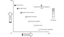
Figure 1
Trade-off between accuracy and explainability. Different machine learning models trade off between accuracy, explainability and complexity. Deep learning and random forests perform better in terms of accuracy, but are less explainable, while decision trees and linear regression models are easier to interpret, but have relatively lower accuracy."
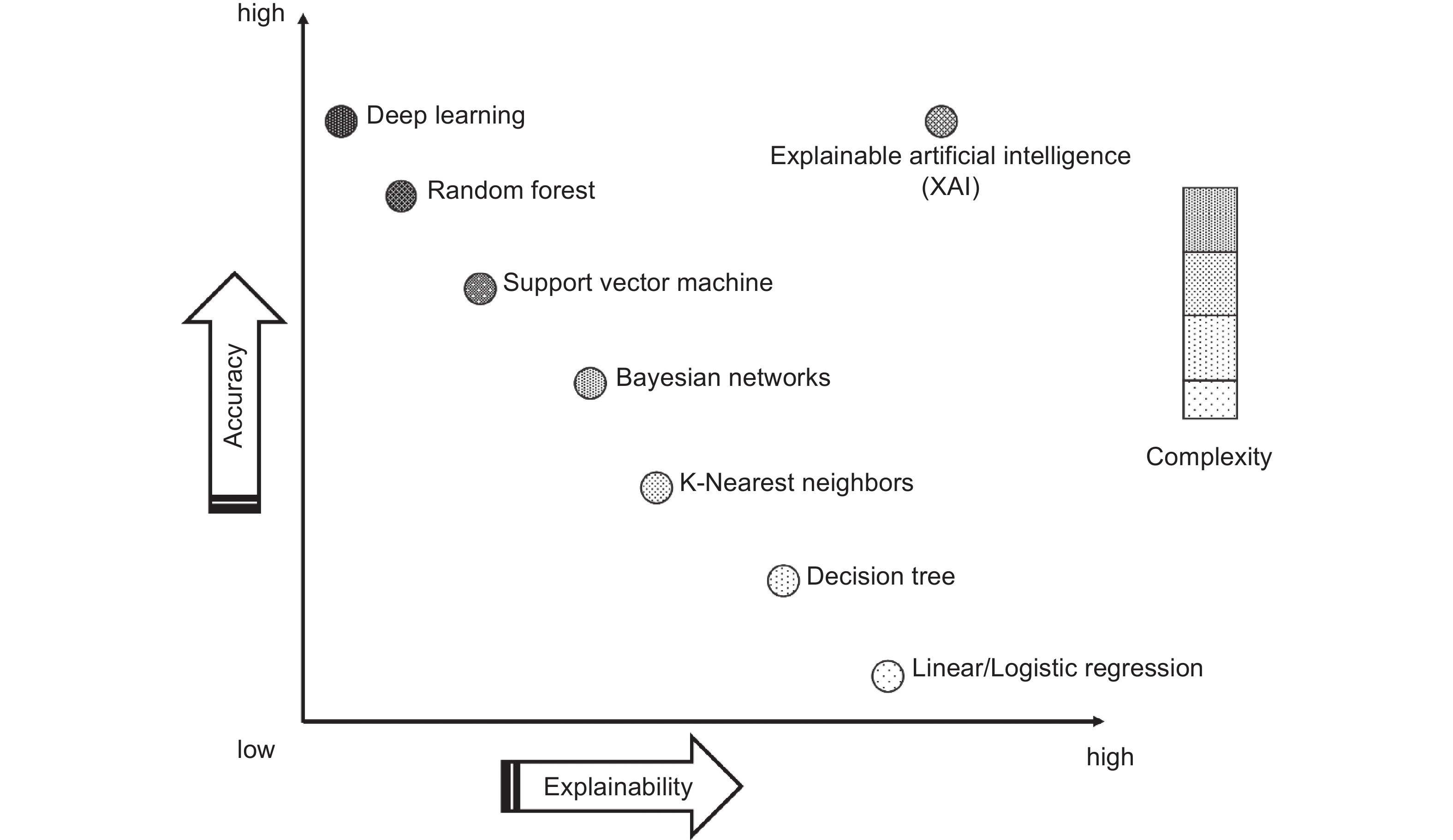
Table 2
Inclusion and exclusion criteria for the review"
| Type | Criteria | Rationale |
| Inclusion | XAI in echocardiography | XAI can be applied to many different applications. Still, the focus of our review is on echocardiography, since this area is one of the most critical applications of AI and XAI. |
| The AI model is built based on human clinical data | In this review, we focus on predictive modeling using human clinical data. | |
| They studied from 2018 to 2025 | Since 2018, the emergence of XAI tools like SHAP and Grad-CAM has helped solve the black box problem. | |
| The study focuses on XAI, so a relevant term is used in the title and/or abstract. | The goal of the study is on XAI, and the relevant terms include: explainable, explainability, interpretable, interpretability, understandable, understandability, comprehensible, comprehensibility, intelligible, machine learning, artificial intelligence, prediction model, predictive model, deep learning, AI, neural network. | |
| Exclusion | Details of the paper are not available. | Abstract papers, or the papers that could not be accessed through the university library or the interlibrary loan, and system demonstrations are not included. |
| Unpublished | We excluded papers uploaded on arXiv or other archiving systems not published in a peer-reviewed venue. | |
| Opinion or other review papers | This review is not a review of reviews, and opinion papers do not fulfill the requirement of delivering an XAI method. | |
| Duplicated | Usually, querying multiple databases returns similar papers. Thus, we removed the duplicates. |
Figure 2
The PRISMA diagram. The PRISMA diagram depicts the number of records identified, included, and excluded and the reasons for exclusions."
Table 3
Key concepts in XAI"
| Term | Concept | Key point | Common method |
| Explainability | After-the-fact explanations | External tools | SHAP, LIME, Grad-CAM, etc. |
| Interpretability | Built-in understandability | Model simplicity | Linear models, decision trees |
| Transparency | System openness | Data/code visibility | Share data, algorithms |
| Trustworthiness | Overall reliability | Ethics/safety compliance | Fairness checks, validation |
| Understandable | User-friendly presentation | Clear communication | Visuals, simplified language techniques |
Table 4
An overview of the most frequently used techniques in echocardiography"
| XAI technique | Input datatype | Visual representations | Classification framework | ||
| phase | scope | model dependency | |||
| CAM, class activation mapping; SHAP, shapley additive explanations; LIME, local interpretable model-agnostic explanations | |||||
| CAM/Grad-CAM | Image | Saliency map | post-hoc | Local | Model-agnostic |
| SHAP/Deep SHAP | Tabular/Text | Bar chart | post-hoc | Local/Global | Model-agnostic |
| Trainable attention | Image | attention map | post-hoc | Local | Model-specific |
| LIME/Deep LIME | Image/Text | Feature weights map | post-hoc | Local | Model-agnostic |
| Occlusion sensitivity | Image | / | post-hoc | Local | Model-agnostic |

Figure 3
Clinical examples and interpretation. (A) Grad-CAM. Original images and the results of EchoV-Net visualization of the most related regions for view recognition [105]; (B) Attention maps. In porcine echocardiographic simulation data, Co-AttentionSTN enhances the interpretability of motion tracking through multi-frame constraints [66]; (C) SHAP analysis. Positive SHAP values indicate that the feature value contributes to a positive classification (MI), while negative SHAP values denote a contribution to a negative classification (Non-MI) [91]; (D) SHAP analysis. SHAP values show their contributions to classification, and the predicted probability of belonging to the MI class for two misclassified patients in the test set [91]. Reproduced with permission from Elsevier © Elsevier"
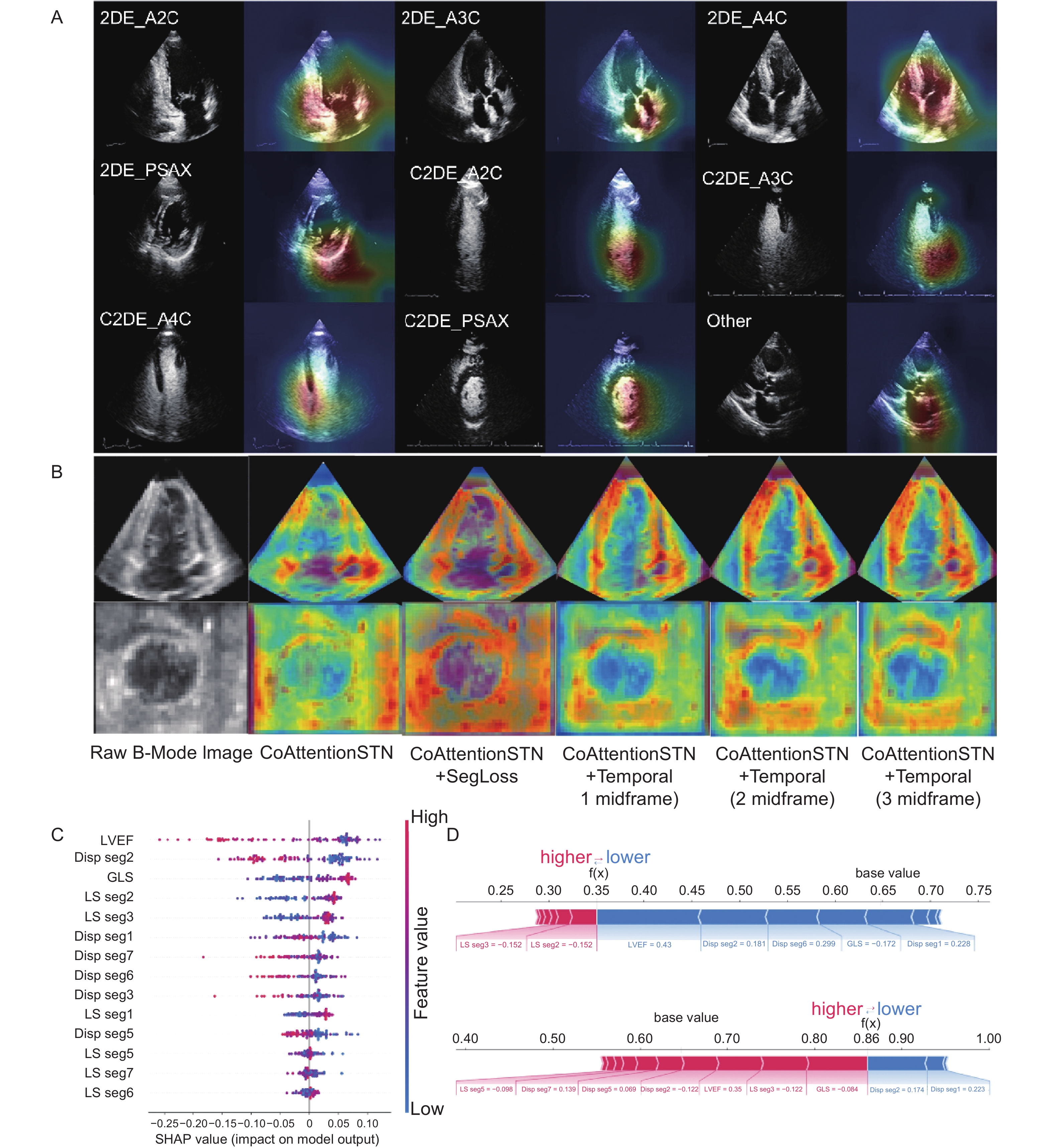
Figure 4
Literature analysis and statistical graphing. (A) Keyword co-occurrence map. The size of the nodes and labels in a cluster is proportional to the frequency of occurrence of the respective keyword. The thickness of the edges denotes the strength of co-occurrence between the connected keywords, while closer nodes represent more relation in context; (B) Pie chart of the share of different XAI techniques in echocardiographic studies. In 59 echocardiography studies utilizing XAI tools, the distribution of various technologies; (C) Bar chart of XAI technologies' distribution in echocardiographic-related scenarios. The distribution of various XAI tools across the four application scenarios of AI-assisted echocardiography images."
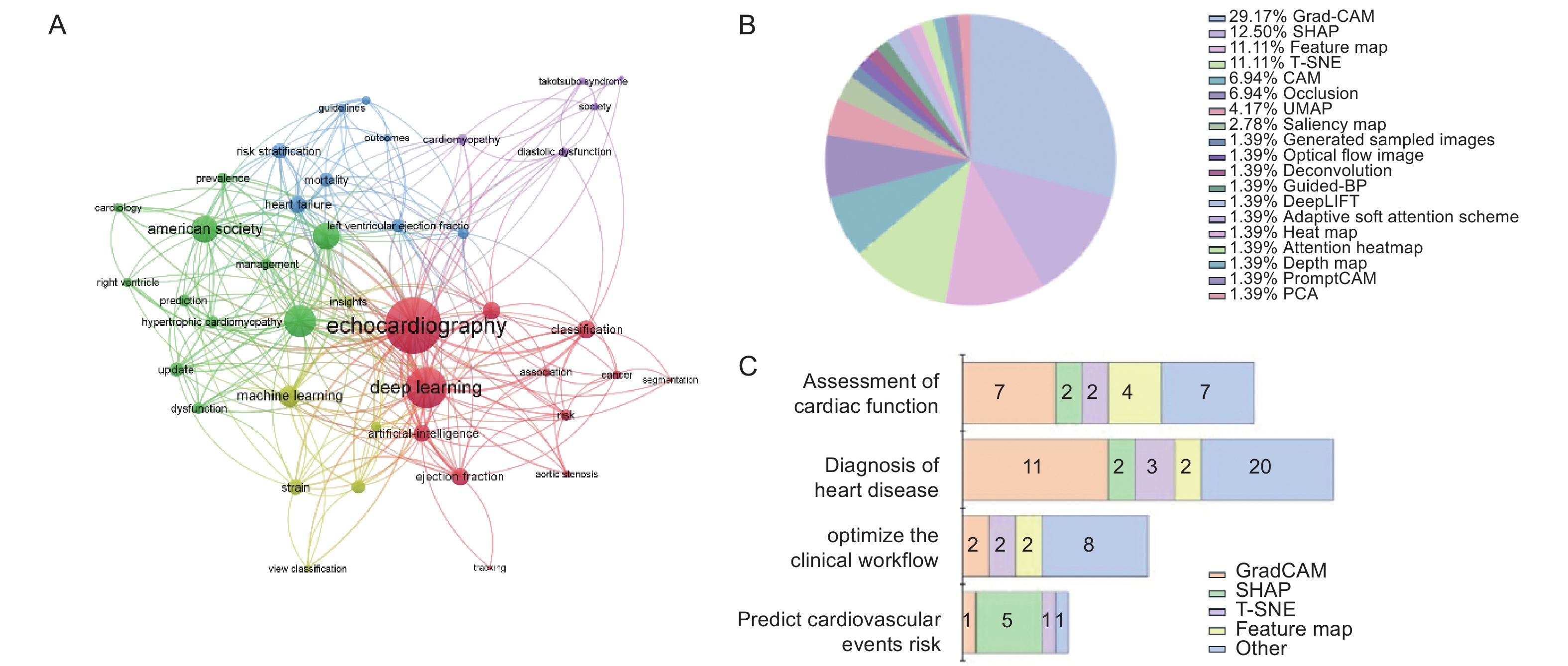
Table 5
Studies of XAI in clinical workflow"
| Study | Objective | Data* | Model | Performance | View | XAI |
| *, The volume of data corresponds to the research phase. QA, quality assessment; LVH, left ventricular hypertrophy; CNN, convolutional neural networks; Acc, accuracy; DL, deep learning; GCN, graph convolutional networks; RNN, recurrent neural networks; TaNet, trilateral attention network; Guided-BP, guided backpropagation | ||||||
| Gao et al.(2017) [ | View classification | 432 images | CNN | Acc. 0.92 | 8 views | the optical flow image |
| Gearhart et al.(2022) [ | View classification | 12,067 images | CNN | Acc. 0.90 | 6 views | UMAP |
| Madani et al.(2018) [ | View classification, LVH classification | 267 studies | CNN | Acc. 0.94 | 15 views | Generated sampled images |
| Huang et al.(2022) [ | View classification | 26,465 images | CNN | Acc. 0.98 | / | deconvolution |
| Madani et al.(2018) [ | View classification | 267 studies | DL | Acc. 0.93 | 15 views | t-SNE, Guided-BP, occlusion |
| Thomas et al.(2023) [ | View classification | 4,258 synthetic images | GCN | Acc. 0.97 | 4 views | GCN explainer |
| Howard et al.(2019) [ | View classification | 9,098 videos | CNN, Two-Stream networks | Acc. 0.96 | 14 views | Saliency map |
| Charton et al.(2023) [ | View classification | 8,292 videos | RNN | Acc. 0.97 | 8 views | decision tree |
| Zhang et al.(2018) [ | View classification, Cardiac function, diagnose | 4035 echocardiograms | CNN | Acc. 0.96 | 23 views | t-SNE, |
| Labs et al.(2023) [ | QA | 11,262 patients | DL | Acc. 0.97 | A4C, PLAX | feature map |
| Zamzmi et al.(2022) [ | View classification, QA, Cardiac function | EchoNet-Dynamic, NIH dataset | TaNet | Acc. 0.97 | 5 views | ablation |
| Hsu et al.(2025) [ | QA | 514 videos | ConvNeXt | Acc. 0.89 | A4C | Grad-CAM |
Table 6
Studies of XAI in quantitative cardiac function parameters"
| Study | Objective | Data* | Model | Performance | View | XAI |
| *, The volume of data corresponds to the research phase. LVDF, left ventricular diastolic function; HFpEF, heart failure with preserved ejection fraction; AUC, area under curve; Corr, correlation; MAE, masked autoencoders; R2, R-Square; LV, left ventricle F1: F1 score; ML, machine learning; LAV, left atrial volume; RWMA, regional wall motion abnormalities; APs, active polynomials; SVM, support vector machine; DT, decision tree; RF, random forest; KNN, k-nearest neighbor; XGB, eXtreme Gradient Boosting; PH, pulmonary hypertension; RVEF, right ventricular ejection fraction; RVOT, right ventricular outflow tract; AO, aorta; RV, right ventricle; NLP, natural language processing | ||||||
| Xu et al.(2023) [ | View classification; Cardiac function -LVDF | 1,304 studies (view); 2,150 studies (LVDF) | CNN | Acc. 0.92 | 5 views | Grad-CAM |
| Akerman Ashley et al.(2023) [ | Cardiac function - HFpEF | 6756 cases | CNN | AUC 0.97 | A4C | Grad-CAM |
| Dai et al.(2023) [ | Cardiac function-LVEF | EchoNet-Dynamic, CAMUS | ML | MAE 4.17 | A4C | attention heatmap |
| Mokhtari et al.(2022) [ | Cardiac function-LVEF | EchoNet-Dynamic | EchoGNN | F1 0.78 | A2C, A4C | learned weights on the echo-graph, average frame distance |
| Zhang et al.(2024) [ | Cardiac function-LVEF | Echonet Dynamic, HMC-QU, CAMUS | DL | Acc. 0.94 | A2C, A3C, A4C | t-SNE, Grad-CAM |
| Duffy et al.(2021) [ | Cardiac function-LV volume | EchoNet-Dynamic | CNN | Acc. 0.93 | A4C | Depth map |
| Barzegar et al.(2021) [ | Cardiac function-LAV | 621 videos | CNN | Acc. 0.94 | A4C | feature map |
| Christensen et al.(2024) [ | Cardiac function-LVEF | 1,032,975 videos | EchoCLIP | AUC 0.86 | A4C | PromptCAM, |
| Sanjeevi et al.(2023) [ | RWMA | HMC-QU | Echo-Cardio 3D Net | AUC 0.82 | A4C | Grad-CAM |
| Huang et al.(2020) [ | RWMA | 10,638 echocardiograms | CNN | AUR 0.91 | 5 views | feature map |
| Gomez et al.(2025) [ | RWMA | CAMUS,HMC-QU | U-Net | Sen. 1.00 | A2C, A3C, A4C | SHAP |
| Ragnarsdottir et al.(2024) [ | Cardiac function -PH | 1,311 videos | DL | Acc. 0.92 | A4C, PSAX, PLAX | Grad-CAM |
| Tokodi et al.(2023) [ | Cardiac function -RVEF | 5,076 videos. | CNN | Acc. 0.78 | A4C | occlusion |
| Hirata et al.(2024) [ | Cardiac function -PH | 885 patients | logistic regression, SVM, RF, XGB | Acc. 0.59 | / | SHAP |
| Zhao et al.(2022) [ | Cardiac function -RVOT, AO | 177 videos, CAMUS | DL | Acc. 0.99 | PSAX, A4C | feature map |
| Anand et al.(2024) [ | Cardiac function -PH | 7,853 patients | XGB | Acc. 0.82 | / | SHAP |
| Hagberg et al.(2022) [ | Cardiac function-RV | 12,684 studies | DL, NLP | Acc. 0.92 | A4C | Saliency map |
| Sun et al.(2024) [ | Cardiac function-PH | 3,912 subjects | chamber attention network | Acc.0.83 | A4C, PLAX | t-SNE, Grad-CAM |
Table 7
Studies of XAI in diagnosis of heart disease"
| Study | Diagnosis | Data* | Model | Performance | View | XAI |
| *, The volume of data corresponds to the research phase. HF, heart failure; CM, cardiomyopathy; AOSAX, short-axis view of the aortic valve; CASA, coronary artery short axis | ||||||
| Cikes et al.(2019) [ | HF | 1,106 patients | ML | a classification of a phenotypically heterogeneous HF cohort | A2C, A4C | intrinsically interpretable model |
| Shad et al.(2021) [ | HF | 723 patients | CNN | AUC 0.73 | A4C | Grad-CAM |
| Ouyang et al.(2020) [ | CM | 10,030 videos | EchoNet-Dynamic | AUC 0.97 | A4C | PromptCAM |
| Morita et al.(2021) [ | CM | 45 patients | CNN | AUC 0.86 | 6 views | Grad-CAM |
| Hwang et al.(2022) [ | CM | 930 subjects | CNN | Acc. 0.92 | 5 views | CAM |
| Liu et al.(2023) [ | CM | 1,807 videos | DL | AUC: ASD 0.99, DCM 0.98, HCM 0.99, prior MI 0.98, Normal 0.98 | A4C | CAM |
| Chao et al.(2024) [ | CM | 381 patients | CNN | AUC 0.97 | A4C | Grad-CAM |
| H et al.(2023 Dec) [ | CM | 91 studies | KNN, LR, MLP, RF, SVM, XGB | Acc. 0.73 | A4C | feature map |
| Peng et al.(2024) [ | CM | 13,575 images | DL | Acc. 0.90 | 5 views | t-SNE, Grad-CAM |
| Vafaeezadeh et al.(2022) [ | Valvular Disease | 1,773 subjects | eight CNNs | Acc. 0.80 | A4C, PLAX | Grad-CAM |
| Cheng et al.(2022) [ | Valvular Disease | 3,554 patients | CNN | Acc. 0.86 | A4C | t-SNE, Deep LIME |
| Vafaeezadeh et al.(2023) [ | Valvular Disease | 1,773 subjects | CNN | Acc. 0.71 | PLAX | Grad-CAM |
| Holste et al.(2023) [ | Valvular Disease | 5,257 studies | CNN | AUC 0.98 | PLAX | Grad-CAM |
| Tang et al.(2024) [ | Valvular Disease | 2,31 patients | DL | Acc. 0.82 | AOSAX | Grad-CAM |
| Gu et al.(2025) [ | Valvular Disease | 2572 studies | ProtoASNet | Acc.0.80 | PLAX,PSAX | PCA, t-SNE, UMAP |
| Wang et al.(2021) [ | Other | 1,308 subjects | CNN | Acc. 0.95(CHD), Acc. 0.92(VSD or ASD) | 5 views | adaptive soft attention scheme, occlusion analysis, relative confidence heat map |
| Zaman et al.(2021) [ | Other | 17,280 images | CNN, RNN | Acc. 0.80 | A4C | Grad-CAM |
| Nurmaini et al.(2022) [ | Other | 76 pregnant women | 4 CNNs | Acc. 1.0 | 4CV fetal | Grad-CAM, Guided-BP |
| Lee et al.(2022) [ | Other | 203 patients | six deep learning networks | Acc. 0.78 | CASA | CAM |
| Zaman et al.(2024) [ | Other | 300 patients | CNN | improve the diagnostic Acc. in 70% of ‘difficult’ TTS cases | A4C | Grad-CAM |
| Jina et al.(2023) [ | Other | 34,368 images | ConvNeXt-V2 | Acc. 0.86 | 4 views | feature map |
| Lee et al.(2025) [ | Other | 203 patients | MLRANet | Sen. 0.93 | PSAX | t-SNE, UMAP, attention mechanism |
Table 8
Studies of XAI in predict the risk of adverse cardiovascular events"
| Study | Objective | Data* | Model | Performance | View | XAI |
| *, The volume of data corresponds to the research phase. BNP, brain natriuretic peptide; BUN, blood urea nitrogen; CCS, chronic coronary syndromes; HCM, hypertrophic cardiomyopathy; LASSO, least absolute shrinkage and selection operator | ||||||
| Ulloa Cerna et al.(2021) [ | predictions of one-year all-cause mortality | 812,278 videos | CNN | improved the sensitivity by 13% | PLAX, A4C | occlusion |
| Valsaraj et al.(2023) [ | identifying patients at high-risk of all-cause mortality | 7,080 videos | CNN | AUC 0.92 | / | Grad-CAM, SHAP |
| Molenaar et al.(2024) [ | predict all-cause 5-year mortality in patients with CCS | 1,253 patients | XGB | AUC 0.79 | / | SHAP |
| Rhee et al.(2024) [ | discriminate major cardiovascular events in patients with HCM | 2,111 patients | Logistic, RF, SVM | AUC 0.80 | / | SHAP |
| Dutta et al.(2020) [ | classify coronary heart disease | the NHANES | CNN | Acc. 0.79 | / | LASSO, t-SNE |
| Wang et al.(2021) [ | prediction of 3-year all-cause mortality in patients with heart failure caused by coronary heart disease | 5,188 patients | XGB | hazard ratio 10.35 | / | SHAP |
| Petmezas et al.(2025) [ | heart failure mortality prediction | 233 patients | Extra-Trees | AUC 0.79 | / | SHAP |
| [1] |
Wenderott K , Krups J , Zaruchas F , Weigl M . Effects of artificial intelligence implementation on efficiency in medical imaging-a systematic literature review and meta-analysis. NPJ Digital Medicine 2024; 7: 265.
doi: 10.1038/s41746-024-01248-9 |
| [2] | G. S , Gopalakrishnan U , Parthinarupothi RK , Madathil T . Deep learning supported echocardiogram analysis: A comprehensive review. Artif Intell Med 2024; 151: 26. |
| [3] |
Narang A , Bae R , Hong H , Thomas Y , Surette S , Cadieu C , et al . Utility of a deep-learning algorithm to guide novices to acquire echocardiograms for limited diagnostic use. JAMA cardiology 2021; 6: 624-632.
doi: 10.1001/jamacardio.2021.0185 |
| [4] |
Leclerc S , Smistad E , Pedrosa J , Østvik A , Cervenansky F , Espinosa F , et al . Deep learning for segmentation using an open large-scale dataset in 2D echocardiography. IEEE transactions on medical imaging 2019; 38: 2198-2210.
doi: 10.1109/TMI.2019.2900516 |
| [5] |
Ouyang D , He B , Ghorbani A , Yuan N , Ebinger J , Langlotz CP , et al . Video-based AI for beat-to-beat assessment of cardiac function. Nature 2020; 580: 252-256.
doi: 10.1038/s41586-020-2145-8 |
| [6] | IEEE approved draft guide for an architectural framework for explainable artificial intelligence. IEEE P2894/D9, August 2023 2024:1-51. |
| [7] |
Gao X , Li W , Loomes M , Wang L . A fused deep learning architecture for viewpoint classification of echocardiography. Information Fusion 2017; 36: 103-113.
doi: 10.1016/j.inffus.2016.11.007 |
| [8] |
Madani A , Ong JR , Tibrewal A , Mofrad MRK . Deep echocardiography: data-efficient supervised and semi-supervised deep learning towards automated diagnosis of cardiac disease. NPJ Digital Medicine 2018; 1: 59.
doi: 10.1038/s41746-018-0065-x |
| [9] |
Madani A , Arnaout R , Mofrad M , Arnaout R . Fast and accurate view classification of echocardiograms using deep learning. NPJ Digital Medicine 2018; 1: 6.
doi: 10.1038/s41746-017-0013-1 |
| [10] |
Christensen M , Vukadinovic M , Yuan N , Ouyang D . Vision–language foundation model for echocardiogram interpretation. Nature Medicine 2024; 30: 1481-1488.
doi: 10.1038/s41591-024-02959-y |
| [11] |
Zamzmi G , Rajaraman S , Hsu L-Y , Sachdev V , Antani S . Real-time echocardiography image analysis and quantification of cardiac indices. Medical Image Analysis 2022; 80: 102438.
doi: 10.1016/j.media.2022.102438 |
| [12] |
Tokodi M , Magyar B , Soós A , Takeuchi M , Tolvaj M , Lakatos BK , et al . Deep learning-based prediction of right ventricular ejection fraction using 2D echocardiograms. JACC: Cardiovascular Imaging 2023; 16: 1005-1018.
doi: 10.1016/j.jcmg.2023.02.017 |
| [13] |
Cikes M , Sanchez‐Martinez S , Claggett B , Duchateau N , Piella G , Butakoff C , et al . Machine learning‐based phenogrouping in heart failure to identify responders to cardiac resynchronization therapy. European Journal of Heart Failure 2019; 21: 74-85.
doi: 10.1002/ejhf.1333 |
| [14] |
Holste G , Oikonomou EK , Mortazavi BJ , Coppi A , Faridi KF , Miller EJ , et al . Severe aortic stenosis detection by deep learning applied to echocardiography. Eur Heart J 2023; 44: 4592-4604.
doi: 10.1093/eurheartj/ehad456 |
| [15] |
Wang J , Liu X , Wang F , Zheng L , Gao F , Zhang H , et al . Automated interpretation of congenital heart disease from multi-view echocardiograms. Medical Image Analysis 2021; 69: 101942.
doi: 10.1016/j.media.2020.101942 |
| [16] |
Ulloa Cerna AE , Jing L , Good CW , vanMaanen DP , Raghunath S , Suever JD , et al . Deep-learning-assisted analysis of echocardiographic videos improves predictions of all-cause mortality. Nat Biomed Eng 2021; 5: 546-554.
doi: 10.1038/s41551-020-00667-9 |
| [17] |
Valsaraj A , Kalmady SV , Sharma V , Frost M , Sun W , Sepehrvand N , et al . Development and validation of echocardiography-based machine-learning models to predict mortality. EBioMedicine 2023; 90: 104479.
doi: 10.1016/j.ebiom.2023.104479 |
| [18] | Page MJ , McKenzie JE , Bossuyt PM , Boutron I , Hoffmann TC , Mulrow CD , et al . The PRISMA 2020 statement: an updated guideline for reporting systematic reviews. BMJ 2021; 372: n71 |
| [19] |
Barredo Arrieta A , Díaz-Rodríguez N , Del Ser J , Bennetot A , Tabik S , Barbado A , et al . Explainable artificial intelligence (XAI): concepts, taxonomies, opportunities and challenges toward responsible AI. Information Fusion 2020; 58: 82-115.
doi: 10.1016/j.inffus.2019.12.012 |
| [20] |
Du M , Liu N , Hu X . Techniques for interpretable machine learning. Commun Acm 2019; 63: 68-77.
doi: 10.1145/3359786 |
| [21] |
Sengupta PP , Shrestha S , Berthon B , Messas E , Donal E , Tison GH , et al . Proposed requirements for cardiovascular imaging-related machine learning evaluation (PRIME): a checklist: reviewed by the american college of cardiology healthcare innovation council. JACC: Cardiovascular Imaging 2020; 13: 2017-2035.
doi: 10.1016/j.jcmg.2020.07.015 |
| [22] | Charton J , Ren H , Khambhati J , DeFrancesco J , Cheng J , Waheed AA , et al . View classification of color doppler echocardiography via automatic alignment between doppler and b-mode imaging. In: Aylward S, Noble JA, Hu Y, Lee S-L, Baum Z, Min Z, editors. Simplifying Medical Ultrasound. Cham: Springer International Publishing; 2022. p. 64-71. |
| [23] | Liao Z , Jafari MH , Girgis H , Gin K , Rohling R , Abolmaesumi P , Tsang T . Echocardiography view classification using quality transfer star generative adversarial networks. In: Shen D, Liu T, Peters TM, Staib LH, Essert C, Zhou S, et al., editors. Medical image computing and computer assisted intervention – MICCAI 2019. Cham: Springer International Publishing; 2019. p. 687-695. |
| [24] |
Zhang J , Gajjala S , Agrawal P , Tison GH , Hallock LA , Beussink-Nelson L , et al . Fully automated echocardiogram interpretation in clinical practice. Circulation 2018; 138: 1623-1635.
doi: 10.1161/CIRCULATIONAHA.118.034338 |
| [25] | Huang M , Lin W , Chen Y , Hsiao T , Liu P , Tsai W . Explainable deep neural network for echocardiography view classification. European Heart Journal - Cardiovascular Imaging 2022; 23. |
| [26] | Charton J , Ren H , Kim S , Gonzalez CM , Khambhati J , Cheng J , et al . Multi-task learning for hierarchically-structured images: study on echocardiogram view classification. In: Kainz B, Noble A, Schnabel J, Khanal B, Müller JP, Day T, editors. Simplifying Medical Ultrasound. Cham: Springer Nature Switzerland; 2023. p. 185-194. |
| [27] |
Abdi AH , Luong C , Tsang T , Allan G , Nouranian S , Jue J , et al . Automatic quality assessment of echocardiograms using convolutional neural networks: feasibility on the apical four-chamber view. IEEE transactions on medical imaging 2017; 36: 1221-1230.
doi: 10.1109/TMI.2017.2690836 |
| [28] |
Huang M-S , Wang C-S , Chiang J-H , Liu P-Y , Tsai W-C . Automated recognition of regional wall motion abnormalities through deep neural network interpretation of transthoracic echocardiography. Circulation 2020; 142: 1510-1520.
doi: 10.1161/CIRCULATIONAHA.120.047530 |
| [29] |
Hsu C-C , Wang Y-W , Lin L-C , Chang R-F . Spatiotemporal feature disentanglement for quality surveillance of left ventricular echocardiographic video using ST-R(2 + 1)D-ConvNeXt. Biomedical Signal Processing and Control 2025; 105: 107671.
doi: 10.1016/j.bspc.2025.107671 |
| [30] |
Tromp J , Seekings PJ , Hung C-L , Iversen MB , Frost MJ , Ouwerkerk W , et al . Automated interpretation of systolic and diastolic function on the echocardiogram: a multicohort study. The Lancet Digital Health 2022; 4: e46-e54.
doi: 10.1016/S2589-7500(21)00235-1 |
| [31] |
Olaisen S , Smistad E , Espeland T , Hu J , Pasdeloup D , Østvik A , et al . Automatic measurements of left ventricular volumes and ejection fraction by artificial intelligence: clinical validation in real time and large databases. European Heart Journal - Cardiovascular Imaging 2024; 25: 383-395.
doi: 10.1093/ehjci/jead280 |
| [32] | Duffy G , Jain I , He B , Ouyang D . Interpretable deep learning prediction of 3d assessment of cardiac function. Biocomputing 2022: WORLD SCIENTIFIC; 2021. p. 231-241. |
| [33] |
Chen X , Yang F , Zhang P , Lin X , Wang W , Pu H , et al . Artificial intelligence–assisted left ventricular diastolic function assessment and grading: multiview versus single view. J Am Soc Echocardiogr 2023; 36: 1064-1078.
doi: 10.1016/j.echo.2023.07.001 |
| [34] |
Barzegar N , Khatibi T , Hosseinsabet A . Proposing novel methods for simultaneous cardiac cycle phase identification and estimating maximal and minimal left atrial volume (LAV) from apical four-chamber view in 2-D echocardiography. Informatics in Medicine Unlocked 2021; 23: 100517.
doi: 10.1016/j.imu.2021.100517 |
| [35] |
Sanjeevi G , Gopalakrishnan U , Pathinarupothi RK , Madathil T . Automatic diagnostic tool for detection of regional wall motion abnormality from echocardiogram. J Med Syst 2023; 47: 13.
doi: 10.1007/s10916-023-01911-w |
| [36] |
Degerli A , Kiranyaz S , Hamid T , Mazhar R , Gabbouj M . Early myocardial infarction detection over multi-view echocardiography. Biomedical Signal Processing and Control 2024; 87: 105448.
doi: 10.1016/j.bspc.2023.105448 |
| [37] |
Konstam MA , Kiernan MS , Bernstein D , Bozkurt B , Jacob M , Kapur NK , et al . Evaluation and management of right-sided heart failure: a scientific statement from the american heart association. Circulation 2018; 137: e578-e622.
doi: 10.1161/CIRCULATIONAHA.117.032207 |
| [38] |
Hassoun Paul M . Pulmonary arterial hypertension. New England Journal of Medicine 2021; 385: 2361-2376.
doi: 10.1056/NEJMra2000348 |
| [39] |
Surkova E , Muraru D , Genovese D , Aruta P , Palermo C , Badano LP . Relative prognostic importance of left and right ventricular ejection fraction in patients with cardiac diseases. Journal of the American Society of Echocardiography 2019; 32: 1407-1415.e1403.
doi: 10.1016/j.echo.2019.06.009 |
| [40] |
Hagberg E , Hagerman D , Johansson R , Hosseini N , Liu J , Björnsson E , et al . Semi-supervised learning with natural language processing for right ventricle classification in echocardiography—a scalable approach. Comput Biol Med 2022; 143: 105282.
doi: 10.1016/j.compbiomed.2022.105282 |
| [41] |
Ragnarsdottir H , Ozkan E , Michel H , Chin-Cheong K , Manduchi L , Wellmann S , Vogt JE . Deep learning based prediction of pulmonary hypertension in newborns using echocardiograms. Int J Comput Vis 2024; 132: 2567-2584.
doi: 10.1007/s11263-024-01996-x |
| [42] |
Sun D , Hu Y , Li Y , Yu X , Chen X , Shen P , et al . Chamber attention network (CAN): towards interpretable diagnosis of pulmonary artery hypertension using echocardiography. J Adv Res 2024; 63: 103-115.
doi: 10.1016/j.jare.2023.10.013 |
| [43] |
Hirata Y , Tsuji T , Kotoku J , Sata M , Kusunose K . Echocardiographic artificial intelligence for pulmonary hypertension classification. Heart 2024; 110: 586-593.
doi: 10.1136/heartjnl-2023-323320 |
| [44] |
Liu L , Duan S , Li Y , Liu R , Wu Y , Zhang L . Development status and prospect of remote diagnosis and treatment of echocardiography worldwide. Advanced Ultrasound in Diagnosis and Therapy 2020; 4: 303-307.
doi: 10.37015/AUDT.2020.200047 |
| [45] |
Moor M , Banerjee O , Abad ZSH , Krumholz HM , Leskovec J , Topol EJ , Rajpurkar P . Foundation models for generalist medical artificial intelligence. Nature 2023; 616: 259-265.
doi: 10.1038/s41586-023-05881-4 |
| [46] |
Webster P . Six ways large language models are changing healthcare. Nature Medicine 2023; 29: 2969-2971.
doi: 10.1038/s41591-023-02700-1 |
| [47] |
Chen RJ , Ding T , Lu MY , Williamson DFK , Jaume G , Song AH , et al . Towards a general-purpose foundation model for computational pathology. Nature Medicine 2024; 30: 850-862.
doi: 10.1038/s41591-024-02857-3 |
| [48] |
Sanchez-Martinez S , Duchateau N , Erdei T , Kunszt G , Aakhus S , Degiovanni A , et al . Machine learning analysis of left ventricular function to characterize heart failure with preserved ejection fraction. Circ Cardiovasc Imaging 2018; 11: e007138.
doi: 10.1161/CIRCIMAGING.117.007138 |
| [49] |
Atsushi Kyodo , Koshiro Kanaoka , Ayaka Keshi , Maki Nogi , Kazutaka Nogi 1 , Satomi Ishihara , et al . Heart failure with preserved ejection fraction phenogroup classification using machine learning. ESC heart failure 2023; 10: 2019-2030.
doi: 10.1002/ehf2.14368 |
| [50] |
Shad R , Quach N , Fong R , Kasinpila P , Bowles C , Castro M , et al . Predicting post-operative right ventricular failure using video-based deep learning. Nat Commun 2021; 12: 5192.
doi: 10.1038/s41467-021-25503-9 |
| [51] |
Chen W , Xie Y , Zhang Z , Zhu Y , Zhang Y , Zhu S , et al . Artificial Intelligence-assisted medical imaging in interventional management of valvular heart disease. Advanced Ultrasound in Diagnosis and Therapy 2023; 7: 217-227.
doi: 10.37015/AUDT.2023.230030 |
| [52] |
Gu AN , Vaseli H , Tsang MY , Wu V , Ahmadi Amiri SN , Kondori N , et al . ProtoASNet: Comprehensive evaluation and enhanced performance with uncertainty estimation for aortic stenosis classification in echocardiography. Medical Image Analysis 2025; 103: 103600.
doi: 10.1016/j.media.2025.103600 |
| [53] |
Tang L , Wang X , Yang J , Wang Y , Qu M , Li H . DLFFNet: A new dynamical local feature fusion network for automatic aortic valve calcification recognition using echocardiography. Comput Methods Programs Biomed 2024; 243: 107882.
doi: 10.1016/j.cmpb.2023.107882 |
| [54] |
Bernard J , Yanamala N , Shah R , Seetharam K , Altes A , Dupuis M , et al . Integrating echocardiography parameters with explainable artificial intelligence for data-driven clustering of primary mitral regurgitation phenotypes. JACC: Cardiovascular Imaging 2023; 16: 1253-1267.
doi: 10.1016/j.jcmg.2023.02.016 |
| [55] | Vafaeezadeh M , Behnam H , Hosseinsabet A , Gifani P . CarpNet: Transformer for mitral valve disease classification in echocardiographic videos. International Journal of Imaging Systems and Technology 2023; 33: 1505-1514 |
| [56] | Hwang I-C , Choi D , Choi Y-J , Ju L , Kim M , Hong J-E , et al . Differential diagnosis of common etiologies of left ventricular hypertrophy using a hybrid CNN-LSTM model. Scientific Reports 2022; 12: 20998 |
| [57] | Chao C-J , Jeong J , Arsanjani R , Kim K , Tsai Y-L , Yu W-C , et al . Echocardiography-based deep learning model to differentiate constrictive pericarditis and restrictive cardiomyopathy. JACC: Cardiovascular Imaging 2024; 17: 349-360 |
| [58] | Li R , Zhang Y , Zhang C , Huang X , Ding S . Contrast echocardiography evaluation of microcirculation of myocardial infarction caused by takotsubo syndrome: case report and literature review. Advanced Ultrasound in Diagnosis and Therapy 2021; 5: 258-261 |
| [59] |
Zaman F , Ponnapureddy R , Wang YG , Chang A , Cadaret LM , Abdelhamid A , et al . Spatio-temporal hybrid neural networks reduce erroneous human “judgement calls” in the diagnosis of Takotsubo syndrome. EClinicalMedicine 2021; 40: 101115.
doi: 10.1016/j.eclinm.2021.101115 |
| [60] |
Zaman F , Isom N , Chang A , Wang YG , Abdelhamid A , Khan A , et al . Deep learning from atrioventricular plane displacement in patients with Takotsubo syndrome: lighting up the black-box. European Heart Journal - Digital Health 2024; 5: 134-143.
doi: 10.1093/ehjdh/ztad077 |
| [61] | Lee H , Eun Y , Hwang JY , Eun LY . Explainable deep learning algorithm for distinguishing incomplete Kawasaki disease by coronary artery lesions on echocardiographic imaging. Comput Methods Programs Biomed 2022; 223: 106970 |
| [62] |
Wang K , Tian J , Zheng C , Yang H , Ren J , Liu Y , et al . Interpretable prediction of 3-year all-cause mortality in patients with heart failure caused by coronary heart disease based on machine learning and SHAP. Comput Biol Med 2021; 137: 104813.
doi: 10.1016/j.compbiomed.2021.104813 |
| [63] |
Park J , Hwang I-C , Yoon YE , Park J-B , Park J-H , Cho G-Y . Predicting long-term mortality in patients with acute heart failure by using machine learning. J Card Fail 2022; 28: 1078-1087.
doi: 10.1016/j.cardfail.2022.02.012 |
| [64] |
Molenaar MA , Bouma BJ , Asselbergs FW , Verouden NJ , Selder JL , Chamuleau SAJ , Schuuring MJ . Explainable machine learning using echocardiography to improve risk prediction in patients with chronic coronary syndrome. Eur Heart J Digit Health 2024; 5: 170-182.
doi: 10.1093/ehjdh/ztae001 |
| [65] | Hirata Y , Kusunose K . AI in echocardiography: state-of-the-art automated measurement techniques and clinical applications. JMA J 2024; 8: 141-150 |
| [66] | Ahn SS , Ta K , Thorn SL , Onofrey JA , Melvinsdottir IH , Lee S , et al . Co-attention spatial transformer network for unsupervised motion tracking and cardiac strain analysis in 3D echocardiography. Medical Image Analysis 2023; 84: 102711 |
| [67] |
Cheng LH , Bosch PBJ , Hofman RFH , Brakenhoff TB , Bruggemans EF , van der Geest RJ , Holman ER . Revealing unforeseen diagnostic image features with deep learning by detecting cardiovascular diseases from apical 4‐chamber ultrasounds. Journal of the American Heart Association 2022; 11: e024168.
doi: 10.1161/JAHA.121.024168 |
| [68] |
Jin W , Li X , Fatehi M , Hamarneh G . Guidelines and evaluation of clinical explainable AI in medical image analysis. Medical Image Analysis 2023; 84: 102684.
doi: 10.1016/j.media.2022.102684 |
| [69] |
Behzad S , Tabatabaei SMH , Lu MY , Eibschutz LS , Gholamrezanezhad A . Pitfalls in interpretive applications of artificial intelligence in radiology. American Journal of Roentgenology 2024; 223: e2431493.
doi: 10.2214/AJR.24.31493 |
| [70] |
Duffy G , Cheng PP , Yuan N , He B , Kwan AC , Shun-Shin MJ , et al . High-throughput precision phenotyping of left ventricular hypertrophy with cardiovascular deep learning. JAMA cardiology 2022; 7: 386-395.
doi: 10.1001/jamacardio.2021.6059 |
| [71] | Kiranyaz S , Degerli A , Hamid T , Mazhar R , Fadil Ahmed RE , Abouhasera R , et al . Left ventricular wall motion estimation by active polynomials for acute myocardial infarction detection. Ieee Access 2020; 8: 210301-210317 |
| [72] |
Alessandrini M , Chakraborty B , Heyde B , Bernard O , De Craene M , Sermesant M , D’Hooge J . Realistic vendor-specific synthetic ultrasound data for quality assurance of 2-D speckle tracking echocardiography: simulation pipeline and open access database. IEEE Transactions on Ultrasonics, Ferroelectrics, and Frequency Control 2018; 65: 411-422.
doi: 10.1109/TUFFC.2017.2786300 |
| [73] | Huang Z , Long G , Wessler B , Hughes MC . A new semi-supervised learning benchmark for classifying view and diagnosing aortic stenosis from echocardiograms. In: Ken J, Serena Y, Mark S, Michael S, Rajesh R, editors. proceedings of the 6th machine learning for healthcare conference. Proceedings of Machine Learning Research: PMLR; 2021. p. 614-647. |
| [74] |
Qiu T , Wang X , Chen C , Atiquzzaman M , Liu L . TMED: a spider-web-like transmission mechanism for emergency data in vehicular ad hoc networks. IEEE Transactions on Vehicular Technology 2018; 67: 8682-8694.
doi: 10.1109/TVT.2018.2841348 |
| [75] | Kaikai L , Yiyu S , Zhuang J , Meiping H , Hongwen F , Boyang L , et al. Enhance regional wall segmentation by style transfer for regional wall motion assessment. 2023. |
| [76] | Yang J , Ding X , Zheng Z , Xu X , Li X . GraphEcho: graph-driven unsupervised domain adaptation for echocardiogram video segmentation. 2023. p. 11844-11853. |
| [77] | Yang J , Lin Y , Pu B , Guo J , Xu X , Li X . CardiacNet: learning to reconstruct abnormalities for cardiac disease assessment from echocardiogram videos. In: Leonardis A, Ricci E, Roth S, Russakovsky O, Sattler T, Varol G, editors. Computer Vision – ECCV 2024. Cham: Springer Nature Switzerland; 2025. p. 293-311. |
| [78] |
Carbonati T , Eslami P , Chaudhari A , Herbst E , Fiorina L , Porquet P , et al . Mimic-iv-ecg & mimic-iv-echo: detection of regional wall motion abnormalities from electrocardiogram using deep learning. Journal of the American College of Cardiology 2024; 83: 1172-1172.
doi: 10.1016/S0735-1097(24)03162-0 |
| [79] | Magyar B , Tokodi M , Soós A , Tolvaj M , Lakatos BK , Fábián A , et al. RVENet: A large echocardiographic dataset for the deep learning-based assessment of right ventricular function. In: Karlinsky L, Michaeli T, Nishino K, editors. Computer Vision – ECCV 2022 Workshops. Cham: Springer Nature Switzerland; 2023. p. 569-583. |
| [80] | Zhao D , Ferdian E , Maso Talou GD , Quill GM , Gilbert K , Wang VY , et al. MITEA: A dataset for machine learning segmentation of the left ventricle in 3D echocardiography using subject-specific labels from cardiac magnetic resonance imaging. Frontiers in Cardiovascular Medicine 2023;9. |
| [81] | Gearhart A , Goto S , Deo RC , Powell AJ . An automated view classification model for pediatric echocardiography using artificial intelligence. Journal of the American Society of Echocardiography 2022; 35: 1238-1246 |
| [82] |
Jwan A Naser , Eunjung Lee , Sorin V Pislaru , Gal Tsaban , Jeffrey G Malins , John I Jackson , et al . Artificial intelligence-based classification of echocardiographic views. European Heart Journal - Digital Health 2024; 5: 260-269.
doi: 10.1093/ehjdh/ztae015 |
| [83] | Thomas S , Tiago C , Andreassen BS , Aase S-A , Šprem J , Steen E , et al. Graph convolutional neural networks for automated echocardiography view recognition: a holistic approach. In: Kainz B, Noble A, Schnabel J, Khanal B, Müller JP, Day T, editors. Simplifying Medical Ultrasound. Cham: Springer Nature Switzerland; 2023. p. 44-54. |
| [84] | Howard JP , Tan J , Shun-Shin MJ , Mahdi D , Nowbar AN , Arnold AD , et al. Improving ultrasound video classification: an evaluation of novel deep learning methods in echocardiography. Journal of medical artificial intelligence 2019;3. |
| [85] |
Labs RB , Vrettos A , Loo J , Zolgharni M . Automated assessment of transthoracic echocardiogram image quality using deep neural networks. Intelligent Medicine 2023; 3: 191-199.
doi: 10.1016/j.imed.2022.08.001 |
| [86] | Shiokawa N , Izumo M , Shimamura T , Kurosaka Y , Sato Y , Okamura T , et al . Accuracy and efficacy of artificial intelligence-derived automatic measurements of transthoracic echocardiography in routine clinical practice. J Clin Med 2024; 13: 1861 |
| [87] |
Akerman Ashley P , Porumb M , Scott Christopher G , Beqiri A , Chartsias A , Ryu Alexander J , et al . Automated echocardiographic detection of heart failure with preserved ejection fraction using artificial intelligence. JACC: Advances 2023; 2: 100452.
doi: 10.1016/j.jacadv.2023.100452 |
| [88] |
Dai W , Li X , Ding X , Cheng KT . Cyclical self-supervision for semi-supervised ejection fraction prediction from echocardiogram videos. IEEE transactions on medical imaging 2023; 42: 1446-1461.
doi: 10.1109/TMI.2022.3229136 |
| [89] | Mokhtari M , Tsang T , Abolmaesumi P , Liao R . EchoGNN: explainable ejection fraction estimation with graph neural networks. In: Wang L, Dou Q, Fletcher PT, Speidel S, Li S, editors. Medical Image Computing and Computer Assisted Intervention – MICCAI 2022. Cham: Springer Nature Switzerland; 2022. p. 360-369. |
| [90] |
Zhang Z , Yu C , Zhang H , Gao Z . Embedding tasks into the latent space: cross-space consistency for multi-dimensional analysis in echocardiography. IEEE transactions on medical imaging 2024; 43: 2215-2228.
doi: 10.1109/TMI.2024.3362964 |
| [91] | Gomez C , Letizia A , Tufano V , Molinari F , Salvi M . A cascade approach for the early detection and localization of myocardial infarction in 2D-echocardiography. Medical Engineering & Physics 2025:104400. |
| [92] |
Zhao C , Chen W , Qin J , Yang P , Xiang Z , Frangi AF , et al . IFT-net: interactive fusion transformer network for quantitative analysis of pediatric echocardiography. Medical Image Analysis 2022; 82: 102648.
doi: 10.1016/j.media.2022.102648 |
| [93] |
Anand V , Weston AD , Scott CG , Kane GC , Pellikka PA , Carter RE . Machine learning for diagnosis of pulmonary hypertension by echocardiography. Mayo Clin Proc 2024; 99: 260-270.
doi: 10.1016/j.mayocp.2023.05.006 |
| [94] |
Morita SX , Kusunose K , Haga A , Sata M , Hasegawa K , Raita Y , et al . Deep learning analysis of echocardiographic images to predict positive genotype in patients with hypertrophic cardiomyopathy. Frontiers in Cardiovascular Medicine 2021; 8: 669860.
doi: 10.3389/fcvm.2021.669860 |
| [95] |
Liu B , Chang H , Yang D , Yang F , Wang Q , Deng Y , et al . A deep learning framework assisted echocardiography with diagnosis, lesion localization, phenogrouping heterogeneous disease, and anomaly detection. Scientific Reports 2023; 13: 3.
doi: 10.1038/s41598-022-27211-w |
| [96] | H T , G H , M S , M P , G H , A B-R , et al . Left ventricular myocardial dysfunction evaluation in thalassemia patients using echocardiographic radiomic features and machine learning algorithms. Journal of digital imaging 2023; 36: 2494-2506 |
| [97] | Peng B , Li X , Li X , Wang Z , Deng H , Luo X , et al. A deep learning-driven pipeline for differentiating hypertrophic cardiomyopathy from cardiac amyloidosis using 2D multi-view echocardiography. ArXiv 2024;abs/2404.16522. |
| [98] | Vafaeezadeh M , Behnam H , Hosseinsabet A , Gifani P . Automatic morphological classification of mitral valve diseases in echocardiographic images based on explainable deep learning methods. Int J CARS 2022; 17: 413-425 |
| [99] | Nurmaini S , Partan RU , Bernolian N , Sapitri AI , Tutuko B , Rachmatullah MN , et al . Deep learning for improving the effectiveness of routine prenatal screening for major congenital heart diseases. Journal of Clinical Medicine 2022; 11: 6454 |
| [100] | Jina L , Youngtaek H , Da Hae J , Yeonggul J , Sihyeon J , Taekgeun J , et al. Self supervised convolutional kernel based handcrafted feature harmonization: Enhanced left ventricle hypertension disease phenotyping on echocardiography. ArXiv 2023;abs/2310.08897. |
| [101] |
Lee H , Lee K , Lee MH , Kim S , Eun Y , Eun LY , Hwang JY . Expert-level differentiation of incomplete Kawasaki disease and pneumonia from echocardiography via multiple large receptive attention mechanisms. Computers in Biology and Medicine 2025; 195: 110478.
doi: 10.1016/j.compbiomed.2025.110478 |
| [102] |
Rhee T-M , Ko Y-K , Kim H-K , Lee S-B , Kim B-S , Choi H-M , et al . Machine learning-based discrimination of cardiovascular outcomes in patients with hypertrophic cardiomyopathy. JACC: Asia 2024; 4: 375-386.
doi: 10.1016/j.jacasi.2023.12.001 |
| [103] | Dutta A , Batabyal T , Basu M , Acton ST . An efficient convolutional neural network for coronary heart disease prediction. Expert Syst Appl 2020; 159: 113408 |
| [104] |
Petmezas G , Papageorgiou VE , Vassilikos V , Pagourelias E , Tachmatzidis D , Tsaklidis G , et al . Enhanced heart failure mortality prediction through model-independent hybrid feature selection and explainable machine learning. Journal of Biomedical Informatics 2025; 163: 104800.
doi: 10.1016/j.jbi.2025.104800 |
| [105] |
Zhu Y , Ma J , Zhang Z , Zhang Y , Zhu S , Liu M , et al . Automatic view classification of contrast and non-contrast echocardiography. Frontiers in Cardiovascular Medicine 2022; 9: 989091.
doi: 10.3389/fcvm.2022.989091 |
| [1] | Li Yanran, Cui Yuanjie, Wu Qingqing, Zhang Na. Current Applications of Artificial Intelligence in Obstetric Ultrasound [J]. Advanced Ultrasound in Diagnosis and Therapy, 2025, 9(4): 449-456. |
| [2] | Zhong Xian, Xie Xiaoyan. Multimodal Ultrasound Radiomics in Liver Disease: Current Status and Future Directions [J]. Advanced Ultrasound in Diagnosis and Therapy, 2025, 9(4): 388-408. |
| [3] | Jin Tong, Yu Xiaohu, Ai Zheng, Guo Hongcheng. Artificial Intelligence in Ultrasound Imaging: A Review of Progress from Machine Learning to Large Language Model [J]. Advanced Ultrasound in Diagnosis and Therapy, 2025, 9(4): 483-496. |
| [4] | Feng Qing, Yang Huihui, Xu Wanting, He Yu. Application of Two-Dimensional Speckle Tracking Echocardiography in Evaluation of Neonatal Pulmonary Hypertension [J]. Advanced Ultrasound in Diagnosis and Therapy, 2025, 9(3): 254-259. |
| [5] | Lohith Kumar Bittugondanahalli Prakash, Shivakumar Neeraj, Gaduputi Jahnavi, Kashif Mohammed S, K Praneethi, Reddy Manda Pranay, S Sampangi Ramaiah, Krishnamurthy Umesh, Prabhakar Suman. Comparative Analysis of Fetal Ventricular Function: AGA vs. SGA Fetuses Using 2D Speckle-Tracking [J]. Advanced Ultrasound in Diagnosis and Therapy, 2025, 9(3): 290-297. |
| [6] | Elkouahy Fatima Ezzahra, Bennis Ahmed, Merke Nicolas, Ouahid Hajar, Malali Hamid El, Taleb Lhoucine Ben, Mouhsen Azeddine. Advanced Diagnosis of Aortic Stenosis Disease Based on Ultrasound Images: A Novel Artificial Intelligence Approach [J]. Advanced Ultrasound in Diagnosis and Therapy, 2025, 9(3): 298-306. |
| [7] | Qin Shuxuan, He Qing, Wu Zhenni, Lin Yixia, Ji Mengmeng, Zhang Li, Xie Mingxing, Li Yuman. Clinical Utility of Speckle Tracking Echocardiography in Heart Transplantation [J]. Advanced Ultrasound in Diagnosis and Therapy, 2025, 9(2): 103-116. |
| [8] | Yang Lan, Li Zhenyi, Chen Ya, Chen Anni, Wang Xinqi, Jin Lin, Li Zhaojun. Clinical Usefulness of Atrioventricular Coupling in Cardiovascular Disease [J]. Advanced Ultrasound in Diagnosis and Therapy, 2025, 9(1): 1-9. |
| [9] | Zhang Xin, Yang Yun, Zhang Ruize, Zhang Linyue, Xie Yuji, Wu Wenqian, Zhang Jing, Lv Qing, Wang Jing, Xie Mingxing. Noninvasive Evaluation of Left Ventricular-Arterial Coupling: Methodologies and Clinical Relevance [J]. Advanced Ultrasound in Diagnosis and Therapy, 2024, 8(4): 149-158. |
| [10] | Chen Anni, Yang Lan, Li Zhenyi, Wang Xinqi, Chen Ya, Jin Lin, Li Zhaojun. Left Ventricular-Arterial Coupling in Cardiovascular Health: Development, Assessment Methods, and Future Directions [J]. Advanced Ultrasound in Diagnosis and Therapy, 2024, 8(4): 159-171. |
| [11] | Yang Yun, Zhang Xin, Zhang Ruize, Jiang Jingrong, Xie Yuji, Fang Lingyun, Zhang Jing, Xie Mingxing, Wang Jing. Current Status and Progress in Arterial Stiffness Evaluation: A Comprehensive Review [J]. Advanced Ultrasound in Diagnosis and Therapy, 2024, 8(4): 172-182. |
| [12] | Li Zhenyi, Chen Ya, Wang Xinqi, Yang Lan, Chen Anni, Li Zhaojun, Jin Lin. Left and Right Ventricular Interaction: Insight from Echocardiography Imaging [J]. Advanced Ultrasound in Diagnosis and Therapy, 2024, 8(4): 195-204. |
| [13] | Wang Xinqi, Chen Anni, Yang Lan, Chen Ya, Li Zhenyi, Li Zhaojun, Jin Lin. Evaluation Methods and Progress of Right Ventricular-pulmonary Artery Coupling [J]. Advanced Ultrasound in Diagnosis and Therapy, 2024, 8(4): 205-216. |
| [14] | Yuhang Zheng, BS, Jianqiao Zhou, MD. Deep Learning in Ultrasound Localization Microscopy [J]. Advanced Ultrasound in Diagnosis and Therapy, 2024, 8(3): 86-92. |
| [15] | Raymond Sutjiadi, MS, Siti Sendari, PhD, Heru Wahyu Herwanto, PhD, Yosi Kristian, PhD. Deep Learning for Segmentation and Classification in Mammograms for Breast Cancer Detection: A Systematic Literature Review [J]. Advanced Ultrasound in Diagnosis and Therapy, 2024, 8(3): 94-105. |
| Viewed | ||||||
|
Full text |
|
|||||
|
Abstract |
|
|||||
Share: WeChat
Copyright ©2018 Advanced Ultrasound in Diagnosis and Therapy
|
 Advanced Ultrasound in Diagnosis and Therapy (AUDT)
is licensed under a Creative Commons Attribution 4.0 International License.
Advanced Ultrasound in Diagnosis and Therapy (AUDT)
is licensed under a Creative Commons Attribution 4.0 International License.