
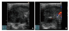


| [1] | Zheng PC, Sang L, Li YY, Wang HP, Chen ZG, Li YM, et al. Ultrasound diagnosis of primary squamous cell carcinoma of thyroid gland: case report and review. Advanced Ultrasound in Diagnosis and Therapy 2021; 01:054-057. |
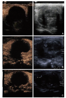
| [2] | Chen S, Peng Q, Zhang Q, Niu C. Contrast-enhanced ultrasound of primary squamous cell carcinoma of the thyroid: a case report. Front Endocrinol (Lausanne) 2020; 11:512. |
| [3] | Sun BH, Yu ST, Ge JN, Lei ST. Primary squamous cell carcinoma (PSCC) of the thyroid: A case report and review of the literature. Gland Surg 2020; 9:474-477. |
| [4] | Ibrahim MI, Jusoh YR, Adam NN, Mohamad I. Primary squamous cell carcinoma of the thyroid gland. Iran J Otorhinolaryngol 2018; 30:65-68. |
| [5] | Zheng RZ, Huang GH, Xu YJ. A primary squamous cell carcinoma of the thyroid presenting as the anaplastic thyroid carcinoma: a case report. Front Surg 2020; 7:590956. |
| [6] |
Shenoy VS, Rao RA, Kamath PM, Prasad V, Haseena S. Primary squamous cell carcinoma of thyroid - a rare malignant goitre. Indian J Surg Oncol 2016; 7:467-469.
doi: 10.1007/s13193-016-0530-4 |
| [7] | Chavan RN, Chikkala B, Biswas C, Biswas S, Sarkar DK. Primary squamous cell carcinoma of thyroid: A rare entity. Case Rep Pathol 2015; 2015:838079. |
| [8] |
Koyama S, Fujiwara K, Nosaka K, Fukuhara T, Morisaki T, Miyake N, et al. Immunohistochemical features of primary pure squamous sell carcinoma in the thyroid: An autopsy case. Case Rep Oncol 2018; 11:418-424.
doi: 10.1159/000490410 |
| [9] | Rausch T, Benhattar J, Sutter M, Andrejevic-Blant S. Thyroid carcinoma with papillary and squamous features: report of a case with histogenetic considerations. Pathol Res Pract 2010; 206:263-269. |
| [10] |
Wang SS, Ye DX, Wang B, Xie C. The expressions of peratins and P63 in primary squamous cell carcinoma of the thyroid gland: an application of raman spectroscopy. Onco Targets Ther 2020; 13:585-91.
doi: 10.2147/OTT |
| [11] | Yang S, Li C, Shi X, Ma B, Xu W, Jiang H, et al. Primary squamous cell carcinoma in the thyroid gland: A population-based analysis using the SEER database. World J Surg 2019; 43:1249-1255. |
| [12] |
Jang JY, Kwon KW, Kim SW, Youn I. Primary squamous cell carcinoma of thyroid gland with local recurrence: ultrasonographic and computed tomographic findings. Ultrasonography 2014; 33:143-148.
doi: 10.14366/usg.13022 pmid: 24936508 |
| [13] | Kondo T, Matsuyoshi A, Matsuyoshi H, Goto R, Ono K, Honda Y, et al. A case of primary thyroid squamous cell cancer: transformation from benign tumour associated with chronic thyroiditis? BMJ Case Rep 2009; 2009:bcr10.2008.1137. |
| [14] |
Daniels SP, Mankowski Gettle L, Blankenbaker DG, Lee KS, Ross AB. Contrast-enhanced ultrasound-guided musculoskeletal biopsies: our experience and technique. Skeletal Radiol 2021; 50:673-681.
doi: 10.1007/s00256-020-03604-8 |
| [15] | Fu Y, Tan S, Cui LG, Mei F. Contrast-enhanced ultrasound improves technical sufficiency of fine-needle aspiration in suspicious thyroid nodules. Advanced Ultrasound in Diagnosis and Therapy 2021; 03:219-225. |
| [16] |
Sparchez Z, Radu P, Kacso G, Eniu D, Hica S, Sparchez M. Contrast-enhanced ultrasound guided biopsy of superficial toraco-abdominal and neck lesions. Initial experience in 20 patients. Med Ultrason 2012; 14:288-293.
pmid: 23243642 |
| [17] |
Colevas AD, Yom SS, Pfister DG, Spencer S, Adelstein D, Adkins D, et al. NCCN guidelines insights: head and neck cancers, Version 1.2018. J Natl Compr Canc Netw 2018; 16:479-490.
doi: 10.6004/jnccn.2018.0026 |
| No related articles found! |
|
||