Review Article
- Advances in Modern Clinical Ultrasound
- Sriharsha Gummadi, MD, John Eisenbrey, PhD, Jingzhi Li, MD, Zhaojun Li, MD, Flemming Forsberg, PhD, Andrej Lyshchik, MD, Ji-Bin Liu, MD
- 2018, 2 (2): 51-63. DOI:10.37015/AUDT.2018.180801
- Abstract ( 1782 ) HTML ( 1011 ) PDF ( 837KB ) ( 2076 )
-
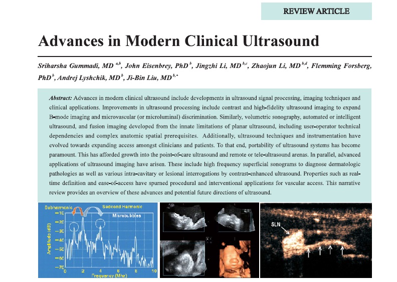
Advances in modern clinical ultrasound include developments in ultrasound signal processing, imaging techniques and clinical applications. Improvements in ultrasound processing include contrast and high-fidelity ultrasound imaging to expand B-mode imaging and microvascular (or microluminal) discrimination. Similarly, volumetric sonography, automated or intelligent ultrasound, and fusion imaging developed from the innate limitations of planar ultrasound, including user-operator technical dependencies and complex anatomic spatial prerequisites. Additionally, ultrasound techniques and instrumentation have evolved towards expanding access amongst clinicians and patients. To that end, portability of ultrasound systems has become paramount. This has afforded growth into the point-of-care ultrasound and remote or tele-ultrasound arenas. In parallel, advanced applications of ultrasound imaging have arisen. These include high frequency superficial sonograms to diagnose dermatologic pathologies as well as various intra-cavitary or lesional interrogations by contrast-enhanced ultrasound. Properties such as realtime definition and ease-of-access have spurned procedural and interventional applications for vascular access. This narrative review provides an overview of these advances and potential future directions of ultrasound.
-
- Progress of In-stent Restenosis after Vertebral Artery Ostium Stenosis and Imaging Assessments
- Jingzhi Li, MD, John R. Eisenbrey, PhD, Yang Hua, MD
- 2018, 2 (2): 64-70. DOI:10.37015/AUDT.2018.180802
- Abstract ( 500 ) HTML ( 11 ) PDF ( 345KB ) ( 455 )
-
Stenting has become one of the primary procedure to treat vertebral artery ostium stenosis patients. Postoperative instent restenosis (ISR) of the stenting is still one of the unsolved issues and requires systematic and further investigation. Through imaging assessments, ISR could be identified and evaluated in clinical practice and researches.
- Ultrasonic Thermal Strain Imaging for Noninvasive Temperature Estimation in Tissue
- Wenlong Zeng, Christopher J Krueger, Zhifei Dai, PhD
- 2018, 2 (2): 71-81. DOI:10.37015/AUDT.2018.180803
- Abstract ( 888 ) HTML ( 12 ) PDF ( 824KB ) ( 1115 )
-
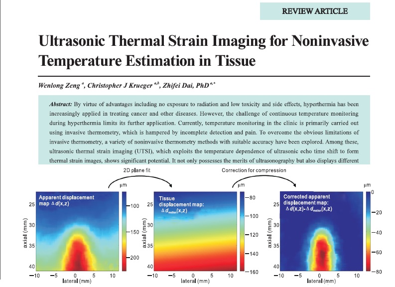
By virtue of advantages including no exposure to radiation and low toxicity and side effects, hyperthermia has been increasingly applied in treating cancer and other diseases. However, the challenge of continuous temperature monitoring during hyperthermia limits its further application. Currently, temperature monitoring in the clinic is primarily carried out using invasive thermometry, which is hampered by incomplete detection and pain. To overcome the obvious limitations of invasive thermometry, a variety of noninvasive thermometry methods with suitable accuracy have been explored. Among these, ultrasonic thermal strain imaging (UTSI), which exploits the temperature dependence of ultrasonic echo time shift to form thermal strain images, shows significant potential. It not only possesses the merits of ultrasonography but also displays different tissue characteristics (thermal properties of tissue and sound velocity) from other ultrasound imaging methods, so it has been investigated extensively over the past few years. This paper reviews recent advances in UTSI for noninvasive thermometry and discusses its main limitations, hoping to show the strong clinical application potential of UTSI from solid basic theory and practical research results.
-
Original Research
- Deep Learning Models for Segmentation of Lesion Based on Ultrasound Images
- Jinlian Ma, PhD, Dexing Kong, PhD
- 2018, 2 (2): 82-93. DOI:10.37015/AUDT.2018.180804
- Abstract ( 843 ) HTML ( 18 ) PDF ( 697KB ) ( 626 )
-
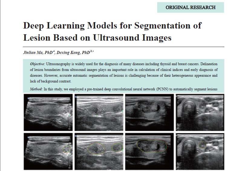
Objective: Ultrasonography is widely used for the diagnosis of many diseases including thyroid and breast cancers. Delineation of lesion boundaries from ultrasound images plays an important role in calculation of clinical indices and early diagnosis of diseases. However, accurate automatic segmentation of lesions is challenging because of their heterogeneous appearance and lack of background contrast.
Method: In this study, we employed a pre-trained deep convolutional neural network (PCNN) to automatically segment lesions from ultrasound images. Specifically, our PCNN based method used whole images of normal tissues and lesions as inputs and then generated the segmentation probability maps as outputs. A pre-training strategy was used to improve the performance of the PCNN based model. Additionally, we compared the performance of our approach with that of the common convolutional neural network segmentation methods on the same dataset.
Results: We validated on ultrasound images of thyroid nodules and breast nodules. The experimental results were shown in true positive rate (TP), false positive rate (FP), overlap metric (OM) and dice ratio (DR). Specifically, for thyroid nodule segmentation, our method could achieve an average of OM, DR, TP, FP as 0.8943, 0.9558, 0.9694, 0.0569 on overall folds, respectively. For breast nodule segmentation, our method could achieve an average of OM, DR, TP, FP as 0.8572, 0.9001, 0.9497, 0.8619, respectively.
Conclusion: Our proposed method is fully automatic without any user interaction and may be good enough to replace the timeconsuming and tedious manual segmentation approach, demonstrating the potential clinical applications. -
- Trans-lymphatic Contrast-Enhanced Ultrasound in Combination with Blue Dye Injection is Feasible for Detection and Biopsy of Sentinel Lymph Nodes in Breast Cancer
- Xiangmei Chen, MD, Jieyu Zhong, MD, Zhengming Hu, MD, Wei Wei, MD, Weihua Yin, MD, Ligang Cui, MD, Ji-Bin Liu, MD, Desheng Sun, MD
- 2018, 2 (2): 94-100. DOI:10.37015/AUDT.2018.180805
- Abstract ( 481 ) HTML ( 5 ) PDF ( 383KB ) ( 776 )
-
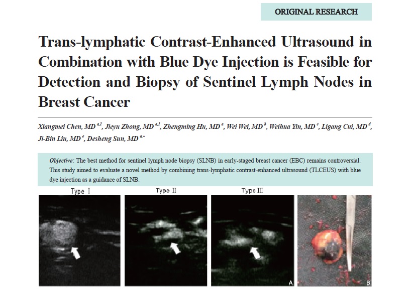
Objective: The best method for sentinel lymph node biopsy (SLNB) in early-staged breast cancer (EBC) remains controversial. This study aimed to evaluate a novel method by combining trans-lymphatic contrast-enhanced ultrasound (TLCEUS) with blue dye injection as a guidance of SLNB.
Methods: TLCEUS was performed in 88 patients with newly diagnosed EBC. Methylene blue dye was percutaneously injected into enhanced sentinel lymph nodes (SLNs) under ultrasound guidance, followed by standard SLNB and axillary lymph node dissection. Enhancement patterns and the arriving time (AT) of contrast agent within SLNs were evaluated. Histopathological examination of dissected nodes was performed to confirm metastasis.
Results: A total of 95 enhanced SLNs were identified and biopsied in 86 of 88 patients with identification rate of 97.7%. The specificity was 75.0%, sensitivity was 83.3%, and false-negative rate was 16.7%. Contrast-enhanced SLNs with type I, type II, and type III patterns had a metastatic positive rate of 11.4% (5/44), 57.1% (12/21) and 80.0% (24/30), respectively. Metastatic positive SLNs showed a mean AT of 61.6 ± 58.7 s while metastatic negative SLNs showed a mean AT of 41.3 ± 19.9 s, which was statistically significantly different.
Conclusion: The TLCEUS/blue dye method can be used as an alternative to the radioisotope/blue dye method for its feasibility and accuracy. -
- Treatment of Calcific Achilles Tendinitis Using the Ultrasound-guided Single-needle Lavage Technique
- Ligang Cui, MD, Chang Liu, MD, Zhiqiang Li, MD, Desheng Sun, MD
- 2018, 2 (2): 101-105. DOI:10.37015/AUDT.2018.180806
- Abstract ( 791 ) HTML ( 11 ) PDF ( 333KB ) ( 538 )
-
Objective: To report our initial experience using the ultrasound (US)-guided single-needle lavage technique for treating calcific Achilles tendinitis.
Methods: Eighteen patients with chronic ankle pain refractory to medical treatment were treated percutaneously using a fine needle lavage technique under US guidance. Patients were evaluated using the visual analogue scale (VAS) pain score both before and 3 months after the procedure. Treatment-related complications were also recorded.
Results: All 18 cases of US-guided single-needle lavage were performed successfully. The range of recovery time was 30-45 days, and there was a significant reduction in the VAS pain score.
Conclusion: The US-guided single-needle lavage technique seems to be an effective and minimally invasive way to treat calcific Achilles tendinitis.
- Assessment of the Cervix Using Strain Elastography in Pregnant Women with Spontaneous Preterm Birth
- Jinghua Li, MD, Yuanyuan An, MD, Lina Zhang, MD, Yinghua Xuan, MD, Qingqing Wu, MD
- 2018, 2 (2): 106-112. DOI:10.37015/AUDT.2018.180807
- Abstract ( 635 ) HTML ( 10 ) PDF ( 420KB ) ( 471 )
-
Objective: Preterm birth is still the leading cause of neonatal mortality. How to identify women who are at a higher risk of preterm birth continues to be one of the main problems in obstetrics. The aim of this study was to estimate the potential value of cervical strain in predicting the risk of preterm birth.
Methods: A total of 152 pregnant women who received prenatal examination in Beijing Obstetrics and Gynecology Hospital during January 2017 and October 2017. On the basis of the clinical diagnostic criteria for preterm birth, all of pregnant women were divided into two groups: There are 50 patients with the diagnosis of threatened premature or preterm labor in the study group and 102 patients without diagnosis of threatened premature or preterm labor in the control group. Measure cervical length by transvaginal ultrasound and obtain three cervical elasticity indexes by strain elastography: Closed internal cervical os strain rate, external cervical os strain rate and midpoint of the cervical canal strain rate. In the study group the pregnant women were divided into three teams: Length of cervix is 2 cm or less (10 cases) for T1, 2 to 3 cm (21 cases) for T2 and 3 cm or more (19 cases) for T3. Independent sample t-test was used to analyze the difference between the two groups. LSD-t test was used in the two groups. The receiver operating characteristic curve was used to analysis these four indicators. Statistical significance was defined as a p value < 0.05.
Results: Cervical length (CL) was lower and cervical os strain rate (CIS) was higher in the study group (2.67 ± 0.76 vs. 3.85 ± 0.65, p < 0.05; 0.53 ± 0.35 vs. 0.27 ± 0.24, p < 0.05). There was a significant difference with CIS in different cervical length team (p = 0.017) and the mean value of CIS in T1 was higher than the last two teams. The cutoff value of CL maximizing the accuracy of diagnosis was 3.2 cm; area under the curves (AUC), sensitivity, specificity, accuracy, positive predictive value (PPV) and negative predictive value (NPV) were 0.877, 73.3%, 87.0%, 82.2%, 73.5%, 86.4% respectively. Combining CL and CIS, the results of AUC, sensitivity, specificity, accuracy, PPV and NPV (0.891, 73.3%, 91.3%, 84.9%, 80.0%, 86.9%) increased in predicting the risk of spontaneous preterm birth, and the cutoff values of the CL and CIS were 3.1 cm and 0.35%.
Conclusion: The shortening of CL and the increasing CIS are closely related to the occurrence of premature birth and can be used as the indexes to predict the preterm labor. CIS can be used as an indicator to monitor early preterm changes. The combination of CL and CIS may have a role in assessing the risk of spontaneous preterm birth.
- The Value of Percutaneous Contrast-Enhanced Ultrasound in the Evaluation of Sentinel Lymph Node of Breast Cancer
- Ping Wang, MM, Jian Liu, MD, Wensheng Yue, MM, Wenyan Li, MM, Yuqun Luo, MM, Jiao He, MM
- 2018, 2 (2): 113-118. DOI:10.37015/AUDT.2018.180808
- Abstract ( 409 ) HTML ( 2 ) PDF ( 408KB ) ( 474 )
-
Objective: To analysis breast cancer sentinel lymph channels (SLCs) and the shape of the sentinel lymph nodes (SLNs) perfusion by contrast-enhanced ultrasound (CEUS) and to explore its value in the diagnosis of sentinel metastasis lymph nodes.
Methods: A retrospective analysis, 66 breast cancer patients with 101 ipsilateral axillary SLNs checking by CEUS, was confirmed by pathology. SLCs and SLNs positions were marked and then the rough needle biopsy pathology was used to confirm the benign or malignant SLNs. According to the different perfusion, SLNs were divided into five groups and evaluated by scores. MecNemar test and receiver operating characteristic (ROC) curve were used to analyze the statistical analysis. The difference of p < 0.05 was statistically significant. The cut-off value was finally determined, and the results of ultrasound screening test were evaluated.
Results: A total of 66 patients with 101 SLNs were successfully evaluated, including 42 cases of metastatic lymph nodes, and 59 cases of benign lymph nodes. The area under the ROC curve was 0.752 (p < 0.05). To ROC curve, the cut off value was 3.5. The scores 4 and 5 of lymph nodes were negative, and scores 1, 2, 3 of lymph nodes were positive. The sensitivity, specificity, false negative rate, false positive rate, and the coincidence rate of the screening test were 44.19%, 93.10%, 55.81%, 6.9%, 72.28% respectively. The value of positive predictive and the negative predictive were 82.61%, 69.23% respectively.
Conclusion: To evaluate SLCs shape and judge benign and malignant SLNs, percutaneous CEUS has a high practical value.
- Use of Transperineal Sonography to Diagnose Vaginal Abnormalities
- Yan Wang, PhD, Zan Sun, PhD, Na Li, MD, Xin Liao, MD, Xiaolu Liang, MD
- 2018, 2 (2): 119-122. DOI:10.37015/AUDT.2018.180809
- Abstract ( 539 ) HTML ( 5 ) PDF ( 371KB ) ( 420 )
-
Objective: Vagina illness is common in young girls recently. Transperineal ultrasonography is not commonly used in evaluation of vagina illness. Here we report the use of the mechanical properties of color Doppler ultrasonography to further characterize vagina illness.
Methods: Eighty patients who came to Shenyang Children Hospital with vaginal discharge, vaginal bleeding, vulva redness and swelling were evaluated with transperineal ultrasonography and four typically cases were chosed which had tumor, vagina foreign body, vaginitis and imperforate hymen. Color Doppler ultrasonography was applied to these abnormities.
Results: When transperineal ultrasonography was applied to these abnormities, the tumor had the sign of occupying lesion and sparse color flow. Vagina foreign body showed high-echo strip object or moniliform in vagina accompany with vaginal dropsy. A 15-ays neonate girl with hydrocolpocele was diagnosed imperforate hymen later.
Conclusion: Transperineal ultrasonography can be used to enhance the diagnosis of vagina illness in young girls.
- Ultrasound Measurement of Aortic Diameter at the Diaphragmatic Hiatus in Normal and Congenital Heart Disease Fetuses
- Zhongshan Gou, MD, Xinxin Yan, MD, Jianfang Ma, MD, Qi Pan, MD, Zhong Yang, MD, Xuedong Deng. MD
- 2018, 2 (2): 123-127. DOI:10.37015/AUDT.2018.180810
- Abstract ( 622 ) HTML ( 4 ) PDF ( 347KB ) ( 493 )
-
Objectives: The size of cardiovascular structures in fetuses with congenital heart disease (CHD) is variable, and current reference structures used for comparative purposes are suboptimal: There are no known cardiovascular structures that are reliably normal in size. We sought to evaluate whether the aortic diameter at the diaphragmatic hiatus (ADDH) is affected in CHD.
Methods: The ADDH was evaluated by sonography in both study subjects (CHD group, n = 506) and age-matched normal controls (normal group, n = 506). The CHD group was divided into different subgroups according to evident anomalies in the cardiovascular system. ADDH differences between CHD subgroups and their controls were calculated with a paired Student's t-test. Possible sex differences were determined by an unpaired Student's t-test on the whole group.
Results: There were no significant differences in the ADDH between all CHD subgroups and age-matched controls (p > 0.05). No difference was found in the ADDH between female and male fetuses (p > 0.05).
Conclusion: The ADDH in fetuses with CHD is similar to that in controls. Hence, ratios between size of involved cardiovascular structures and ADDH may be a useful parameter for monitoring fetuses with CHD .
- Ultrasound Characteristics of Kimura Disease: Retrospective Analysis of 30 cases
- Bingyan Liu, MD, Shaoqing Fu, MD, Dongni Luo, MD, Shengxin Fu, MD, Donglin Wang, MD, Wei Liao, MD
- 2018, 2 (2): 128-132. DOI:10.37015/AUDT.2018.180811
- Abstract ( 548 ) HTML ( 4 ) PDF ( 337KB ) ( 520 )
-
Objective: To demonstrate the characteristics of the lesion in Kimura disease on ultrasound imaging and compare with clinical features in patients with pathologically-approved Kimura disease.
Methods: A total of 30 patients (29 men and 1 woman; a mean age of 35.4 years) with Kimura disease confirmed by surgery and pathology were enrolled in the study. Ultrasonographic findings were analyzed retrospectively and compared with clinical features and laboratory examinations to better understand the relationship among them.
Results: All patients presented with painless, progressively enlarged soft tissue masses in head and neck, axillary, inguinal regions or limb extremities. Laboratory results showed elevation of eosinophils in both peripheral blood and serum IgE in 12 of 30 cases. Ultrasound findings of the lesions in Kimura disease were divided into three types: Type I: characterized by simple lymph node enlargement; Type II: characterized by flake-like thickening of the lesion without mass effect, and enlargement of nearby lymph nodes; Type III: a tumor-like lesion associate with enlargement of nearby lymph nodes.
Conclusion: The Kimura disease has specific ultrasonographic characteristics of lymph node and adjacent structures. When combined with patient’s clinical information and laboratory tests, ultrasound examination can provide useful information for making clinical diagnosis and management.
- Sonographic Characteristics of Congenital Pulmonary Airway Malformations in Children: A Report of 21 Cases and Review of Literature
- Hongkui Yu, MD, Zhihui Li, MD, Xiao Liu, MD, Weiling Chen, MD, Yuanxiang Wang, MD, Bei Xia, MD
- 2018, 2 (2): 133-138. DOI:10.37015/AUDT.2018.180812
- Abstract ( 425 ) HTML ( 2 ) PDF ( 406KB ) ( 463 )
-
Objective: This study discusses the diagnostic value of ultrasonography for congenital pulmonary airway malformations (CPAM) in children.
Methods: A retrospective analysis was conducted for 21 pathologically confirmed paediatric cases of CPAM by reviewing the ultrasonography images and comparing them with the clinical and pathologic results.
Results: All 21 paediatric cases of CPAM had lesions in unilateral lobes, with five cases involving the left lung and 16 cases involving the right lung. There were six cases with type 1 ultrasonography manifestations, 12 cases with type 2 ultrasonography manifestations and one case with a type 4 ultrasonography manifestation. One case was misdiagnosed as pulmonary sequestration (PS) by ultrasonography, and another case had no abnormalities based on ultrasonography. With respect to the pathologic results, six type 1 (28.6%), 13 type 2 (61.9%) and one type 4 (4.8%) were found, and one type 2 was complicated with PS (4.8%). The ultrasonography diagnoses agreed with surgical pathology reports in 18 cases (85.7%).
Conclusion: Ultrasonography is feasible and accurate in identifying subtype within postnatally diagnosed pediatric CPAM lesions.
Special Report
- Imaging Diagnosis of Tubular Adenoma of Common Bile Duct: Case Report and Review of the Literature
- Yi Dong, MD, Daohui Yang, MD, Qi Zhang, MD, Jiaying Cao, MD, Feng Mao, MD, Wen-Ping Wang, MD
- 2018, 2 (2): 139-142. DOI:10.37015/AUDT.2018.180813
- Abstract ( 412 ) HTML ( 9 ) PDF ( 336KB ) ( 478 )
-
Tubular adenoma of common bile duct (CBD) is an extremely rare benign extrahepatic bile duct adenoma. We report a case of surgery and histopathological proved CBD tubular adenoma. A 69-year-old man was admitted to the hospital for yellow urine for one month. Clinical, radiological and laboratory results were analyzed. Medical literature in PubMed pertaining to similar cases was reviewed. A solid mass was detected in the distant part of CBD, with dilation of the extrahepatic and intrahepatic bile duct by ultrasound, contrast enhanced ultrasound (CEUS), computer tomography (CT) and magnetic resonance imaging (MRI). It showed heterogeneous enhancement both in contrast-enhanced CT, MRI and CEUS. After operation, pathological findings indicated a tubular adenoma of CBD. Our case highlighted the fact that tubular adenoma of CBD was demonstrated as a homogeneous mass in the distant part of CBD.
Review Article
- Estimation of Clinical Outcomes of Irreversible Electroporation Use During Locally Advanced Pancreatic Cancer: A Systematic Review and Meta-analysis
- Tian’an Jiang, PhD, Guo Tian, MD, Liming Wu, PhD, Qiyu Zhao, PhD
- 2018, 2 (2): 143-149. DOI:10.37015/AUDT.2018.180814
- Abstract ( 411 ) HTML ( 2 ) PDF ( 581KB ) ( 1017 )
-
Objective: Irreversible electroporation (IRE) is a novel nonthermal ablative technique that transmits pulsatile electricity to enable nanoscale damages of the cellular membrane and induce cellular apoptosis. To assess the safety and efficacy of IRE for locally advanced pancreatic cancer (LAPC).
Methods: Electronic databases of PubMed, Embase, Web of Science, Scopus were searched up to June 2018 for studies comparing the standardized mean differences (SMD) of size, amylase and carbohydrate antigen 19-9 (CA199) levels between pre- and post-operation for patients with pancreatic cancer. Sensitivity and stratified analyses were conducted. Quality was estimated using Newcastle-Ottawa Scale (NOS).
Results: We finally identified 10 studies including 203 participants during a mean 7.06 months of follow-up (range 1 to 29 months). The meta-analyses showed the declined tumor size at 6 months post-IRE but unchanged at 1 month, and increased amylase level at 1-day post-IRE while unchanged at the 1 week. No significant difference of CA199 level was observed between pre-IRE and post-IRE at 1 week and 1 month. No risk of publication bias was detectable, and the favorable quality and validity of all outcomes were assessed based on NOS.
Conclusions: IRE may be a relatively state-of-the-art therapy option for most patients with LAPC if imaging or explorative laparotomy indicated that LAPC was unable to be successfully resected.
- The Ultrasonographic Findings of Appendiceal Diseases: Benign and Malignant
- Yixuan Wang, Bei Wang, Menene NKONIKA, Hongyu Ding, Hongjun Sun
- 2018, 2 (2): 150-154. DOI:10.37015/AUDT.2018.180815
- Abstract ( 534 ) HTML ( 9 ) PDF ( 316KB ) ( 486 )
-
The appendix is a small pouch-like sac with no fixed anatomical position, narrowing entrance to caecum predisposes to both benign and malignant obstruction in nature. In recent years, ultrasonog-raphy plays an important part in diagnosis of appendiceal diseases. This article aims to describe ul-trasonic assessment of different kinds of appendiceal diseases. We provide an overview of the liter-ature about the ultrasonographic features of appendiceal diseases.
Technical Development
- Application and Development of Handheld Ultrasound in the Field of Medicine and Healthcare
- Xing Yu, Yaoyao Cui, Yuankai Xuan, Tingyi Jiang, Ligang Cui
- 2018, 2 (2): 155-160. DOI:10.37015/AUDT.2018.180816
- Abstract ( 1184 ) HTML ( 623 ) PDF ( 303KB ) ( 2612 )
-
With the rapid development of microelectronics, handheld ultrasound devices emerged and have a widespread use in the field of medicine and healthcare. The handheld ultrasound is a modality of medical ultrasonic device that is portable (i.e., carried by hand), easy to operate and in some cases even combine the scanner with the host system. It normally has the characteristics of light weight, small size and low power consumption. With the wireless imaging transmission technique, remote and intelligent diagnosis is possible for the doctors using handheld ultrasound devices. As soon as the low-cost, highperformance handheld ultrasound device was launched, it attracted clinical attention and received a warm welcome from primary doctors and clinicians, thereby accelerating the development of ultrasonic clinical visualization. In this paper, we will illustrate the status and roles of handheld ultrasound for its clinical settings and applications in the past, current, and future.
Case Report
- Subacute Right Ventricular Pacing Lead Perforation Diagnosed by Echocardiography
- Jinjie Xie, MD, Rongjuan Li, MD, Taiyang Luo, MD, Ya Yang, MD
- 2018, 2 (2): 161-163. DOI:10.37015/AUDT.2018.180817
- Abstract ( 418 ) HTML ( 4 ) PDF ( 318KB ) ( 404 )
-
A 69-year-old woman with sick sinus syndrome underwent permanent pacemaker implantation in our hospital. A mild and persistent chest pain occurred 2 days after device implantation, and myocardial perforation without pericardial effusion was found at the tip of the right ventricular lead by two-dimensional transthoracic echocardiography. The diagnosis was then confirmed by fluoroscopy, and the lead was repositioned under fluoroscopic guidance.
- Hepatic Angiomyolipoma: A Case Report
- Meng Li, MD, Zhiyan Li, MD, Yuejuan Gao, MD
- 2018, 2 (2): 164-166. DOI:10.37015/AUDT.2018.180818
- Abstract ( 600 ) HTML ( 14 ) PDF ( 336KB ) ( 550 )
-
Hepatic angiomyolipoma (AML) is a rare benign tumor with heterogeneous components composed of vascular, fat, and smooth muscle elements which is often misdiagnosed as other neoplasms such as hepatocellular carcinoma due to nonspecific clinical and image features. A typical case of hepatic AML located in liver with overall clinic and ultrasonography information was analyzed and relative references were reviewed in this article.


