CONTENTS
Review Articles
- Renal Contrast-enhanced Ultrasound: Clinical Applications and Emerging Research
- Felipe Velasquez-Botero, MD, Ananya Balasubramanya, Ying Tang, MD, Qiang Lu, MD, Ji-Bin Liu, MD, John R. Eisenbrey, PhD
- 2022, 6 (4): 129-146. DOI:10.37015/AUDT.2022.220036
- Abstract ( 1019 ) HTML ( 561 ) PDF ( 7061KB ) ( 671 )
-
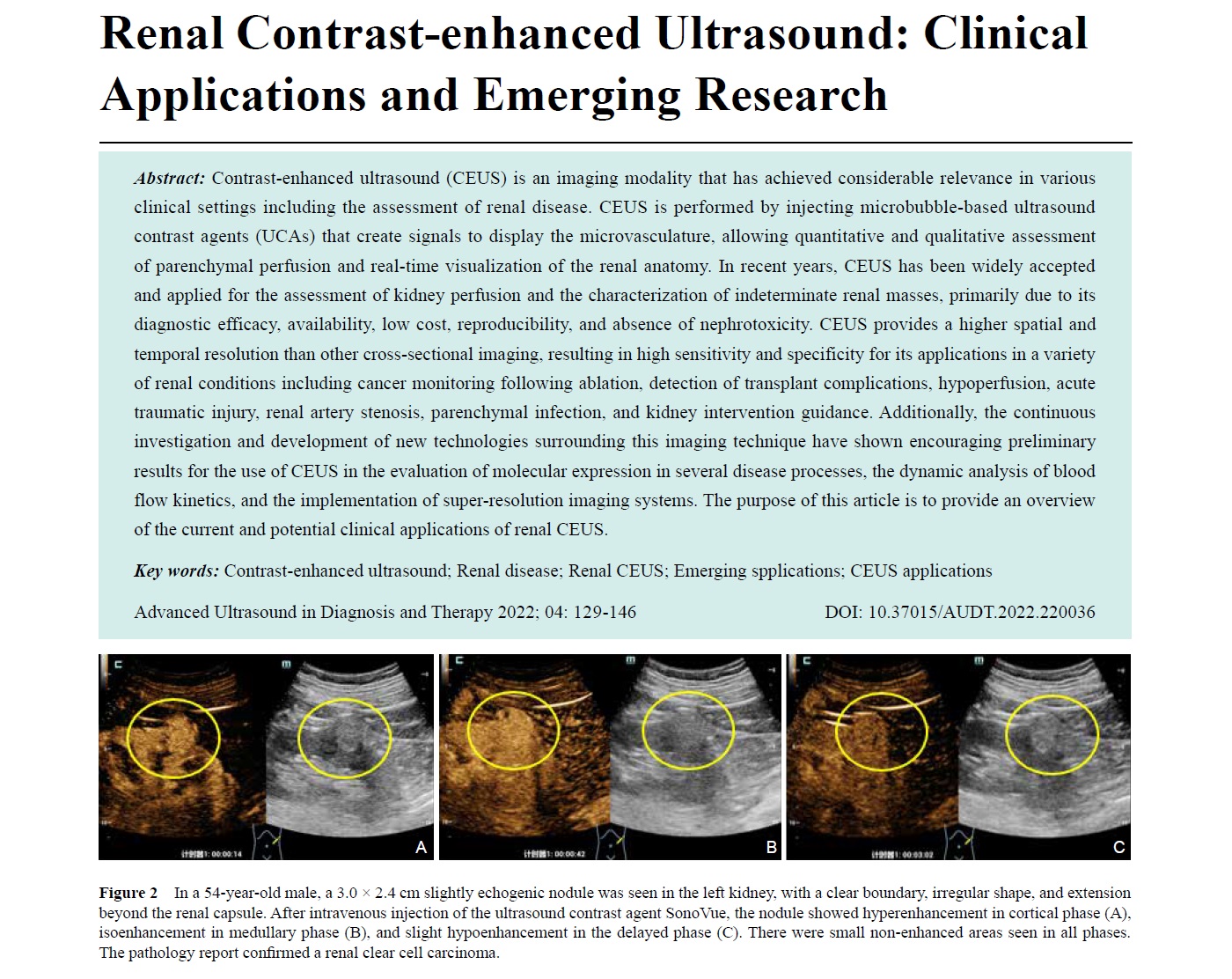
Contrast-enhanced ultrasound (CEUS) is an imaging modality that has achieved considerable relevance in various clinical settings including the assessment of renal disease. CEUS is performed by injecting microbubble-based ultrasound contrast agents (UCAs) that create signals to display the microvasculature, allowing quantitative and qualitative assessment of parenchymal perfusion and real-time visualization of the renal anatomy. In recent years, CEUS has been widely accepted and applied for the assessment of kidney perfusion and the characterization of indeterminate renal masses, primarily due to its diagnostic efficacy, availability, low cost, reproducibility, and absence of nephrotoxicity. CEUS provides a higher spatial and temporal resolution than other cross-sectional imaging, resulting in high sensitivity and specificity for its applications in a variety of renal conditions including cancer monitoring following ablation, detection of transplant complications, hypoperfusion, acute traumatic injury, renal artery stenosis, parenchymal infection, and kidney intervention guidance. Additionally, the continuous investigation and development of new technologies surrounding this imaging technique have shown encouraging preliminary results for the use of CEUS in the evaluation of molecular expression in several disease processes, the dynamic analysis of blood flow kinetics, and the implementation of super-resolution imaging systems. The purpose of this article is to provide an overview of the current and potential clinical applications of renal CEUS.
-
- Research Progress in Ultrasonic Regulation of Cholinergic Anti-inflammatory Pathway
- Wuqi Zhou, MD, Yishu Song, MD, Rui Wang, MD, Qiaofeng Jin, MD, Mingxing Xie, PhD, Li Zhang, PhD
- 2022, 6 (4): 147-152. DOI:10.37015/AUDT.2022.200034
- Abstract ( 718 ) HTML ( 151 ) PDF ( 521KB ) ( 747 )
-
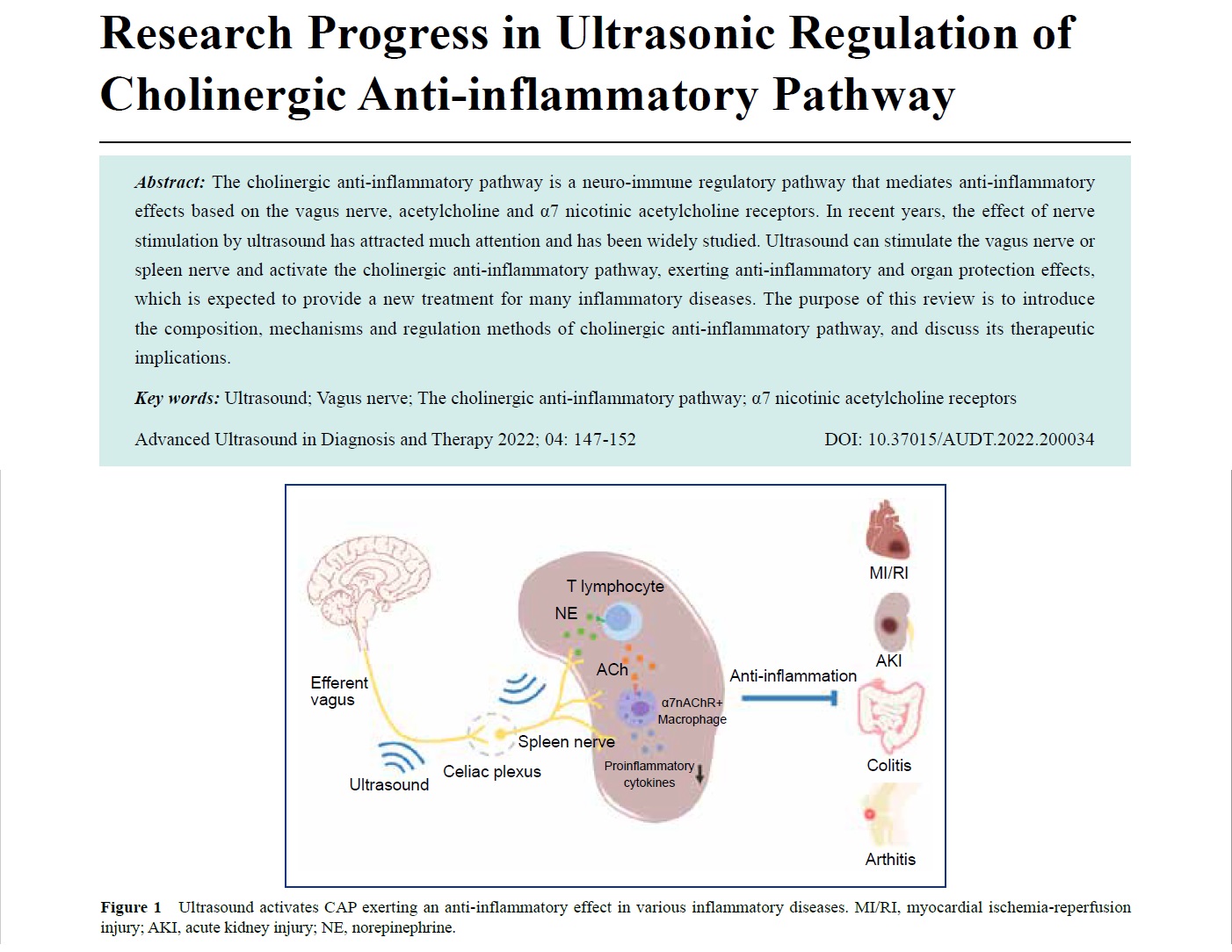
The cholinergic anti-inflammatory pathway is a neuro-immune regulatory pathway that mediates anti-inflammatory effects based on the vagus nerve, acetylcholine and α7 nicotinic acetylcholine receptors. In recent years, the effect of nerve stimulation by ultrasound has attracted much attention and has been widely studied. Ultrasound can stimulate the vagus nerve or spleen nerve and activate the cholinergic anti-inflammatory pathway, exerting anti-inflammatory and organ protection effects, which is expected to provide a new treatment for many inflammatory diseases. The purpose of this review is to introduce the composition, mechanisms and regulation methods of cholinergic anti-inflammatory pathway, and discuss its therapeutic implications.
-
- Functional Brain Imaging Based on the Neurovascular Unit for Evaluating Neural Networks after Stroke
- Yongyue Zhang, MM, Yang Sun, MM, Li Zhang, MM, Rongjin Zhang, MM, Shumin Wang, PhD
- 2022, 6 (4): 153-164. DOI:10.37015/AUDT.2022.210033
- Abstract ( 623 ) HTML ( 727 ) PDF ( 681KB ) ( 727 )
-
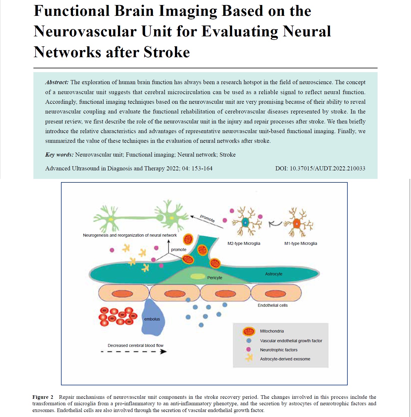
The exploration of human brain function has always been a research hotspot in the field of neuroscience. The concept of a neurovascular unit suggests that cerebral microcirculation can be used as a reliable signal to reflect neural function. Accordingly, functional imaging techniques based on the neurovascular unit are very promising because of their ability to reveal neurovascular coupling and evaluate the functional rehabilitation of cerebrovascular diseases represented by stroke. In the present review, we first describe the role of the neurovascular unit in the injury and repair processes after stroke. We then briefly introduce the relative characteristics and advantages of representative neurovascular unit-based functional imaging. Finally, we summarized the value of these techniques in the evaluation of neural networks after stroke.
-
- Diagnostic Values of CEUS, CECT and CEMRI for Renal Cystic Lesions on the Current Bosniak Criterion-A Meta-analysis
- Xiaojuan Yang, MD, Huihui Yang, MD, Yu He,MD, PhD
- 2022, 6 (4): 165-173. DOI:10.37015/AUDT.2022.210037
- Abstract ( 566 ) HTML ( 6 ) PDF ( 1102KB ) ( 821 )
-
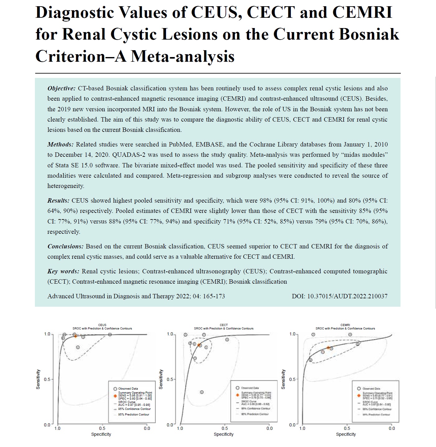
Objective: CT-based Bosniak classification system has been routinely used to assess complex renal cystic lesions and also been applied to contrast-enhanced magnetic resonance imaging (CEMRI) and contrast-enhanced ultrasound (CEUS). Besides, the 2019 new version incorporated MRI into the Bosniak system. However, the role of US in the Bosniak system has not been clearly established. The aim of this study was to compare the diagnostic ability of CEUS, CECT and CEMRI for renal cystic lesions based on the current Bosniak classification.
Methods: Related studies were searched in PubMed, EMBASE, and the Cochrane Library databases from January 1, 2010 to December 14, 2020. QUADAS-2 was used to assess the study quality. Meta-analysis was performed by “midas modules” of Stata SE 15.0 software. The bivariate mixed-effect model was used. The pooled sensitivity and specificity of these three modalities were calculated and compared. Meta-regression and subgroup analyses were conducted to reveal the source of heterogeneity.
Results: CEUS showed highest pooled sensitivity and specificity, which were 98% (95% CI: 91%, 100%) and 80% (95% CI: 64%, 90%) respectively. Pooled estimates of CEMRI were slightly lower than those of CECT with the sensitivity 85% (95% CI: 77%, 91%) versus 88% (95% CI: 77%, 94%) and specificity 71% (95% CI: 52%, 85%) versus 79% (95% CI: 70%, 86%), respectively.
Conclusions: Based on the current Bosniak classification, CEUS seemed superior to CECT and CEMRI for the diagnosis of complex renal cystic masses, and could serve as a valuable alternative for CECT and CEMRI.
-
- Peripheral Nerve Lipomatosis: Pathology, Clinical Features, Imaging Diagnosis and Treatment
- Ping Xu, MM, Heping Deng, MD, Bo Lu, MD, Yaru Mi, MM
- 2022, 6 (4): 174-179. DOI:10.37015/AUDT.2022.210039
- Abstract ( 549 ) HTML ( 6 ) PDF ( 2703KB ) ( 610 )
-
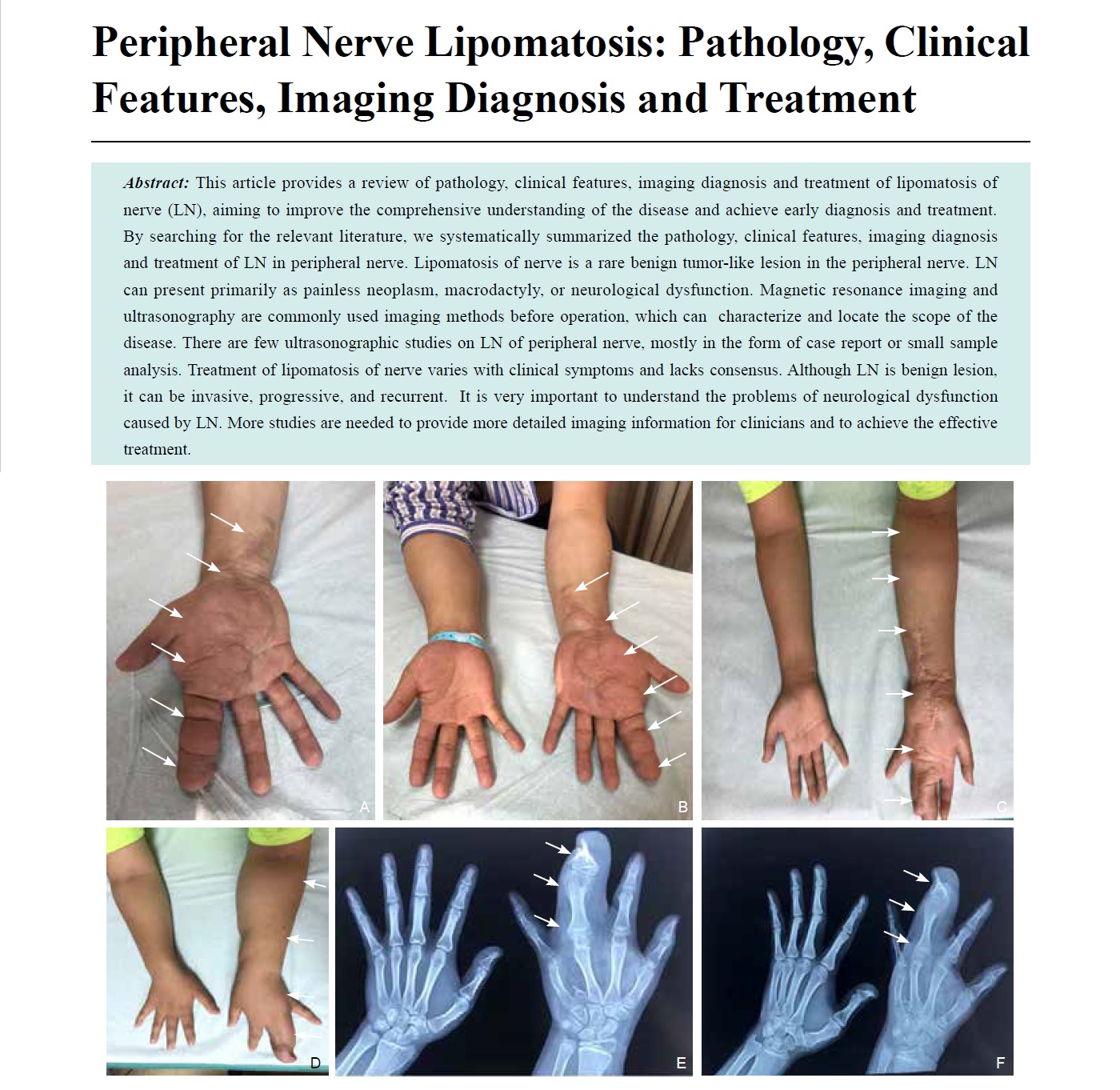
This article provides a review of pathology, clinical features, imaging diagnosis and treatment of lipomatosis of nerve (LN), aiming to improve the comprehensive understanding of the disease and achieve early diagnosis and treatment. By searching for the relevant literature, we systematically summarized the pathology, clinical features, imaging diagnosis and treatment of LN in peripheral nerve. Lipomatosis of nerve is a rare benign tumor-like lesion in the peripheral nerve. LN can present primarily as painless neoplasm, macrodactyly, or neurological dysfunction. Magnetic resonance imaging and ultrasonography are commonly used imaging methods before operation, which can characterize and locate the scope of the disease. There are few ultrasonographic studies on LN of peripheral nerve, mostly in the form of case report or small sample analysis. Treatment of lipomatosis of nerve varies with clinical symptoms and lacks consensus. Although LN is benign lesion, it can be invasive, progressive, and recurrent. It is very important to understand the problems of neurological dysfunction caused by LN. More studies are needed to provide more detailed imaging information for clinicians and to achieve the effective treatment.
-
Original Research
- Using S-Detect to Improve Breast Ultrasound: The Different Combined Strategies Based on Radiologist Experience
- Ying Zhu, MD, Xiaohong Jia, MD, Yijie Dong, MD, Juan Liu, MD, Yilai Chen, MD, Congcong Yuan, MD, Weiwei Zhan, MD, Jianqiao Zhou, MD
- 2022, 6 (4): 180-187. DOI:10.37015/AUDT.2022.220007
- Abstract ( 526 ) HTML ( 3 ) PDF ( 1224KB ) ( 813 )
-
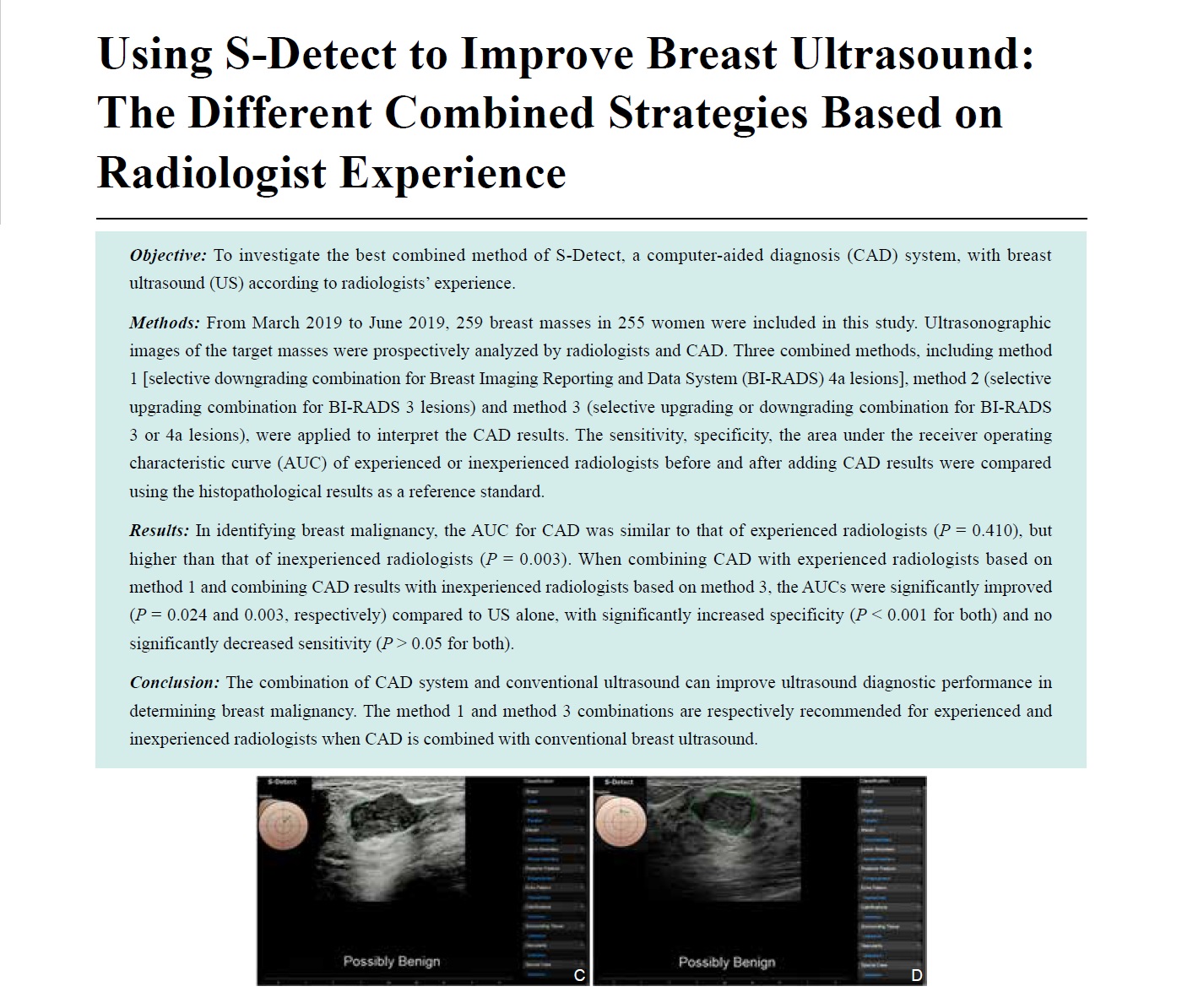
Objective: To investigate the best combined method of S-Detect, a computer-aided diagnosis (CAD) system, with breast ultrasound (US) according to radiologists’ experience.
Methods: From March 2019 to June 2019, 259 breast masses in 255 women were included in this study. Ultrasonographic images of the target masses were prospectively analyzed by radiologists and CAD. Three combined methods, including method 1 [selective downgrading combination for Breast Imaging Reporting and Data System (BI-RADS) 4a lesions], method 2 (selective upgrading combination for BI-RADS 3 lesions) and method 3 (selective upgrading or downgrading combination for BI-RADS 3 or 4a lesions), were applied to interpret the CAD results. The sensitivity, specificity, the area under the receiver operating characteristic curve (AUC) of experienced or inexperienced radiologists before and after adding CAD results were compared using the histopathological results as a reference standard.
Results: In identifying breast malignancy, the AUC for CAD was similar to that of experienced radiologists (P= 0.410), but higher than that of inexperienced radiologists (P= 0.003). When combining CAD with experienced radiologists based on method 1 and combining CAD results with inexperienced radiologists based on method 3, the AUCs were significantly improved (P= 0.024 and 0.003, respectively) compared to US alone, with significantly increased specificity (P< 0.001 for both) and no significantly decreased sensitivity (P> 0.05 for both).
Conclusion: The combination of CAD system and conventional ultrasound can improve ultrasound diagnostic performance in determining breast malignancy. The method 1 and method 3 combinations are respectively recommended for experienced and inexperienced radiologists when CAD is combined with conventional breast ultrasound.
-
- Graphene Oxide/Polylactic Acid Microbubbles for Efficient Removal of Lead Ions from Aqueous Solution
- Meng Han, MD, Ruirui Kang, MD, Juanjuan Chen, MD
- 2022, 6 (4): 188-194. DOI:10.37015/AUDT.2022.210030
- Abstract ( 528 ) HTML ( 1 ) PDF ( 733KB ) ( 658 )
-
Objective: Heavy metal pollution has become one of the environmental contamination problems in today's world. Adsorption materials can effectively remove heavy metal ions from the water. There are some shortcomings for traditional adsorbents, such as difficult separation after adsorption, long separation time, and may cause secondary pollution in the environment without recycling. The aim of this study was to seek new materials with effective ways to absorb heavy metal ions in the water.
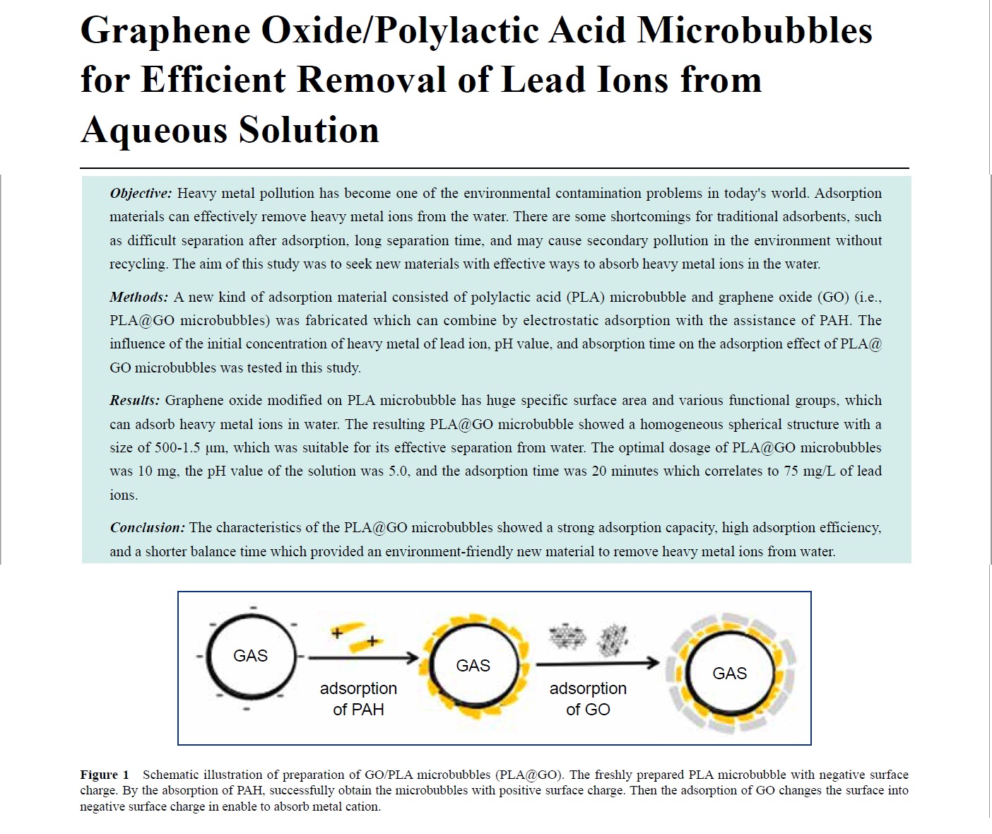
Methods: A new kind of adsorption material consisted of polylactic acid (PLA) microbubble and graphene oxide (GO) (i.e., PLA@GO microbubbles) was fabricated which can combine by electrostatic adsorption with the assistance of PAH. The influence of the initial concentration of heavy metal of lead ion, pH value, and absorption time on the adsorption effect of PLA@GO microbubbles was tested in this study.
Results: Graphene oxide modified on PLA microbubble has huge specific surface area and various functional groups, which can adsorb heavy metal ions in water. The resulting PLA@GO microbubble showed a homogeneous spherical structure with a size of 500-1.5 μm, which was suitable for its effective separation from water. The optimal dosage of PLA@GO microbubbles was 10 mg, the pH value of the solution was 5.0, and the adsorption time was 20 minutes which correlates to 75 mg/L of leadions.
Conclusion: The characteristics of the PLA@GO microbubbles showed a strong adsorption capacity, high adsorption efficiency, and a shorter balance time which provided an environment-friendly new material to remove heavy metal ions from water.
-
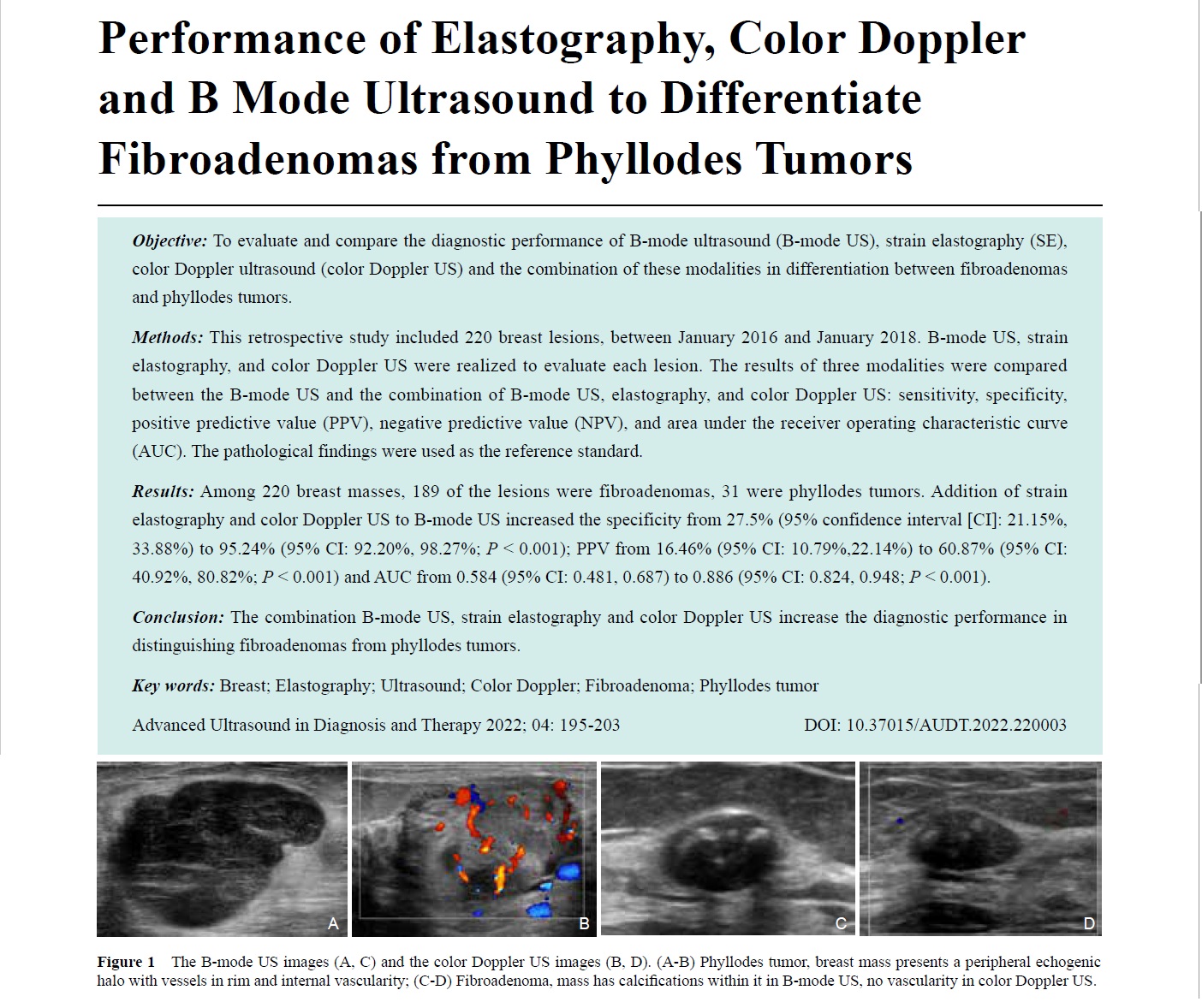
- Performance of Elastography, Color Doppler and B Mode Ultrasound to Differentiate Fibroadenomas from Phyllodes Tumors
- Lynda Aoudia, Amal Kouchkar, Salah Eddine Bendib
- 2022, 6 (4): 195-203. DOI:10.37015/AUDT.2022.220003
- Abstract ( 660 ) HTML ( 112 ) PDF ( 2261KB ) ( 553 )
-
Objective: To evaluate and compare the diagnostic performance of B-mode ultrasound (B-mode US), strain elastography (SE), color Doppler ultrasound (color Doppler US) and the combination of these modalities in differentiation between fibroadenomas and phyllodes tumors.
Methods: This retrospective study included 220 breast lesions, between January 2016 and January 2018. B-mode US, strain elastography, and color Doppler US were realized to evaluate each lesion. The results of three modalities were compared between the B-mode US and the combination of B-mode US, elastography, and color Doppler US: sensitivity, specificity, positive predictive value (PPV), negative predictive value (NPV), and area under the receiver operating characteristic curve (AUC). The pathological findings were used as the reference standard.
Results: Among 220 breast masses, 189 of the lesions were fibroadenomas, 31 were phyllodes tumors. Addition of strain elastography and color Doppler US to B-mode US increased the specificity from 27.5% (95% confidence interval [CI]: 21.15%, 33.88%) to 95.24% (95% CI: 92.20%, 98.27%; P < 0.001); PPV from 16.46% (95% CI: 10.79%,22.14%) to 60.87% (95% CI: 40.92%, 80.82%; P < 0.001) and AUC from 0.584 (95% CI: 0.481, 0.687) to 0.886 (95% CI: 0.824, 0.948; P < 0.001).
Conclusion: The combination B-mode US, strain elastography and color Doppler US increase the diagnostic performance in distinguishing fibroadenomas from phyllodes tumors.
-
Case Reports
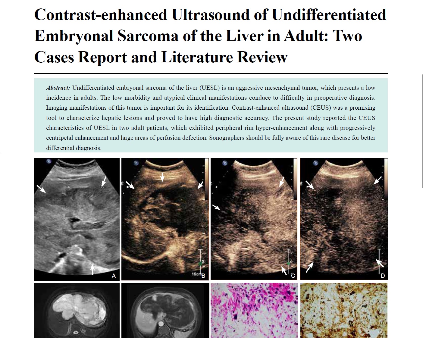
- Contrast-enhanced Ultrasound of Undifferentiated Embryonal Sarcoma of the Liver in Adult: Two Cases Report and Literature Review
- Yanling Chen, MM, Hantao Wang, MM, Hong Han, PhD, Yi Dong, PhD, Wen-ping Wang, MD
- 2022, 6 (4): 204-209. DOI:10.37015/AUDT.2022.220014
- Abstract ( 496 ) HTML ( 8 ) PDF ( 2507KB ) ( 760 )
-
Undifferentiated embryonal sarcoma of the liver (UESL) is an aggressive mesenchymal tumor, which presents a low incidence in adults. The low morbidity and atypical clinical manifestations conduce to difficulty in preoperative diagnosis. Imaging manifestations of this tumor is important for its identification. Contrast-enhanced ultrasound (CEUS) was a promising tool to characterize hepatic lesions and proved to have high diagnostic accuracy. The present study reported the CEUS characteristics of UESL in two adult patients, which exhibited peripheral rim hyper-enhancement along with progressively centripetal enhancement and large areas of perfusion defection. Sonographers should be fully aware of this rare disease for better differential diagnosis.
-
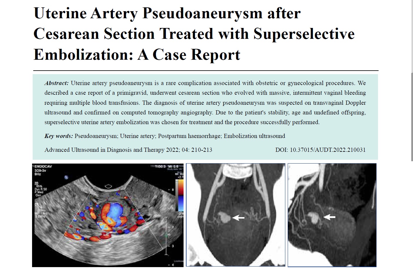
- Uterine Artery Pseudoaneurysm after Cesarean Section Treated with Superselective Embolization: A Case Report
- Jullie Anne Chiste, Larissa Cavalli de Oliveira, Liziane Lorusso, Anna Luisa Aranha Nunes, João Vitor Bacarin
- 2022, 6 (4): 210-213. DOI:10.37015/AUDT.2022.210031
- Abstract ( 602 ) HTML ( 82 ) PDF ( 818KB ) ( 814 )
-
Uterine artery pseudoaneurysm is a rare complication associated with obstetric or gynecological procedures. We described a case report of a primigravid, underwent cesarean section who evolved with massive, intermittent vaginal bleeding requiring multiple blood transfusions. The diagnosis of uterine artery pseudoaneurysm was suspected on transvaginal Doppler ultrasound and confirmed on computed tomography angiography. Due to the patient's stability, age and undefined offspring, superselective uterine artery embolization was chosen for treatment and the procedure successfully performed.
-
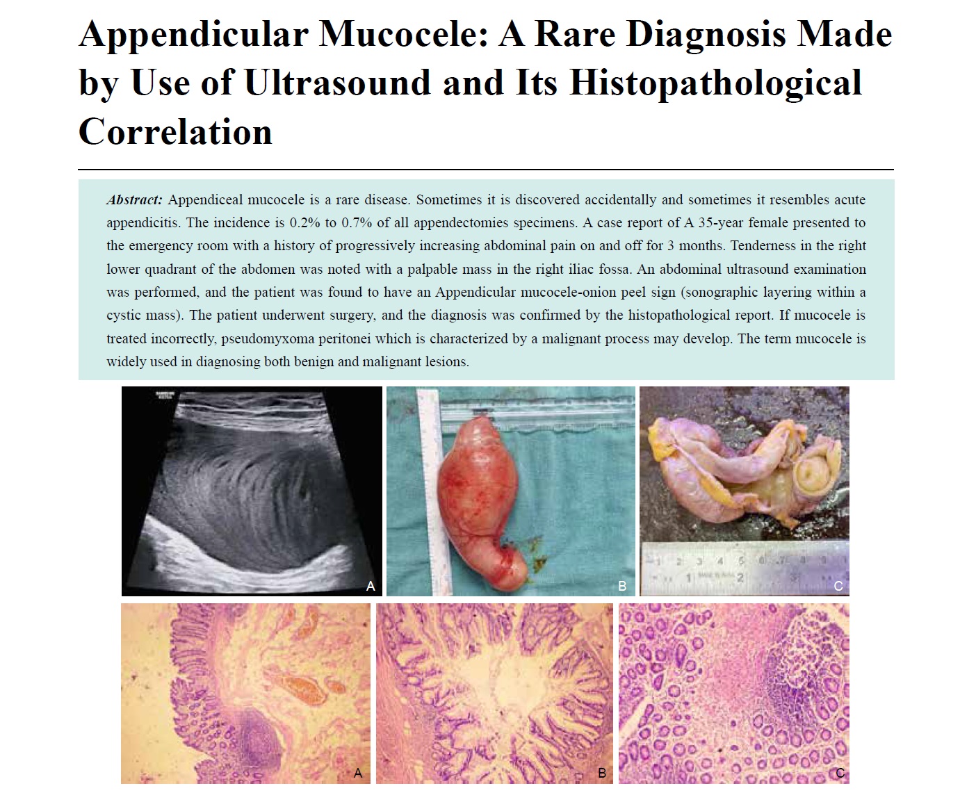
- Appendicular Mucocele: A Rare Diagnosis Made by Use of Ultrasound and Its Histopathological Correlation
- Sachin Khanduri, Surbhi , Ashkrit Gupta, Harleen Chawla, Asif Khan, Vaibhav Pathak, Saim Ali Siddiqui, Shahnawaz , Shreya Chitravanshi, Rohit
- 2022, 6 (4): 214-216. DOI:10.37015/AUDT.2022.220019
- Abstract ( 720 ) HTML ( 8 ) PDF ( 1277KB ) ( 822 )
-
Appendiceal mucocele is a rare disease. Sometimes it is discovered accidentally and sometimes it resembles acute appendicitis. The incidence is 0.2% to 0.7% of all appendectomies specimens. A case report of A 35-year female presented to the emergency room with a history of progressively increasing abdominal pain on and off for 3 months. Tenderness in the right lower quadrant of the abdomen was noted with a palpable mass in the right iliac fossa. An abdominal ultrasound examination was performed, and the patient was found to have an Appendicular mucocele-onion peel sign (sonographic layering within a cystic mass). The patient underwent surgery, and the diagnosis was confirmed by the histopathological report. If mucocele is treated incorrectly, pseudomyxoma peritonei which is characterized by a malignant process may develop. The term mucocele is widely used in diagnosing both benign and malignant lesions.
-
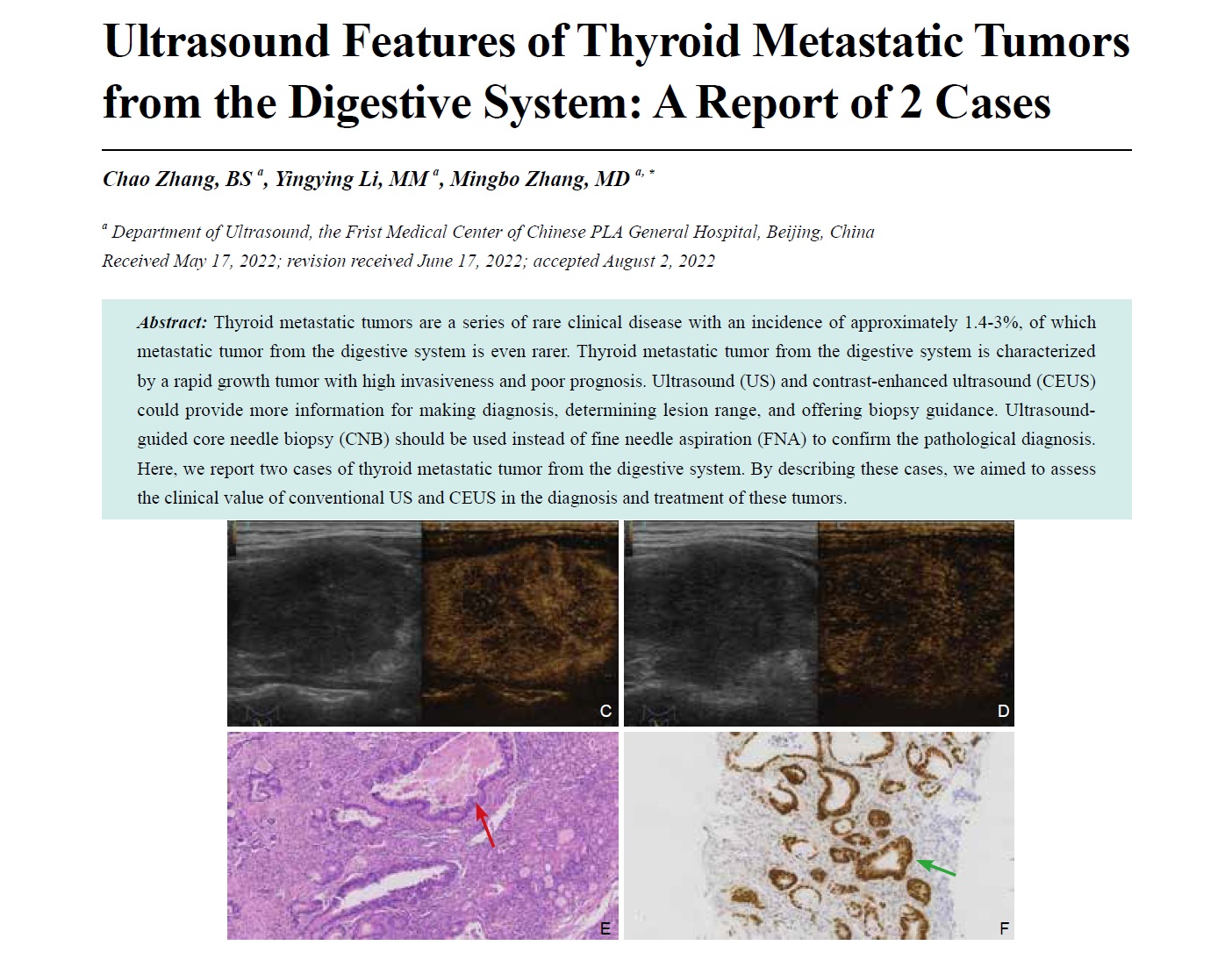
- Ultrasound Features of Thyroid Metastatic Tumors from the Digestive System: A Report of 2 Cases
- Chao Zhang, BS, Yingying Li, MD, Mingbo Zhang, MD
- 2022, 6 (4): 217-222. DOI:10.37015/AUDT.2022.220021
- Abstract ( 438 ) HTML ( 0 ) PDF ( 2472KB ) ( 748 )
-
Thyroid metastatic tumors are a series of rare clinical disease with an incidence of approximately 1.4-3%, of which metastatic tumor from the digestive system is even rarer. Thyroid metastatic tumor from the digestive system is characterized by a rapid growth tumor with high invasiveness and poor prognosis. Ultrasound (US) and contrast-enhanced ultrasound (CEUS) could provide more information for making diagnosis, determining lesion range, and offering biopsy guidance. Ultrasound-guided core needle biopsy (CNB) should be used instead of fine needle aspiration (FNA) to confirm the pathological diagnosis. Here, we report two cases of thyroid metastatic tumor from the digestive system. By describing these cases, we aimed to assess the clinical value of conventional US and CEUS in the diagnosis and treatment of these tumors.
-
Meeting Abstracts
- CSUM2022 ABSTRACTS
- 2022, 6 (4): 223-237.
- Abstract ( 217 ) HTML ( 5 ) PDF ( 383KB ) ( 181 )
-
The Annual National Congress of the Chinese Society of Ultrasound in Medicine (CSUM) is one of the most prestigious and influential academic conferences in China, with the largest number of participants. The 22th Annual Meeting of Chinese Society of Ultrasound in Medicine (CSUM 2022) will be held from November 3 to November 6 in Chengdu, Sichuan, China. The scientific program of the meeting received 4,625 proceeding papers which covers the latest developments and trends in basic and clinical ultrasound research, continuing education and promotion of ultrasound-related guidelines and consensus, clinical application of new technologies such as interventional ultrasound, elastography, real-time three-dimensional ultrasound, and contrast-enhanced ultrasound.
The conference will hold a Young English-Presentation Forum to provide a stage for early career doctors to display academic achievement and cultivate the international vision of young physicians. A total of 271 papers were submitted for this forum, including 20 on Ob/Gyn ultrasound, 75 on abdominal ultrasound, 55 on interventional ultrasound, 45 on superficial organs and vascular ultrasound, and 76 on echocardiography. Under the recommendation of the Academic Committee of the General Assembly, the journal of AUDT selected 25 abstracts from the oral presentation group to be published online and in print. Following publication in AUDT, these abstracts can be cited as scientific publications and shared with the international community.
Technical Papers
- Intel® oneAPI Base Toolkit Helps SonoScape Optimize the Performance of Its S-Fetus 4.0 Obstetric Screening Assistant
- Intel; Naizhang Feng, Guoyi Zhou
- 2022, 6 (4): 238-244.
- Abstract ( 366 ) HTML ( 14 ) PDF ( 823KB ) ( 790 )
- Diagnosis And Management of Carotid Atherosclerosis with 3D Duplex Ultrasonography
- Muhammad Hasan, MBBCh , RPVI , RVT , RDCS , RDMS
- 2022, 6 (4): 245-246.
- Abstract ( 299 ) HTML ( 4 ) PDF ( 348KB ) ( 647 )
- Automated Cardiac Measurements
- Isabella Braun, Matthias Friedrichs, Sean Lucas, Hendrik Wiebel
- 2022, 6 (4): 247-248.
- Abstract ( 279 ) HTML ( 3 ) PDF ( 340KB ) ( 470 )


