CONTENTS
Review Article
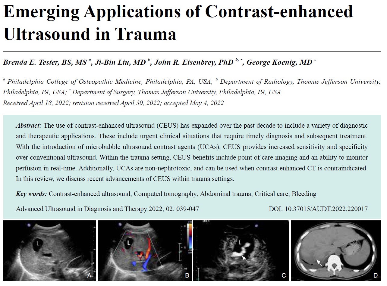
- Emerging Applications of Contrast-enhanced Ultrasound in Trauma
- Brenda E. Tester, BS, MS, Ji-Bin Liu, MD, John R. Eisenbrey, PhD, George Koenig, MD
- 2022, 6 (2): 39-47. DOI:10.37015/AUDT.2022.220017
- Abstract ( 472 ) HTML ( 35 ) PDF ( 22516KB ) ( 486 )
-
The use of contrast-enhanced ultrasound (CEUS) has expanded over the past decade to include a variety of diagnostic and therapeutic applications. These include urgent clinical situations that require timely diagnosis and subsequent treatment. With the introduction of microbubble ultrasound contrast agents (UCAs), CEUS provides increased sensitivity and specificity over conventional ultrasound. Within the trauma setting, CEUS benefits include point of care imaging and an ability to monitor perfusion in real-time. Additionally, UCAs are non-nephrotoxic, and can be used when contrast enhanced CT is contraindicated. In this review, we discuss recent advancements of CEUS within trauma settings.
-
Original Research
- Follicular Thyroid Neoplasmon Conventional and Contrast-enhanced Ultrasound
- Xuehong Diao, MD, Lin Chen, MD, Bo Yu, MS, Jiamei Jin, MS, Jia Zhan, MD, Yue Chen, BS
- 2022, 6 (2): 48-57. DOI:10.37015/AUDT.2022.210026
- Abstract ( 463 ) HTML ( 23 ) PDF ( 23436KB ) ( 228 )
-
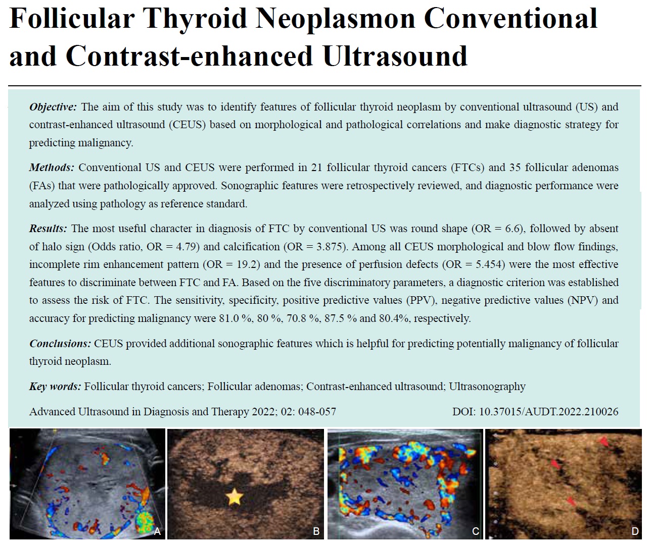
Objective: The aim of this study was to identify features of follicular thyroid neoplasm by conventional ultrasound (US) and contrast-enhanced ultrasound (CEUS) based on morphological and pathological correlations and make diagnostic strategy for predicting malignancy.
Methods: Conventional US and CEUS were performed in 21 follicular thyroid cancers (FTCs) and 35 follicular adenomas (FAs) that were pathologically approved. Sonographic features were retrospectively reviewed, and diagnostic performance were analyzed using pathology as reference standard.
Results: The most useful character in diagnosis of FTC by conventional US was round shape (OR=6.6), followed by absent of halo sign (Odds ratio, OR = 4.79) and calcification (OR = 3.875). Among all CEUS morphological and blow flow findings, incomplete rim enhancement pattern (OR = 19.2) and the presence of perfusion defects (OR = 5.454) were the most effective features to discriminate between FTC and FA. Based on the five discriminatory parameters, a diagnostic criterion was established to assess the risk of FTC. The sensitivity, specificity, positive predictive values (PPV), negative predictive values (NPV) and accuracy for predicting malignancy were 81.0 %, 80 %, 70.8 %, 87.5 % and 80.4%, respectively.
Conclusions: CEUS provided additional sonographic features which is helpful for predicting potentially malignancy of follicular thyroid neoplasm.
-
- Ultrasonographic Features of Intrathyroidal Thymic Carcinoma: Review and Analysis of 10 Cases
- Yanhai Wang, MD, Hua Yang, MD, Hanqing Liu, MD, Xiaoli Luo, MD, Luying Liu, BS, Pingting Zhou, BS
- 2022, 6 (2): 58-63. DOI:10.37015/AUDT.2022.220013
- Abstract ( 481 ) HTML ( 20 ) PDF ( 2979KB ) ( 426 )
-
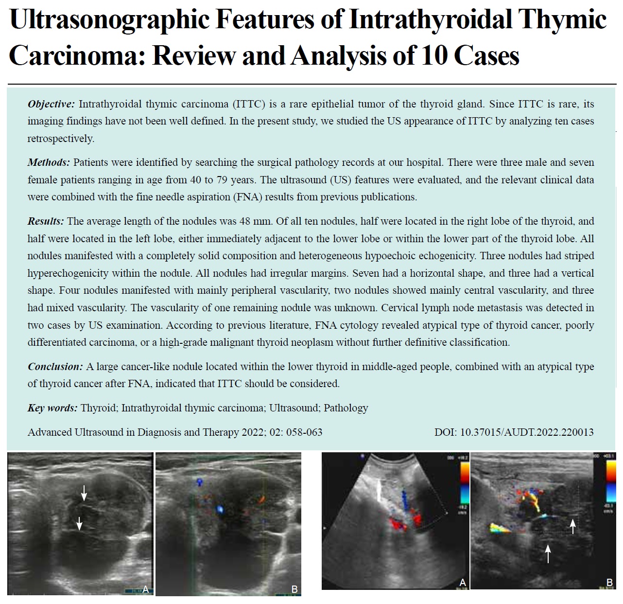
ObjectiveIntrathyroidal thymic carcinoma (ITTC) is a rare epithelial tumor of the thyroid gland. Since ITTC is rare, its imaging findings have not been well defined. In the present study, we studied the US appearance of ITTC by analyzing ten cases retrospectively.
Methods Patients were identified by searching the surgical pathology records at our hospital. There were three male and seven female patients ranging in age from 40 to 79 years. The ultrasound (US) features were evaluated, and the relevant clinical data were combined with the fine needle aspiration (FNA) results from previous publications.
Results The average length of the nodules was 48 mm. Of all ten nodules, half were located in the right lobe of the thyroid, and half were located in the left lobe, either immediately adjacent to the lower lobe or within the lower part of the thyroid lobe. All nodules manifested with a completely solid composition and heterogeneous hypoechoic echogenicity. Three nodules had striped hyperechogenicity within the nodule. All nodules had irregular margins. Seven had a horizontal shape, and three had a vertical shape. Four nodules manifested with mainly peripheral vascularity, two nodules showed mainly central vascularity, and three had mixed vascularity. The vascularity of one remaining nodule was unknown. Cervical lymph node metastasis was detected in two cases by US examination. According to previous literature, FNA cytology revealed atypical type of thyroid cancer, poorly differentiated carcinoma, or a high-grade malignant thyroid neoplasm without further definitive classification.
ConclusionA large cancer-like nodule located within the lower thyroid in middle-aged people, combined with an atypical type of thyroid cancer after FNA, indicated that ITTC should be considered.
-
Case Reports
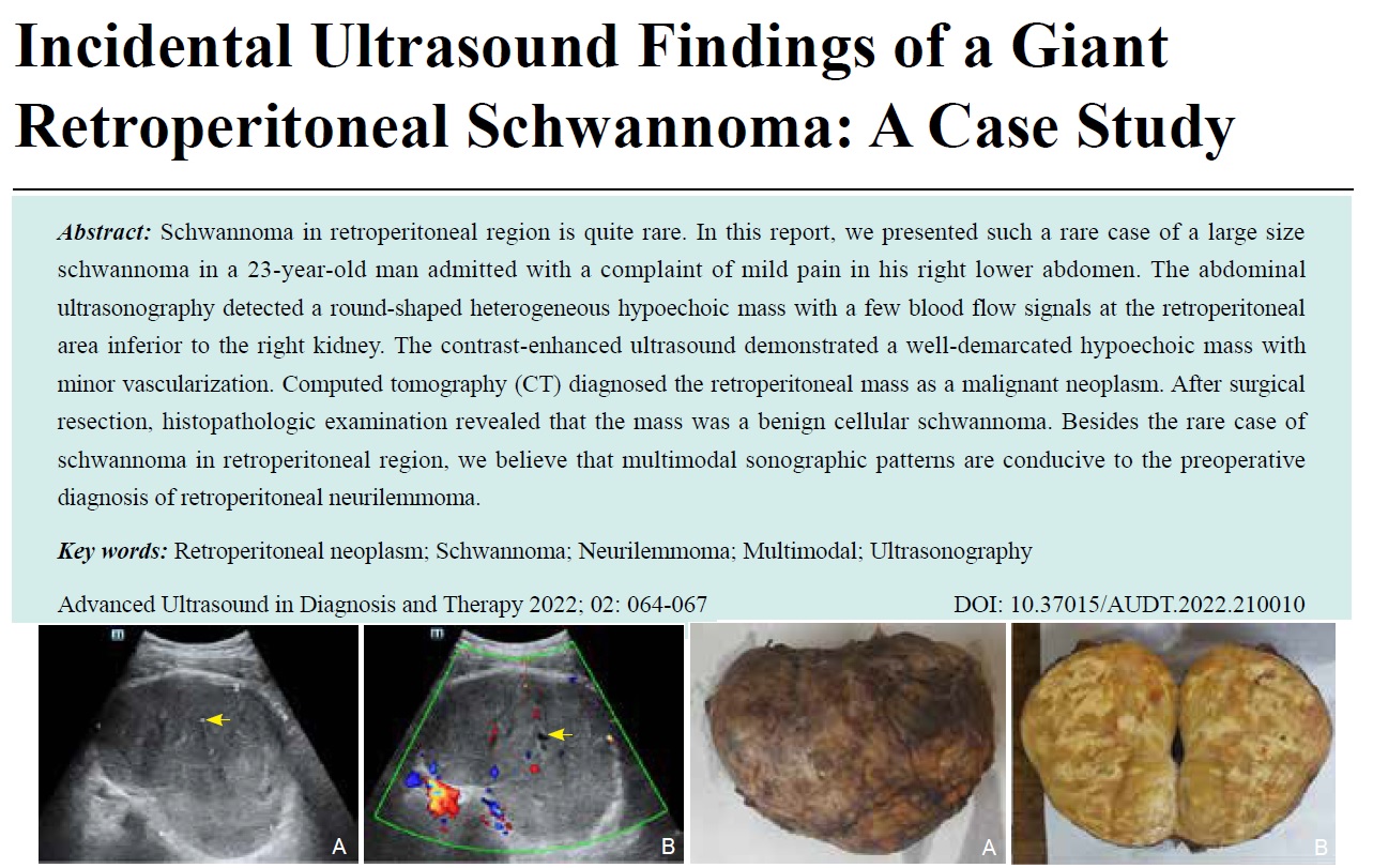
- Incidental Ultrasound Findings of a Giant Retroperitoneal Schwannoma: A Case Study
- Jiaqi Zhao, MD, Weiqing Li, MD, Xiaolin Ma, MD, Rui Chen, MD, Lin Chen, MD
- 2022, 6 (2): 64-67. DOI:10.37015/AUDT.2022.210010
- Abstract ( 396 ) HTML ( 8 ) PDF ( 23925KB ) ( 115 )
-
Schwannoma in retroperitoneal region is quite rare. In this report, we presented such a rare case of a large size schwannoma in a 23-year-old man admitted with a complaint of mild pain in his right lower abdomen. The abdominal ultrasonography detected a round-shaped heterogeneous hypoechoic mass with a few blood flow signals at the retroperitoneal area inferior to the right kidney. The contrast-enhanced ultrasound demonstrated a well-demarcated hypoechoic mass with minor vascularization. Computed tomography (CT) diagnosed the retroperitoneal mass as a malignant neoplasm. After surgical resection, histopathologic examination revealed that the mass was a benign cellular schwannoma. Besides the rare case of schwannoma in retroperitoneal region, we believe that multimodal sonographic patterns are conducive to the preoperative diagnosis of retroperitoneal neurilemmoma.
-
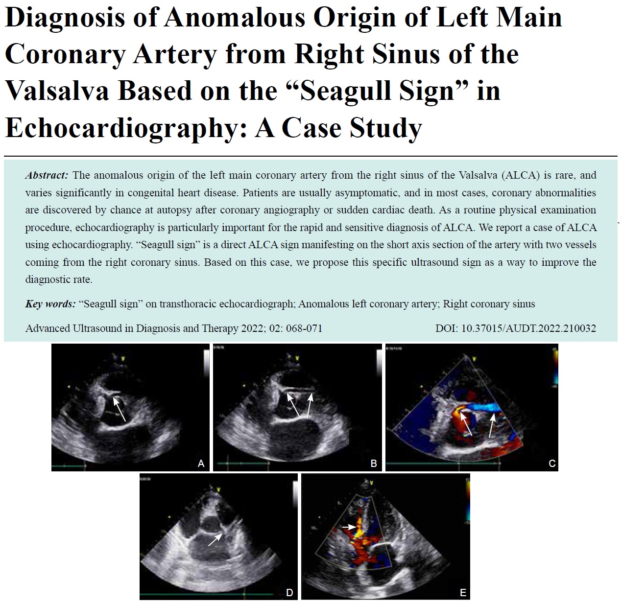
- Diagnosis of Anomalous Origin of Left Main Coronary Artery from Right Sinus of the Valsalva Based on the “Seagull Sign” in Echocardiography: A Case Study
- Yun Zheng, MM, Shiwen Fu, MM, Wei Miao, MM, Shanshan Qu, MM, Junhua Wang, MM, Liping Guo, MM, Xihe Sun, MM
- 2022, 6 (2): 68-71. DOI:10.37015/AUDT.2022.210032
- Abstract ( 384 ) HTML ( 5 ) PDF ( 2735KB ) ( 665 )
-
The anomalous origin of the left main coronary artery from the right sinus of the Valsalva (ALCA) is rare, and varies significantly in congenital heart disease. Patients are usually asymptomatic, and in most cases, coronary abnormalities are discovered by chance at autopsy after coronary angiography or sudden cardiac death. As a routine physical examination procedure, echocardiography is particularly important for the rapid and sensitive diagnosis of ALCA. We report a case of ALCA using echocardiography. “Seagull sign” is a direct ALCA sign manifesting on the short axis section of the artery with two vessels coming from the right coronary sinus. Based on this case, we propose this specific ultrasound sign as a way to improve the diagnostic rate.
-
Consensus and Guidelines
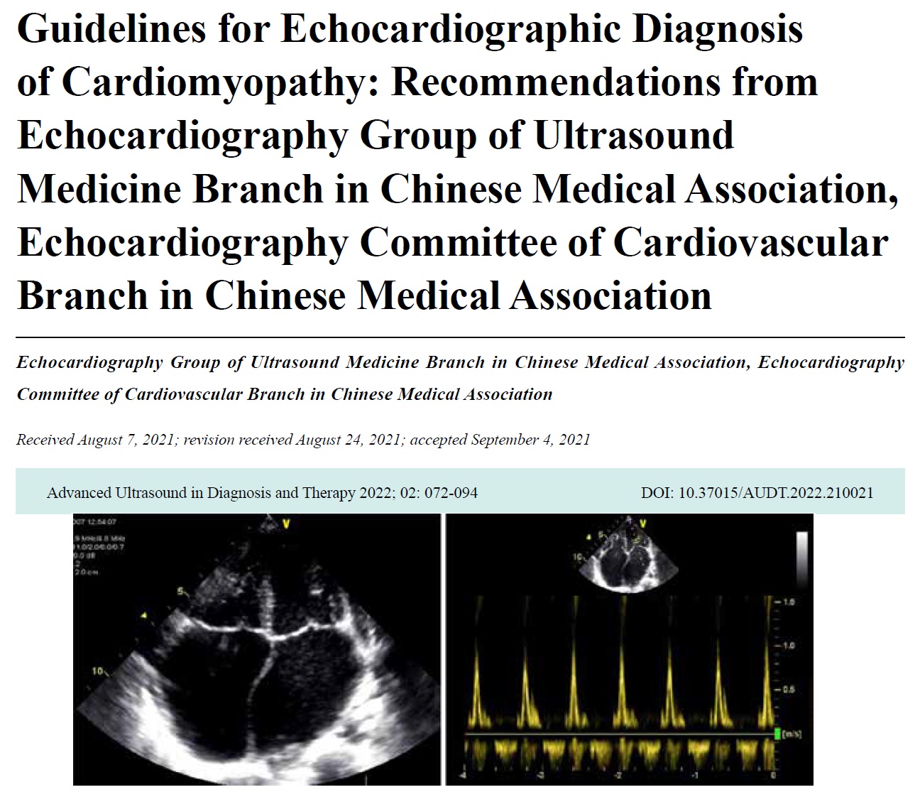
- Guidelines for Echocardiographic Diagnosis of Cardiomyopathy: Recommendations from Echocardiography Group of Ultrasound Medicine Branch in Chinese Medical Association, Echocardiography Committee of Cardiovascular Branch in Chinese Medical Association
- Echocardiography Group of Ultrasound Medicine Branch in Chinese Medical Association, Echocardiography Committee of Cardiovascular Branch in Chinese Medical Association
- 2022, 6 (2): 72-94. DOI:10.37015/AUDT.2022.210021
- Abstract ( 641 ) HTML ( 26 ) PDF ( 22607KB ) ( 628 )
-