Review Articles
- Micro/Nanobubbles Driven Multimodal Imaging and Theragnostics of Cancer
- Xiaoting Zhang, BS, Zhifei Dai, PhD
- 2021, 5 (3): 163-172. DOI:10.37015/AUDT.2021.200053
- Abstract ( 658 ) HTML ( 69 ) PDF ( 2261KB ) ( 929 )
-
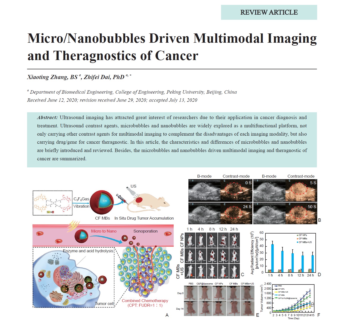
Ultrasound imaging has attracted great interest of researchers due to their application in cancer diagnosis and treatment. Ultrasound contrast agents, microbubbles and nanobubbles are widely explored as a multifunctional platform, not only carrying other contrast agents for multimodal imaging to complement the disadvantages of each imaging modality, but also carrying drug/gene for cancer theragnostic. In this article, the characteristics and differences of microbubbles and nanobubbles are briefly introduced and reviewed. Besides, the microbubbles and nanobubbles driven multimodal imaging and theragnostic of cancer are summarized.
-
- Targeted Delivery of Therapeutic Gas by Microbubbles
- Lingling Xu, MM, Yihan Chen, MM, Qiaofeng Jin, PhD, Li Zhang, MD, Wenpei Fu, BS, Shan Lin, MM, Ling Lin, BS, Rui Wang, BS, Dandan Chen, MM, Zhengyang Han, MM, Mingxing Xie, MD, Yali Yang, MD
- 2021, 5 (3): 173-182. DOI:10.37015/AUDT.2021.200059
- Abstract ( 534 ) HTML ( 37 ) PDF ( 2138KB ) ( 541 )
-
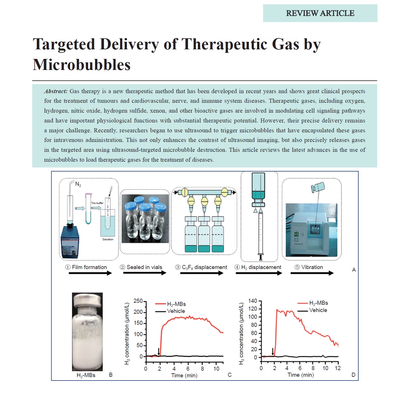
Gas therapy is a new therapeutic method that has been developed in recent years and shows great clinical prospects for the treatment of tumours and cardiovascular, nerve, and immune system diseases. Therapeutic gases, including oxygen, hydrogen, nitric oxide, hydrogen sulfide, xenon, and other bioactive gases are involved in modulating cell signaling pathways and have important physiological functions with substantial therapeutic potential. However, their precise delivery remains a major challenge. Recently, researchers began to use ultrasound to trigger microbubbles that have encapsulated these gases for intravenous administration. This not only enhances the contrast of ultrasound imaging, but also precisely releases gases in the targeted area using ultrasound-targeted microbubble destruction. This article reviews the latest advances in the use of microbubbles to load therapeutic gases for the treatment of diseases.
-
- Doppler-based Renal Resistive Index for Prediction of Acute Kidney Injury in Critically Ill Patients: A Systematic Review and Meta-analysis
- Jianing Zhu, MD, Ying Zhang, MD, Xiaoming Li, MD, Qiuyang Li, MD, PHD, Yukun Luo, MD, PHD
- 2021, 5 (3): 183-196. DOI:10.37015/AUDT.2021.210013
- Abstract ( 661 ) HTML ( 33 ) PDF ( 491KB ) ( 1146 )
-
Objectives: To determine the efficacy of Doppler-based renal resistive index (RRI) in the prediction of acute kidney injury (AKI) in critically ill patients.
Methods: A systematic review and meta-analysis of cohort studies was conducted. Relevant studies were identified in PubMed, Embase and Cochrane Library from inception to November 1, 2020, and reference lists of identified primary studies. Prospective studies that examined the diagnostic accuracy of RRI in AKI were included.
Results: Among the 126 articles identified, 18 were included, with a total of 1656 patients. Bivariate analysis yielded pooled sensitivity and specificity of 0.81 (95% CI 0.74-0.86) and 0.75 (95% CI 0.65-0.83), respectively. The summary positive likelihood ratio was 3.2 (95% CI 2.2-4.6), and negative likelihood ratio was 0.26 (95% CI 0.19-0.36).
Conclusion: Elevated RRI may be an early predictor of AKI in critically ill patients. Further large-scale prospective studies are needed to confirm the predictive efficacy and determine the performance and optimal cutoff value of RRI among the included studies.
- Ultrasound Elastography in Liver Tissue: Current Status
- Mingzhu Zhang, MD, Zhaoyan Ding, MD, Xiaoyan Niu, MD, Yuxiu Gao, MD, Cheng Zhao, MD
- 2021, 5 (3): 197-203. DOI:10.37015/AUDT.2021.210014
- Abstract ( 624 ) HTML ( 44 ) PDF ( 1075KB ) ( 496 )
-
Chronic liver disease is common in China and worldwide, with liver fibrosis as the primary pathological finding. Any chronic liver disease can lead to hepatic fibrosis and gradually develop into cirrhosis. Complications, such as portal hypertension, gastrointestinal bleeding, and liver failure can occur. Some patients even develop hepatocellular carcinoma. Thus, timely diagnosis of hepatic fibrosis is vital for the assessment of etiology, treatment, and prognosis. Conventional ultrasound imaging shows low sensitivity with its subjectivity for preliminary diagnosis of liver fibrosis, creating limitations in qualitative and quantitative evaluation. Ultrasound elastography is a recently developed technique that can help overcome these limitations. The elastic imaging method combines conventional ultrasound to assess liver stiffness along with routine examination. This "one-stop" check for liver disease opens new prospects for clinical and scientific research and improves the accuracy of disease diagnosis for broad clinical application. This article will review the current status of ultrasound elastography for its applications in chronic liver diseases.
Original Researchs
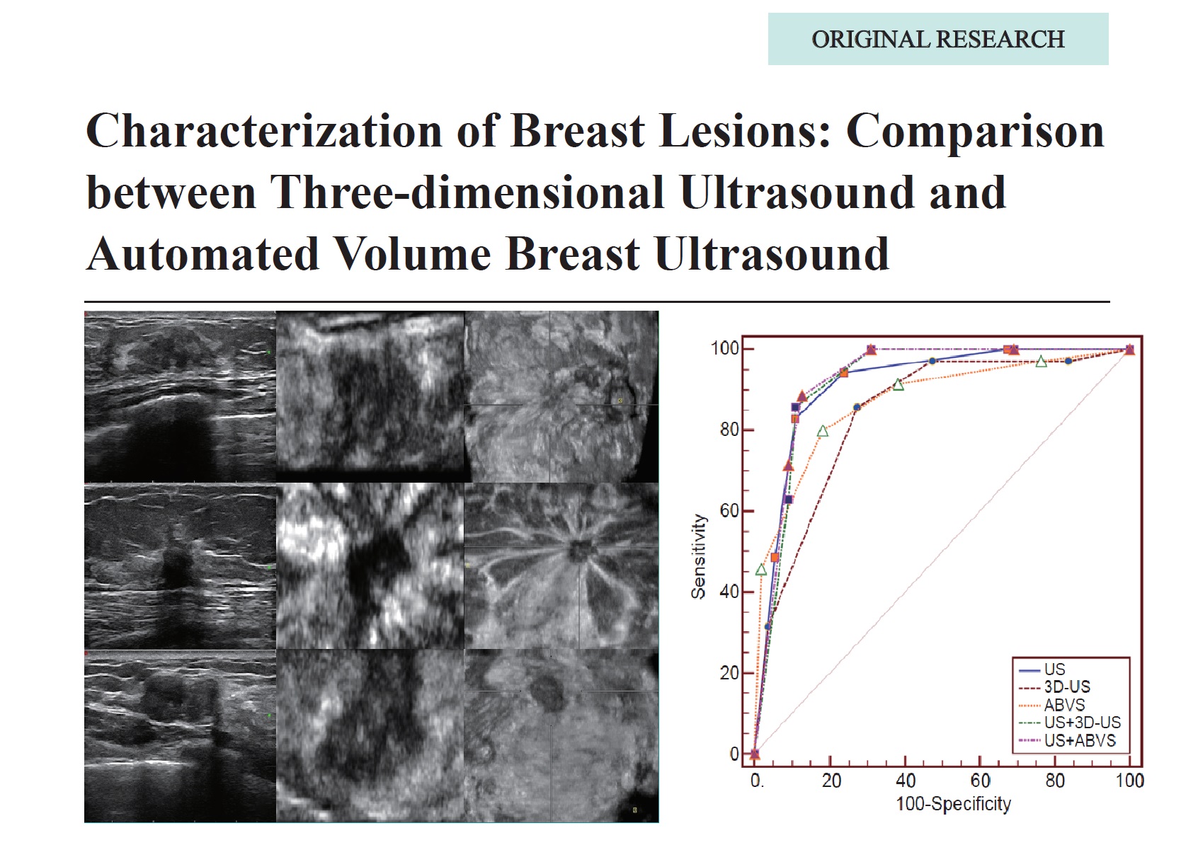
- Characterization of Breast Lesions: Comparison between Three-dimensional Ultrasound and Automated Volume Breast Ultrasound
- Wanru Jia, MD, Jingwen, Zhang, MD, Yijie Dong, MD, Ying Zhu, MD, Xiaohong Jia, MD, Weiwei Zhan, MD, Jianqiao Zhou, MD
- 2021, 5 (3): 204-211. DOI:10.37015/AUDT.2021.210007
- Abstract ( 580 ) HTML ( 21 ) PDF ( 1133KB ) ( 548 )
-
Objective: This study aimed to compare the diagnostic performance of three-dimensional ultrasound (3D-US) and automated breast volume scanner (ABVS) for the characterization of benign and malignant breast lesions.
Methods: Ninety patients who underwent surgery and preoperative conventional ultrasound (US), 3D-US, and ABVS examinations were enrolled in this study. The image quality and adjacent structures of the lesions in the coronal plane were compared. The combination of US, 3D-US, and ABVS for retraction phenomenon of the lesion was compared and the diagnostic performance of each combination was analyzed.
Results: ABVS displayed better image quality and adjacent structures than 3D-US (P < 0.001). The area under the curve (AUC) was 0.913, 0.842, and 0.871 for US, 3D-US, and ABVS, respectively. The AUC of the retraction phenomenon of the lesion was 0.732 and 0.810 for 3D-US and ABVS, respectively. When they were combined, US+ABVS showed the highest AUC of 0.924. No significant difference of diagnostic performances was found among conventional US, US+3D-US, and US+ABVS(P > 0.05).
Conclusions: Compared with 3D-US, ABVS seems to be superior in showing the retraction phenomenon of breast lesions and in the characterization of breast lesions alone or in combination with conventional US. Although no significant difference was observed between them, both ABVS and 3D-US provided valuable information in the coronal plane and improved our confidence level in breast lesion characterization, especially when combined with the conventional US. -
- Feasibility and Efficacy of the Segmental Localization of Lumbar Vertebrae by Ultrasound vs X-ray Examination: A Prospective Comparative Study
- Bo Yu, MD, Peng Huang, MD, Yukun Luo, MD, Mingbo Zhang, MD
- 2021, 5 (3): 212-218. DOI:10.37015/AUDT.2021.200062
- Abstract ( 430 ) HTML ( 12 ) PDF ( 1803KB ) ( 423 )
-
Objectives: To explore the feasibility and efficacy of the segmental localization of lumbar vertebrae by ultrasound (US) compared with X-ray.
Methods: From February 2019 to May 2019, 43 patients (24 males and 19 females), with an average age of 46±15 years, were prospectively enrolled in this study. Longitudinal paramedian sagittal and transverse process sections were used to determine the lumbar segments by US scan. X-ray examination was used to verify the segmentation. The time for segmentation was recorded, and the learning curve of the average localization time was analyzed.
Results: Of all the enrolled patients, 5 had lumbar segmental and alignment abnormalities, and 38 had normal lumbar vertebrae. US accurately located vertebrae in 38 normal cases and 5 abnormal cases, with a 100% accuracy rate, as verified by X-ray examination. The localization time was significantly less for US than for X-ray examination, both in normal cases and in cases with segmental or alignment abnormalities (all P < 0.001). The learning curve of US-guided segmental localization continuously decreased with an increasing number of operations and entered the plateau stage after the third operation day.
Conclusions: The US-guided segmental localization of lumbar vertebrae is an accurate new method that is efficient and easy to learn and does not require radiation.
- Contrast-enhanced Ultrasound Improves Technical Sufficiency of Fine-needle Aspiration in Suspicious Thyroid Nodules
- Ying Fu, MD, Shi Tan, MD, LiGang Cui, MD, Fang Mei, MD
- 2021, 5 (3): 219-225. DOI:10.37015/AUDT.2021.200063
- Abstract ( 335 ) HTML ( 10 ) PDF ( 2065KB ) ( 758 )
-
Objectives: To evaluate contrast-enhanced ultrasound (CEUS) for guiding fine-needle aspiration (FNA) of suspicious thyroid nodules to obtain sufficient biopsy specimens.
Methods: A total of 236 uncertain thyroid nodules detected in 228 patients from October 2016 to March 2017 were retrospectively reviewed and analyzed in this study. Overall, 117 patients underwent CEUS-guided FNA, and 111 patients underwent ultrasound (US)-guided FNA. The target for aspiration was only at the enhanced area in the CEUS group. In the US-guided group, aspiration was conducted within the nodule at various angles and areas. The final cytopathologic findings were reported using the Bethesda criteria. The inadequacy, indeterminacy, malignancy, and benignity rates of FNA specimens were compared between two groups.
Results: There were no significant differences in age, sex, or nodule size between the two groups. The inadequacy rate in the CEUS group was significantly lower than that in the US group (P = 0.008). Twenty-two benign nodules were diagnosed using CEUS-guided FNA, whereas seven were diagnosed using US-guided FNA (P = 0.006). The indeterminacy and malignancy rates were similar for both groups.
Conclusions: CEUS-guided FNA improves the diagnostic success rate and reduces uncertainty by facilitating accurate biopsy of suspected thyroid nodules with microcirculation perfusion imaging.
- Automated Measurements of Left Ventricular Ejection Fraction and Volumes Using the EchoPAC System
- Xiaoxue Chen, MD, Shaoling Yang, PhD, Qianqian He, MD, Yin Wang, PhD, Linyan Fan, MD, Fengling Wang, MD, Kun Zhao, MD, Jing Hu, MD
- 2021, 5 (3): 226-235. DOI:10.37015/AUDT.2021.200072
- Abstract ( 544 ) HTML ( 14 ) PDF ( 2632KB ) ( 565 )
-
Objective: To evaluate the clinical value of automated measurements by AutoEF (GE EchoPAC system, version 113) in left ventricular (LV) volumes and ejection fraction (EF) estimation based on the biplane Simpson’s method (manual method) in different clinical subsets.
Methods: A total of 322 subjects participated in this study (the common group). In the common group, 112 patients with coronary heart disease (CHD) were divided into the CHD group, and 34 CHD patients with LV wall motion abnormalities (WMA) comprising the CHD group, renamed the WMA group. LV volumes and EF were assessed using both manual tracing and automated estimation. Time spent on each method was documented. The agreements in echocardiographic measurements by different methods were assessed by intraclass correlation coefficients (ICC) and Bland-Altman analysis.
Results: The average analysis time of the automated method was 12 ± 1 s/patient with excellent repeatability. ICC revealed good consistency between manual and automated EF in all groups, especially in the CHD and WMA groups, although Bland-Altman analysis showed non-negligible bias in EF estimation between the two methods. ICC analysis showed a good correlation between automated and manual EF in all the good and poor image quality subgroups.
Conclusion: Automated method by AutoEF was a time-saving, excellent reproducible, and resistant to image interference approach, with a strong potential in left ventricular function measurements, especially for patients with CHD and/or WMA.
- Utilization of Ultrasound for Management of Surgical Intervention of Secondary Hyperparathyroidism and Prolonged Hypocalcemia Post-Parathyroidectomy
- Ying Liu, MM, Yang Zhou, MD, Hong Zhou, BS, Yuanyuan Chen, MM, Jian Wu, MD, Juan Wang, BS, Bin Wang, MM, Changyu Chen, MM, Ming Ye, MM
- 2021, 5 (3): 236-244. DOI:10.37015/AUDT.2021.200060
- Abstract ( 354 ) HTML ( 10 ) PDF ( 1843KB ) ( 536 )
-
Objective: To evaluate the application of ultrasound (US) for the surgical intervention in patients with moderate and severe secondary hyperparathyroidism (SHPT), and to identify the risk of prolonged hypocalcemia after parathyroidectomy (PTX).
Methods: A consecutive series of moderate and severe SHPT patients (n = 64) underwent ultrasound evaluation of parathyroid glands. Among the 64 patients who received 6-month medication therapies, ten patients with parathyroid hormone (PTH) 300~500 pg/mL were excluded from the study while 32 patients unresponsive to medication therapy (PTH > 500 pg/mL) received surgical interventions and 22 patients with PTH < 300 pg/mL received medication treatment alone. The correlations between the number, location, volume, sonographic features of parathyroid glands (PTGs), laboratory examinations, the duration of dialysis and the surgical necessity were analyzed. Total parathyroidectomy with synchronous auto-transplantation (PTX + AT) was performed in the surgical group. In both the surgical and medication group, patients with hyper-vascularity of the PTGs dominated (≥50%) were classified as a hyper-vascular subgroup, and the others as a hypo-vascular subgroup. The differences of post-operative calcium (Ca2+) levels and the incidence of prolonged hypocalcemia between hyper- and hypo- vascular subgroups were assessed.
Results: Sonographic evaluations revealed that the numbers of detectable PTGs were higher in the surgical group than that of the medication group (p < 0.05). The detection of supernumerary PTGs was higher in the surgical group than that in the medication group (13/121, 10.7% vs. 2/71, 2.8%, p < 0.05). Baseline PTH, >2 detectable PTGs, detection of supernumerary PTGs, patients with hyper-vascular, and the duration of dialysis were positively associated with the necessity of surgical intervention. For patients in the hyper-vascular subgroup, the serum Ca2+ level was lower than that in the hypo-vascularity subgroup (p < 0.01).
Conclusion: Ultrasonic features can provide useful information for management of surgical intervention of SHPT and prediction of the risk of prolonged hypocalcemia after PXT.
Educational Article
- Value of Ultrasound Images and Reports Scoring System in Quality Control
- Li Qiu, MD, Yulan Peng, MD, Qiang Lu, MD, Yan Luo, MD
- 2021, 5 (3): 245-248. DOI:10.37015/AUDT.2021.210003
- Abstract ( 594 ) HTML ( 14 ) PDF ( 317KB ) ( 558 )
-
Objective: To assess the value of ultrasound images and reports scoring system for quality control in clinical ultrasound practice.
Methods: Ultrasound images and reports scoring system was established and implemented in the Department of Medical Ultrasound at West China Hospital of Sichuan University in 2014. The scoring system along with formulated corresponding management measures was used for quality control of ultrasound examination and clinical practice assessment. The quantitative scoring results were summarized and analyzed.
Results: Through the quantitative assessment of the quality of the images and reports of ultrasound examinations, the total report score in our department had risen from 4.93 in 2014 to 4.98 in 2018. “Inconsistency between description and conclusion” accounted for 46.47% of all report errors, which was the most common report error. The total image score also increased slightly to ≥ 4.98 during the past three years. The most common image error was “not saving color Doppler images”, which accounted for 84.48% of all image errors. The total score of images was higher than that of text reports.
Conclusion: It is important to establish the ultrasound images and reports scoring system for quality control of ultrasound practice. Using quantitative scores for ultrasound images and reports can improve the quality control of ultrasound examination and strengthen the management of clinical operation.
Case Reports
- Echocardiography of Marfan's Syndrome Patient with New Gene Mutation of FBN1 with 13-year Follow-up
- Jianping Xu, MS, Faping Cui, MS, Shuixiu Dou, MS, Jiafu Ou, MD
- 2021, 5 (3): 249-253. DOI:10.37015/AUDT.2021.200070
- Abstract ( 333 ) HTML ( 5 ) PDF ( 719KB ) ( 415 )
-
A case of Marfan syndrome was followed up by echocardiography for 13 years to observe the evolution of cardiovascular disease. The initial cardiovascular manifestations of this patient were "mitral myxoid degeneration, chordal rupture, leaflet prolapse, and massive regurgitation". Subsequently, after several years of development, the aortic sinus and ascending aortic aneurysm dilatation appeared and a new gene mutation site G4331A of FBN1 was found by genetic testing in this patient. Whether the new gene mutation site is related to the initial manifestation of the patient's cardiovascular disease with "mitral valve disease" remains to be further verified.
- Contrast-enhanced Ultrasound Assessment of Treatment Response in a Patient with Multifocal Hepatocellular Carcinoma Treated with Transarterial Chemo and Radioembolization
- Esika Savsani, Mohamed Tantawi, MD, Corinne E. Wessner, MBA, RDMS, RVT, Philip Lee, MD, Andrej Lyshchik, MD, PhD, Kevin Anton, MD, PhD, Colette M. Shaw, MD, Ji-Bin Liu, MD, John R. Eisenbrey, PhD
- 2021, 5 (3): 254-257. DOI:10.37015/AUDT.2021.210018
- Abstract ( 408 ) HTML ( 10 ) PDF ( 2462KB ) ( 348 )
-
Minimally invasive locoregional therapies have become important treatment options for patients with intermediate or late-stage hepatocellular carcinoma (HCC) who are ineligible for surgical resection or liver transplantation. Imaging modalities are essential for procedural guidance and for assessing treatment response thereafter. We report a unique finding of a patient with multifocal HCC treated with transarterial radioembolization (TARE) with yttrium-90 (Y90) and transarterial chemoembolization (TACE). We compared contrast-enhanced ultrasound (CEUS) with contrast-enhanced magnetic resonance imaging (CE-MRI) in the evaluation of treatment response to demonstrate advantages of CEUS imaging technique and its early detection of viable tumor.
- Contrast Echocardiography Evaluation of Microcirculation of Myocardial Infarction Caused by Takotsubo Syndrome: Case Report and Literature Review
- Rifei Li, MM, Yuanmei Zhang, MM, Chengkai Zhang, MM, Xuenian Huang, MM, Shangwei Ding, MD
- 2021, 5 (3): 258-261. DOI:10.37015/AUDT.2021.210016
- Abstract ( 319 ) HTML ( 7 ) PDF ( 495KB ) ( 505 )
-
Takotsubo syndrome (TTS) is also known as stress cardiomyopathy (SCM) or apical ballooning syndrome (ABS). TTS is reversible cardiomyopathy with a good prognosis. Although the patients have chest pain and ECG changes with typical acute myocardial infarction, they have angiographically normal coronary arteries or mild atherosclerosis. TTS can cause microcirculation dysfunction, leading to myocardial infarction with non-obstructive coronary atherosclerosis (MINOCA). Myocardial Contrast Echocardiography (MCE) can detect MINOCA caused by TTS by evaluating myocardial microcirculation. We report a case of MINOCA caused by TTS using MCE to evaluate myocardial ischemia in the subendocardial myocardium.
- Sonographic Features of Borderline Phyllodes Tumor of the Breast: A Case Report
- Yaqin Sun, MD, Shumin Wang, MD, PhD, Zhanfei Zhe, MD, Xin Qi, MD, Li Li, MD, Qin Liu, MD
- 2021, 5 (3): 262-264. DOI:10.37015/AUDT.2021.210006
- Abstract ( 565 ) HTML ( 28 ) PDF ( 1184KB ) ( 620 )
-
Phyllodes tumors of the breast are rare fibroepithelial neoplasm which are generally divided into benign, borderline, or malignant. In this case study, we reported a 40-year-old female presented with an asymptomatic right breast mass underwent sonographic evaluation. The surgical histopathology was diagnosed as borderline phyllodes tumor. Sonographic features of the tumor are described in detail, and the relevant literatures are reviewed and discussed.
- Contrast-enhanced Ultrasound for Diagnosing Pancreatic Solitary Fibrous Tumor: A Case Report
- Lei Chen, MM, Cong Chen, MM, Qingfu Qian, MM, Zhikui Chen, PhD
- 2021, 5 (3): 265-267. DOI:10.37015/AUDT.2021.210004
- Abstract ( 470 ) HTML ( 18 ) PDF ( 1123KB ) ( 500 )
-
Solitary fibrous tumors (SFTs) are rare, especially in the pancreas. Here, we present a case of pancreatic SFT in a 45-year-old woman for its imaging characteristics on contrast-enhanced ultrasound (CEUS). On gray-scale imaging, the lesion was a regular, well-defined, and extremely hypoechoic mass in the body of the pancreas. On CEUS, it manifested as a slightly “slow wash-in and quick wash-out” heterogeneous enhancement. The patient underwent laparoscopic partial pancreatectomy, and the pathological findings confirmed the diagnosis of SFT with malignant potential. CEUS enabled real-time observation of the microcirculatory perfusion of the lesion, which is very useful when a differential diagnosis of pancreatic SFT is suspected.
Technical Paper
- Point-of-Care Ultrasound: New Concepts and Future Trends
- Yaoting Wang, MD, Huihui Chai, MD, Ruizhong Ye, MD, Jingzhi Li, MD, PhD, Ji-Bin Liu, MD, Chen Lin, Chengzhong Peng, MD
- 2021, 5 (3): 268-276. DOI:10.37015/AUDT.2021.210023
- Abstract ( 1242 ) HTML ( 54 ) PDF ( 1653KB ) ( 928 )
-
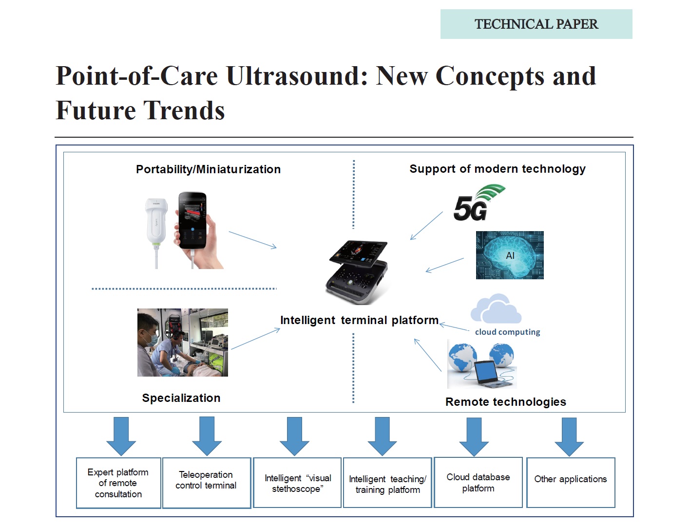
Ultrasound (US) technology, with major advances and new developments, has become an essential and first-line imaging modality for clinical diagnosis and interventional treatment. US imaging has evolved from one-dimensional, two-dimensional to three-dimensional display, and from static to real-time imaging, as well as from structural to functional imaging. Based on its portability and advanced digital imaging technique, US was first adopted by emergency medicine in the 1980s and gradually gained popularity among other specialists for clinical diagnosis and interventional treatment. Point-of-Care Ultrasound (POCUS) was then proposed as a new concept and developed for new uses, which greatly extended clinical US applications. Nowadays, artificial intelligence (AI), cloud computing, 5G network, robotics, and remote technologies are starting to be integrated into US equipment. US systems have gradually evolved to an intelligent terminal platform with powerful imaging and communication tools. In addition, specialized US machines tend to be more suitable and important to meet increasing demands and requirements by various clinical specialties and departments. In this article, we review current US technology and POCUS as new concepts and its future trends, as well as related technological developments and clinical applications.
-