Review Articles
- Advances in Targeted Tumor Diagnosis and Therapy Based on Ultrasound-Responsive Nanodroplets
- Yaqiong Li, PhD, Ruiqing Liu, MD, Shaobo Duan, MD, Lianzhong Zhang, MD
- 2020, 4 (4): 273-283. DOI:10.37015/AUDT.2020.200043
- Abstract ( 677 ) HTML ( 37 ) PDF ( 532KB ) ( 1050 )
-
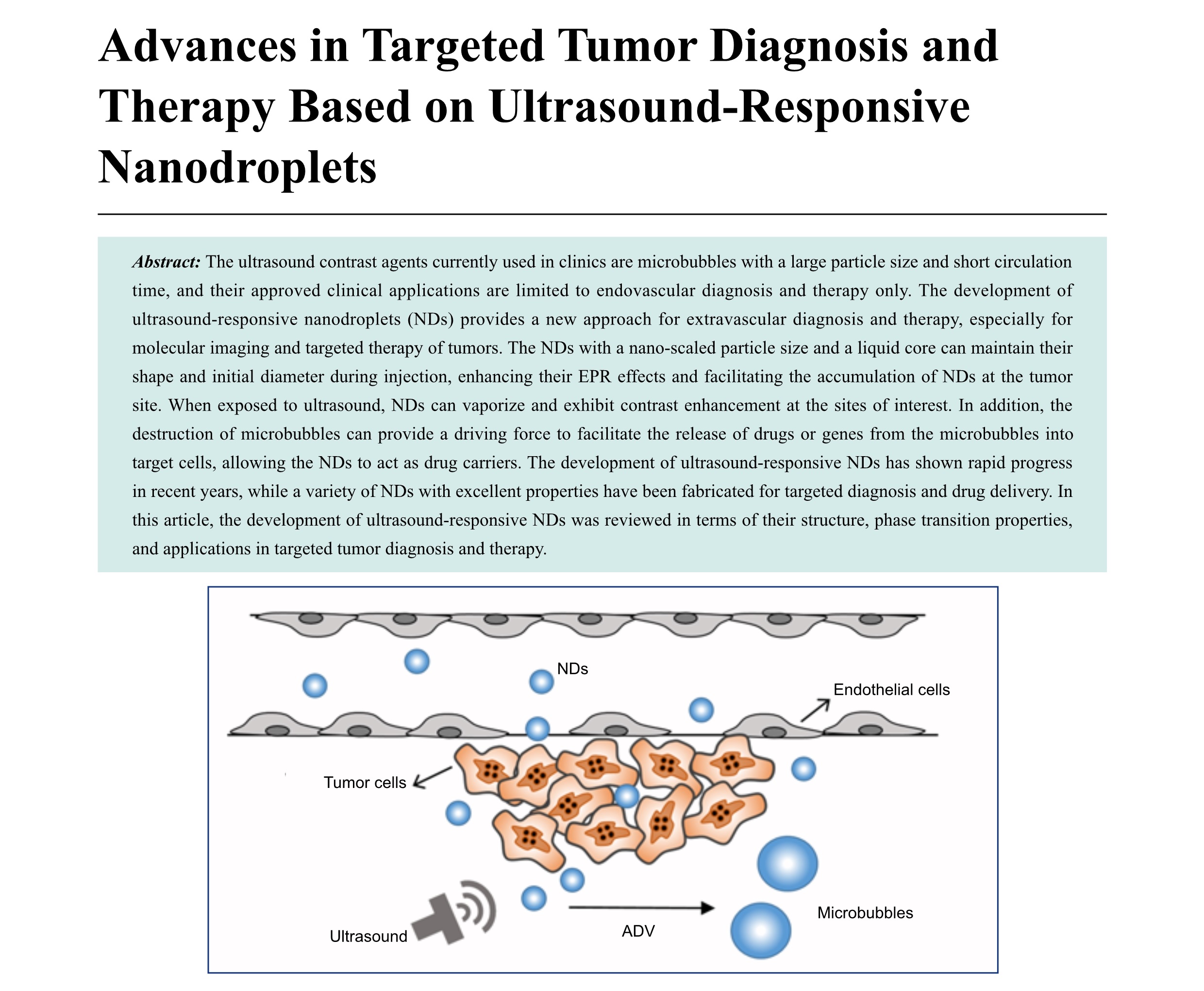
The ultrasound contrast agents currently used in clinics are microbubbles with a large particle size and short circulation time, and their approved clinical applications are limited to endovascular diagnosis and therapy only. The development of ultrasound-responsive nanodroplets (NDs) provides a new approach for extravascular diagnosis and therapy, especially for molecular imaging and targeted therapy of tumors. The NDs with a nano-scaled particle size and a liquid core can maintain their shape and initial diameter during injection, enhancing their EPR effects and facilitating the accumulation of NDs at the tumor site. When exposed to ultrasound, NDs can vaporize and exhibit contrast enhancement at the sites of interest. In addition, the destruction of microbubbles can provide a driving force to facilitate the release of drugs or genes from the microbubbles into target cells, allowing the NDs to act as drug carriers. The development of ultrasound-responsive NDs has shown rapid progress in recent years, while a variety of NDs with excellent properties have been fabricated for targeted diagnosis and drug delivery. In this article, the development of ultrasound-responsive NDs was reviewed in terms of their structure, phase transition properties, and applications in targeted tumor diagnosis and therapy.
-
- Application of Ultrasonography in the Diagnosis and Management of Papillary Thyroid Microcarcinoma
- Kun Huang, MD, Ji-Bin Liu, MD
- 2020, 4 (4): 284-290. DOI:10.37015/AUDT.2020.200001
- Abstract ( 495 ) HTML ( 24 ) PDF ( 446KB ) ( 1419 )
-
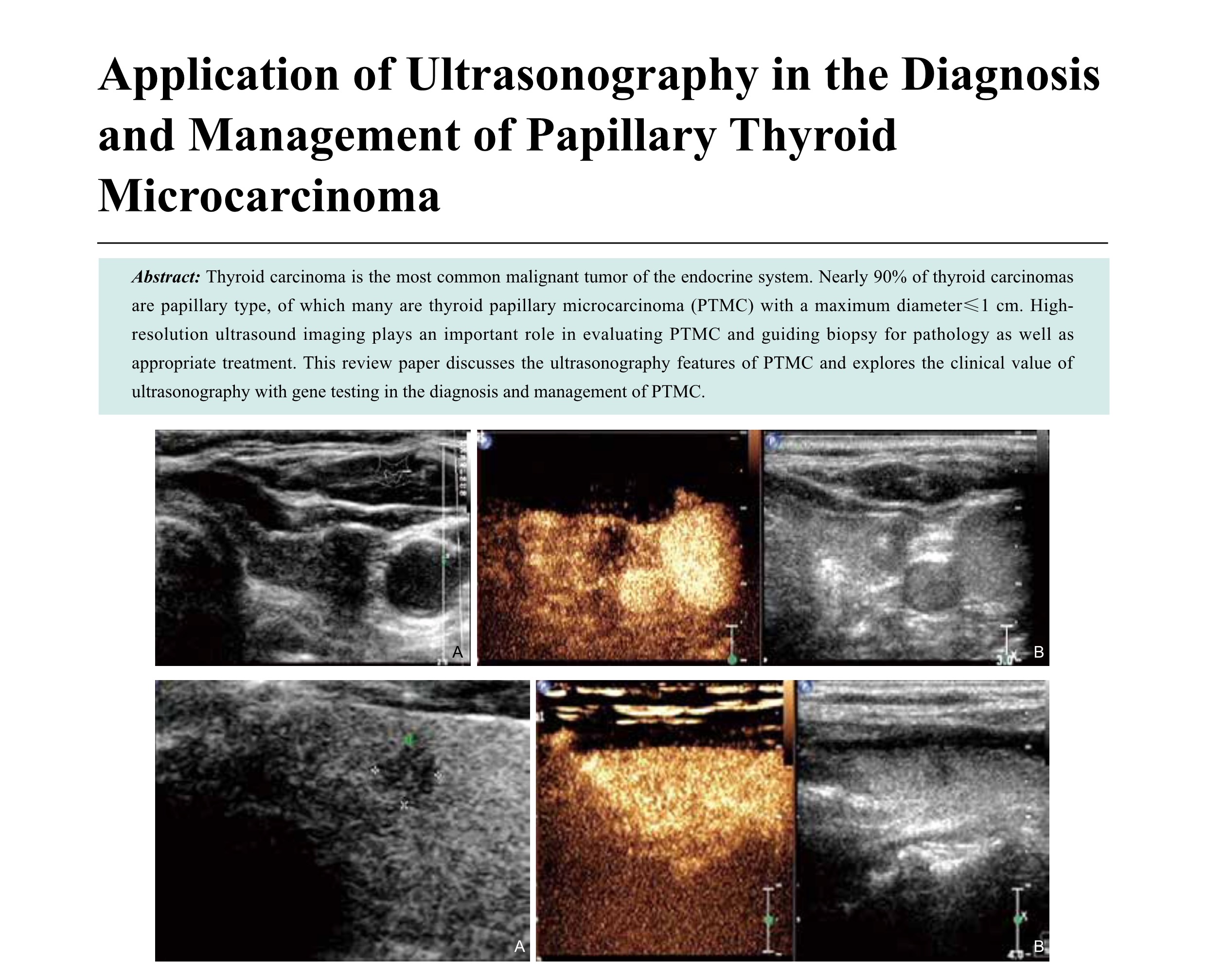
Thyroid carcinoma is the most common malignant tumor of the endocrine system. Nearly 90% of thyroid carcinomas are papillary type, of which many are thyroid papillary microcarcinoma (PTMC) with a maximum diameter≤1 cm. Highresolution ultrasound imaging plays an important role in evaluating PTMC and guiding biopsy for pathology as well as appropriate treatment. This review paper discusses the ultrasonography features of PTMC and explores the clinical value of ultrasonography with gene testing in the diagnosis and management of PTMC.
-
- The Roles of Ultrasound-Based Radiomics In Precision Diagnosis and Treatment of Different Cancers: A Literature Review
- Bing Mao, MD, Shaobo Duan, MD, Ruiqing Liu, MD, Na Li, PhD, Yaqiong Li, PhD, Lianzhong Zhang, MD
- 2020, 4 (4): 291-296. DOI:10.37015/AUDT.2020.200051
- Abstract ( 517 ) HTML ( 15 ) PDF ( 250KB ) ( 797 )
-
The study aims to review literatures on ultrasound-based radiomics, including ultrasound modalities, and discusses basic methods, applications, and limitations of ultrasound-based radiomics. The search strategy was conducted in form of “Radiomics [Title/Abstract] and Ultrasound [Title/Abstract]”in PubMed. The retrieved articles were initially screened via abstracts. Then, the main objectives, methods, and achievements of selected articles were summarized. Finally, twenty articles focused on malignancies of different organs, such as liver, rectum, breast, and thyroid were included into this review. The multiparametric features exhibited a superior diagnostic performance compared with a single modality. Ultrasound-based radiomics can assist radiologists to improve the accuracy of diagnosis, and it may promote the development of precision diagnosis and treatment of various types of cancer.
-
- Inter-ventricular Septum Ablation for the Treatment of Hypertrophic Obstructive Cardiomyopathy
- Yuejin Wu, MS, Shaobo Duan, MD, Luwen Liu, MS, Shuaiyang Wang, MS, Shuang Xu, MS, Liuwei Hao, BS, Lianzhong Zhang, MD
- 2020, 4 (4): 297-302. DOI:10.37015/AUDT.2020.200048
- Abstract ( 535 ) HTML ( 10 ) PDF ( 333KB ) ( 746 )
-
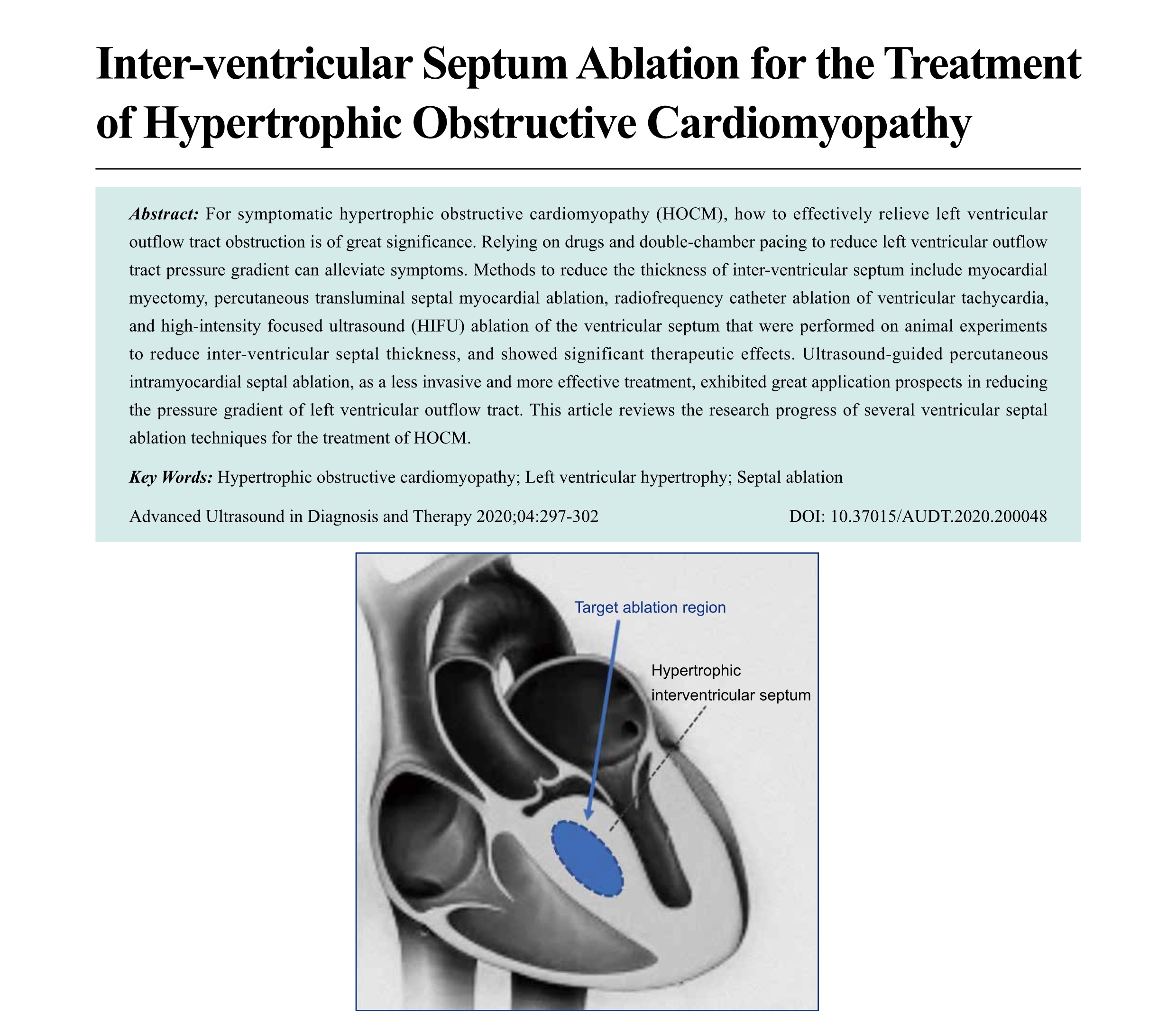
For symptomatic hypertrophic obstructive cardiomyopathy (HOCM), how to effectively relieve left ventricular outflow tract obstruction is of great significance. Relying on drugs and double-chamber pacing to reduce left ventricular outflow tract pressure gradient can alleviate symptoms. Methods to reduce the thickness of inter-ventricular septum include myocardial myectomy, percutaneous transluminal septal myocardial ablation, radiofrequency catheter ablation of ventricular tachycardia, and high-intensity focused ultrasound (HIFU) ablation of the ventricular septum that were performed on animal experiments to reduce inter-ventricular septal thickness, and showed significant therapeutic effects. Ultrasound-guided percutaneous intramyocardial septal ablation, as a less invasive and more effective treatment, exhibited great application prospects in reducing the pressure gradient of left ventricular outflow tract. This article reviews the research progress of several ventricular septal ablation techniques for the treatment of HOCM.
-
- Development Status and Prospect of Remote Diagnosis and Treatment of Echocardiography Worldwide
- Luwen Liu, MS, Shaobo Duan, MD, Yaqiong Li, PhD, Ruiqing Liu, MD, Yuejin Wu, MS, Lianzhong Zhang, MD
- 2020, 4 (4): 303-307. DOI:10.37015/AUDT.2020.200047
- Abstract ( 381 ) HTML ( 10 ) PDF ( 344KB ) ( 631 )
-
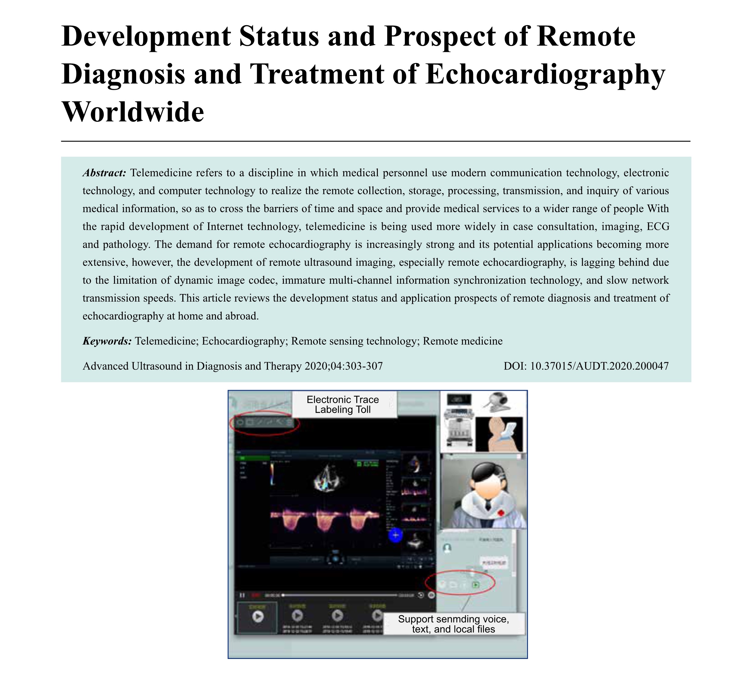
Telemedicine refers to a discipline in which medical personnel use modern communication technology, electronic technology, and computer technology to realize the remote collection, storage, processing, transmission, and inquiry of various medical information, so as to cross the barriers of time and space and provide medical services to a wider range of people With the rapid development of Internet technology, telemedicine is being used more widely in case consultation, imaging, ECG and pathology. The demand for remote echocardiography is increasingly strong and its potential applications becoming more extensive, however, the development of remote ultrasound imaging, especially remote echocardiography, is lagging behind due to the limitation of dynamic image codec, immature multi-channel information synchronization technology, and slow network transmission speeds. This article reviews the development status and application prospects of remote diagnosis and treatment of echocardiography at home and abroad.
-
- Focal Ablation Therapy for Prostate Cancer: A Literature Review
- Ruiqing Liu, MD, Yaqiong Li, PhD, Bing Mao, MD, Na Li, PhD, Shaobo Duan, MD, Zhiyang Chang, MS, Ye Zhang, MS, Shuaiyang Wang, MS, Lianzhong Zhang, MD
- 2020, 4 (4): 308-314. DOI:10.37015/AUDT.2020.200045
- Abstract ( 524 ) HTML ( 9 ) PDF ( 258KB ) ( 785 )
-
The incidence and mortality of prostate cancer (PCa) are gradually increasing. Traditional treatments for PCa may result numerous side effects and complications that affect patients’ quality of life. Furthermore, older patients frequently cannot tolerate conventional treatments, such as surgery, chemotherapy, and radiotherapy. Thus, minimally invasive and effective therapeutic approaches for PCa need to be developed for clinical practice. Focal ablation therapy, which uses high temperature to destroy tumors, holds promise as one such approach. It has been applied to PCa in several countries with gradual success and clinical practice This review briefly discusses the application of high-intensity focused ultrasound ablation (HIFU), cryoablation, laser ablation (LA), and radiofrequency and microwave ablation (RFA, MWA) for PCa, including the principles of treatment, clinical effects, and complications. The aim of this review is to provide a reliable reference for the application of focal ablation therapy to PCa.
-
Original Research
- Liver and Spleen Stiffness Measurements by Sound Touch Elastography and Sound Touch Quantification
- Jian Zheng, MD, Manli Wu, MD, Qingjuan Wang, MD, Rizhen Gu, MD, Xiaohong Yao, MD, Yuansen Chen, MD, Jing Huang, MD, Lexiang Long, MD, Rongqin Zheng, MD
- 2020, 4 (4): 315-321. DOI:10.37015/AUDT.2020.190038
- Abstract ( 564 ) HTML ( 12 ) PDF ( 488KB ) ( 818 )
-
Objective: To evaluate the technical success rate and reproducibility of sound touch elastography (STE) and sound touch quantification (STQ) in liver and spleen stiffness measurement and the reference ranges of normal liver and spleen stiffness. We also compared with a previous validated acoustic radiation force impulse (ARFI) technique.
Methods: Two hundred and fifty-three healthy adults and 40 chronic hepatitis B patients were recruited. All patients underwent liver and spleen stiffness measurements using STE, STQ, and ARFI. A hundred and five patients (36 patients with chronic hepatitis and 69 healthy adults) were examined twice, by two trained sonographers who are familiar with STE and STQ techniques independently. Another 36 healthy adults were examined twice by ARFI imaging. The technical success rates and reproducibility were evaluated.
Results: The success rates of STE, STQ, and ARFI were 96.5%, 95.1%, and 94.8% in liver, and 87.5%, 84.0%, and 78.0% in spleen, respectively. The inter-observer reproducibility of STE, STQ and ARFI were 0.914, 0.896, and 0.845 in liver, and 0.629, 0.601, and 0.543 in spleen, respectively. When the thickness of spleen was greater than 30mm, the reproducibility was 0.704 in STE and 0.668 in STQ. The normal ranges of liver stiffness were 5.80-6.04 kPa measured by STE and 5.87-6.13 kPa measured by STQ, and normal spleen stiffness ranged from 14.83-15.54 kPa measured by STE and 15.85-16.62 kPa measured by STQ.
Conclusion: Our study showed STE and STQ in liver and spleen stiffness measurement had a high success rate with good reproducibility, which were comparable to ARFI. The inter-observer reproducibility of spleen was barely satisfactory, but was good when the thickness of spleen was greater than 30mm.
- Real-Time Tissue Elastography: A Noninvasive Technique to Evaluate Liver Damage after Brain Death in Animal Mode
- Ningning Niu, MD, Ying Tang, MD, Jingwen Zhao, MD
- 2020, 4 (4): 322-328. DOI:10.37015/AUDT.2020.190028
- Abstract ( 416 ) HTML ( 11 ) PDF ( 348KB ) ( 558 )
-
Objective: Using brain-dead donors to obtain organs for transplant is an effective way to overcome the shortage of organ donors. The purpose of this study is to investigate ultrasound flow imaging and elastography as a simple, feasible, accurate and effective method for evaluation of liver damage after brain death using an animal model.
Methods: We established a brain-death model using 15 pigs. Brain death was induced by progressively increasing the intracranial pressure, and the brain-death state was maintained for 9 hours. Spectral Doppler imaging and elastography was used to evaluate hepatic hemodynamic parameters and tissue stiffness over the period of brain death.
Results: Electron microscopy of the liver over the progress of brain death revealed gradual mitochondrial swelling (with rupture), expansion of the endoplasmic reticulum, and increased collagen in the extracellular matrix. Spectral Doppler imagin demonstrated that the HA-RI increased over time, which had statistically significant difference between the Bef-BD measurement and measurements at 3, 6, and 9 hours after brain death. Real time elastography of the liver revealed a gradual decrease of the mean relative strain value (MEAN) over the time, and a gradual increase in standard deviation of the relative strain value, complexity of low strain area, and skewness, suggesting that brain-death induced liver damage increases with time. The ROC curves showed that MEAN had the highest sensitivity, specificity, and accuracy for assessing liver injury.
Conclusion: Hepatic tissue damage induced by brain death increased over time. The HA-RI and liver stiffness index changes can be assessed by Doppler ultrasound and elastographic imaging. Our results showed that elastography is a useful method to evaluate liver damage after brain death.
- Transthoracic Echocardiography for Evaluation of an Intrapulmonary Artery Mass
- Ting Sun, MD, Guoliang Lu, MD, Jian Fang, MD, Shaobo Xie, MD
- 2020, 4 (4): 329-334. DOI:10.37015/AUDT.2020.190022
- Abstract ( 388 ) HTML ( 7 ) PDF ( 390KB ) ( 816 )
-
Objective: Intrapulmonary artery mass is rare and prone to be misdiagnosed. The purpose of this study was to retrospectively review the evaluation of intrapulmonary artery masses by ultrasound imaging, summarize their characteristics, and suggest a standardized approach for clinical management.
Methods: Sixteen patients were enrolled in the study. Transthoracic echocardiography (TTE) showed a mass attached to the major pulmonary artery (MPA) trunk, straddling the bilateral pulmonary arteries or pulmonary valve (PV). The masses were diagnosed based on the site of the attachment, shape, size, mobility, and other morphological characteristics on ultrasound imaging examination. The pathological data were collected and analyzed from medical records.
Results: TTE images showed that the intrapulmonary artery mass was most frequently located in the MPA trunk. Eight patients had a pathological diagnosis and underwent complete mass resection. Five patients were suspected as having pulmonary thromboembolism (PTE) and were prescribed anticoagulation therapy, after which the masses decreased or disappeared on follow-up TTE. Three patients with suspected metastatic tumors died during hospitalization and had no pathological data. The 16 patients had the following distribution of diagnoses: thrombus (32%; 5/16), vegetations (12%; 2/16), primary benign lesions (12%; 2/16), primary malignant tumors (19%; 3/16), and metastatic tumors (25%; 4/16).
Conclusion: The majority of intrapulmonary artery masses were thrombi or primary pulmonary artery sarcomas. Primary tumors are much more common than metastatic tumors in the intrapulmonary artery.
- Identification of Key Genes Between Lung Adenocarcinoma and Lung Squamous Cell Carcinoma by Bioinformatics Analysis
- Jinru Xue, MS, Yu Yang, MS, Bo Jiang, MD, Linxue Qian, MD, Xian-Quan Shi, PhD
- 2020, 4 (4): 335-342. DOI:10.37015/AUDT.2020.200011
- Abstract ( 516 ) HTML ( 11 ) PDF ( 487KB ) ( 493 )
-
Objective: To explore differentially expressed genes (DEGs) between lung adenocarcinoma (LUAD) and lung squamous cell carcinoma (LUSC).
Methods: Based on GEO database, we used R software to identify the DEGs and conducted the bioinformatics analysis to explore the molecular mechanisms of DEGs and constructed PPI network to find the key DEGs. Then we assessed the effect of the eligible key DEGs on survival in LUAD by Kaplan-Meier plotter online tool.
Results: GSE10245 was downloaded from the GEO database, which contained a total of 58 tissue samples, including 18 LUSC and 40 LUAD. We identified 784 DEGs between LUAD and LUSC. DEGs were enriched in statistical significant GO annotation 201 items and KEGG pathways 17 items. By constructed PPI network, we obtained 10 hub genes. Of which, five genes were significantly correlated with the overall survival of LUAD.
Conclusions: P2RY1, CHRM3, LPAR3, NMU, and S1PR5 may be the potential prognostic markers and therapeutic targets for LUAD.
Case Reports
- US-CT Fusion Image-Guided Microwave Ablation of Lung Cancer----A New Mode of Image Guidance in Lung Cancer Ablation
- Tong Zhang, MD, Wenzhao Liang, MD, Yuanyuan Song, MD, Zhengmin Wang, MD, Dezhi Zhang, MD
- 2020, 4 (4): 343-348. DOI:10.37015/AUDT.2020.190025
- Abstract ( 422 ) HTML ( 10 ) PDF ( 454KB ) ( 505 )
-
Image-guided thermal ablation has become an ideal choice for treating lung tumors, including curative ablation for patients with primary peripheral lung cancer and solitary lung metastasis who are not candidates for surgery and palliative ablation. Previously, image guidance was directed by computed tomography (CT), magnetic resonance imaging (MRI) or ultrasound (US), all of which have certain limitations. Here, we report on the use of US-CT fusion image-guided microwave ablation (MWA) of a lung metastasis. Our experience indicated that this approach to can play an important role in lung cancer treatment by combining the individual advantages of US and CT and reducing their individual disadvantages. As a result, US-CT fusion imaging should be considered for image guidance mode in lung cancer ablation treatment.
- Epidermoid Cyst with Foreign Body Granuloma Formation in Hip: A Case Misdiagnosed by Ultrasound
- Shanyong Mao, MD, Yining Xiang, MD, Haiyan Zhou, MD, Linkun Zhang, MD, Tingting Chen, MD, Bei Zhang, MD
- 2020, 4 (4): 349-351. DOI:10.37015/AUDT.2020.200004
- Abstract ( 415 ) HTML ( 5 ) PDF ( 301KB ) ( 776 )
-
Epidermoid cysts are common benign lesions of the skin. High-frequency ultrasound is a good method for evaluation of skin soft-tissue mass. Here, we reported a case of epidermoid cyst which was misdiagnosed by ultrasound imaging as a dermoid cyst. With retrospective analysis of the ultrasound imaging, we realized that edidermoid cysts may have following features, in which the lesion originated from the skin, and have expansive hair follicle-like structure at the skin junction, as well as have onion-like or mixed echogenic changes. Ultrasound imaging can improve diagnostic accuracy through these characteristics along with clinical information.
- Silent Embolization Following Hybrid Device Closure of Atrial Septal Defect
- Ting Sun, MD, Guoliang Lu, MD, Jian Fang, MD, Shaobo Xie, MD
- 2020, 4 (4): 352-353. DOI:10.37015/AUDT.2020.190021
- Abstract ( 388 ) HTML ( 8 ) PDF ( 264KB ) ( 568 )
-
Device-induced embolization is a potential complication of every attempted atrial septal defect (ASD) device closure. Transesophageal echocardiography (TEE) plays an important role in diagnosing ASD and guiding the closure procedure. We report a rare complication of silent embolization of an ASD occlusion that occurred 3 months after a hybrid technique was used to repair the aortic valve.
- Ultrasound and CT Evolution of Severe Anal Injury with the Abscess Caused by a Small Chicken Bone: A Case Report
- Yuming Jin, BS, Ting Huang, BS, Jian Lu, BS
- 2020, 4 (4): 354-356. DOI:10.37015/AUDT.2020.190003
- Abstract ( 500 ) HTML ( 7 ) PDF ( 328KB ) ( 729 )
-
This report presents the case of a 59-year-old man with the anal abscess caused by fragments of chicken bones. Transrectal ultrasound (TRUS) was performed first to evaluate the anal and perianal region for detection of the lesion and then CT scan was used to show the entire region of the pelvis and anal structures. CT first identified the foreign body within the abscess and then TRUS was able to show hyperechoic foci with acoustic shadowing in the abscess indicated foreign bodies. Diagnosed of abscess with chicken bones was made based on both TRUS and CT and confirmed during the open surgery. CT scan can have a global view of the lesion and surrounding structures while TRUS can provide detail information of the perianal lesion and guide the clinical management.
Consensus and Guidelines
- Chinese Expert Consensus and Guidelines on Oral Contrast Gastric Ultrasonography for Scanning Technique and Imaging Acquisition
- Writing Group of the Gastroenterology Group of China Medical Education Association Ultrasound Committee , Tingting Li, MD, Xiaoyan Li, MD, Man Lu, MD, Ji-Bin Liu, MD, Wenming Lu, MD, Lixue Yin, MD, Changjun Wu, MD, Zhiqing Cai, MD, Guangxia Wang, MD, Liying Miao, MD, Jingyu Wang, MD, Dong Xu, MD, Jianqiang Mao, MD, Jin Yan, MD, Kang Wang, MD, Zhi Ding, MD, Ling Guan, MD, Lina Tang, MD, Shengjun Ma, MD, Yinrong Cheng, MD, Kairong Lei, MD, Yanfang Zhou, MD, Xingxing Duan, MD, Xuemei He, MD, Xiaoxia Dou, MD, Yuan Li, MD, Lu Wang, MD
- 2020, 4 (4): 357-365. DOI:10.37015/AUDT.2020.200061
- Abstract ( 568 ) HTML ( 21 ) PDF ( 622KB ) ( 247 )
-
Upper gastrointestinal (GI) abnormalities are one of the most common disease in clinical practice, and among them, gastric cancer is one of the most common causes of cancer-related death in China. However, conventional trans-abdominal ultrasound is difficult to evaluate GI diseases due to gas filled in the GI tract. With the development of oral contrast agents in China, ultrasonography with oral contrast agent has been used to visualize upper GI tract (i.e., stomach and duodenum) as well as its surrounding structures. The primary purposes of this consensus and guideline written by Chinese experts is to provide a coherent and clinical perspectives and practical protocol for using oral contrast in upper GI ultrasound, including four components: (1) indications and contraindications of gastric contrast ultrasound; (2) patients and instruments preparation; (3) scanning technique and imaging acquisition; (4) diagnosis of upper GI abnormalities.
Meeting Abstracts
- HNUS2020 Conference Abstracts
- 2020, 4 (4): 367-378.
- Abstract ( 142 ) HTML ( 11 ) PDF ( 331KB ) ( 29 )
-
The Annual Congress of the Henan Ultrasound Society (HNUS) of the Henan Medical Association is one of the most influential academic conferences in ultrasound medicine in Henan. The 2020 conference will be held in Zhengzhou, Henan from October 17 to 18, 2020. The scientific program of the meeting received 602 proceeding papers which covers the latest developments and trends in basic and clinical ultrasound research, such as interventional ultrasound, elastography, real-time three-dimensional ultrasound, remote ultrasound consultation, and contrast-enhanced ultrasound. The conference will arrange a scientific presentation forum to provide a stage for doctors to display academic achievement and cultivate international vision of physicians. A total of 58 papers were submitted for this forum, including 15 on Ob/Gyn ultrasound, 10 on abdominal ultrasound, 2 on interventional ultrasound, 17 on superficial organs and vascular ultrasound, and 14 on echocardiography. Under the recommendation of the Academic Committee of the General Assembly, 29 abstracts from oral presentations were selected and published in AUDT online and in print. These abstracts can be cited as scientific publications and shared with the international community.
Technical Papers
- Development of 4G CMUT (CMUT Linear SML44 probe)
- Tsuyoshi Otake, Hiroki Tanaka, Akifumi Sako, Makoto Fukada, Kengo Imagawa, Masahiro Sato
- 2020, 4 (4): 379-382.
- Abstract ( 1137 ) HTML ( 44 ) PDF ( 496KB ) ( 1018 )
-
In 2009, Hitachi commercialized “Mappie*1, the world’s first Capacitive Micro-machined Ultrasound Transducer (CMUT) using semiconductor based technology. It generated high quality diagnostic images of mammary glands, thanks to its broad-band characteristics[1]. This year, the 4th generation CMUT (4G CMUT) “SML44” has been brought to the market, achieved using advanced design and precise control of the fabrication process. When combined with new imaging technologies avail-able with the ARIETTA*2 850, the SML44, in addition to excellent image quality, offers commonly used modalities and func- tions such as Tissue Harmonic Imaging (THI), Color Flow Mapping (CFM), Real-time Tissue Elastography*3 (RTE), and Real-time Virtual Sonography*4 (RVS). This report introduces the latest technology adopted in the 4G CMUT design.
- Evaluation of Liver Fibrosis and Inflammation Using Ultrasound Elastography
- Nobuharu Tamaki, MD
- 2020, 4 (4): 383-384.
- Abstract ( 329 ) HTML ( 14 ) PDF ( 1033KB ) ( 519 )
-
There are two ultrasound elastography methods currently available for evaluating liver fibrosis: Strain Imaging and Shear Wave Elastography. Strain imaging can estimate the degree of liver fibrosis without the influence of inflammation whilst the Shear Wave Elastography measurement of fibrosis will be influenced by inflammation, congestion, and jaundice. The two elastography methods use different physical properties, so we examined whether a more accurate diagnosis of liver fibrosis and degree of inflammation is possible by using Real-time Tissue Elastography (RTE, a strain imaging method) and Shear Wave Measurement (SWM, a point Shear Wave Elastography method) simultaneously, and combining the results.


