Review Article
- Artificial Intelligence in Ultrasound Imaging: Current Research and Applications
- Shuo Wang, BS, Ji-Bin Liu, MD, Ziyin Zhu, MD, John Eisenbrey, PhD
- 2019, 3 (3): 53-61. DOI:10.37015/AUDT.2019.190811
- Abstract ( 2430 ) HTML ( 154 ) PDF ( 2242KB ) ( 1626 )
-
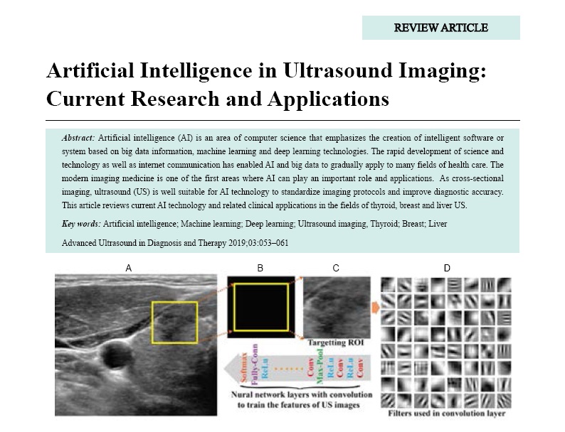
Artificial intelligence (AI) is an area of computer science that emphasizes the creation of intelligent software or system based on big data information, machine learning and deep learning technologies. The rapid development of science and technology as well as internet communication has enabled AI and big data to gradually apply to many fields of health care. The modern imaging medicine is one of the first areas where AI can play an important role and applications. As cross-sectional imaging, ultrasound (US) is well suitable for AI technology to standardize imaging protocols and improve diagnostic accuracy. This article reviews current AI technology and related clinical applications in the fields of thyroid, breast and liver US.
-
- Applications in Molecular Ultrasound Imaging: Present and Future
- Vishal Thumar, MD, Ji-Bin Liu, MD, John Eisenbrey, PhD
- 2019, 3 (3): 62-75. DOI:10.37015/AUDT.2019.190812
- Abstract ( 686 ) HTML ( 48 ) PDF ( 5408KB ) ( 831 )
-
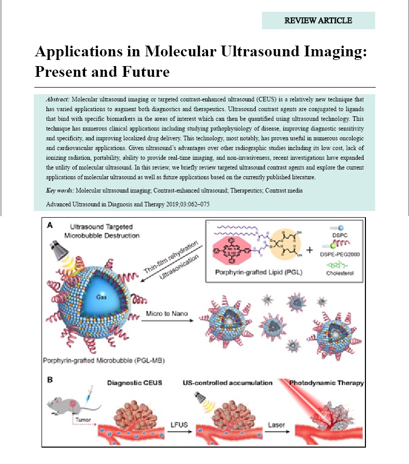
Molecular ultrasound imaging or targeted contrast-enhanced ultrasound (CEUS) is a relatively new technique that has varied applications to augment both diagnostics and therapeutics. Ultrasound contrast agents are conjugated to ligands that bind with specific biomarkers in the areas of interest which can then be quantified using ultrasound technology. This technique has numerous clinical applications including studying pathophysiology of disease, improving diagnostic sensitivity and specificity, and improving localized drug delivery. This technology, most notably, has proven useful in numerous oncologic and cardiovascular applications. Given ultrasound’s advantages over other radiographic studies including its low cost, lack of ionizing radiation, portability, ability to provide real-time imaging, and non-invasiveness, recent investigations have expanded the utility of molecular ultrasound. In this review, we briefly review targeted ultrasound contrast agents and explore the current applications of molecular ultrasound as well as future applications based on the currently published literature.
-
- Coronary Heart Disease Concomitant with Atherosclerotic Cerebrovascular Disease
- Yumei Liu, MD, Beibei Liu, MD, MS, Boyu Li, MD, PhD, Yang Hua, MD
- 2019, 3 (3): 76-80. DOI:10.37015/AUDT.2019.190813
- Abstract ( 548 ) HTML ( 30 ) PDF ( 1403KB ) ( 728 )
-
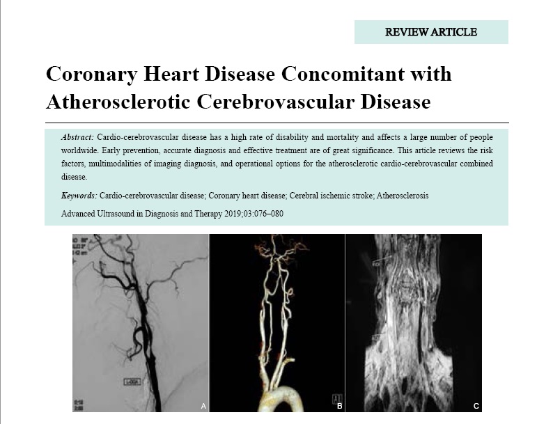
Cardio-cerebrovascular disease has a high rate of disability and mortality and affects a large number of people worldwide. Early prevention, accurate diagnosis and effective treatment are of great significance. This article reviews the risk factors, multimodalities of imaging diagnosis, and operational options for the atherosclerotic cardio-cerebrovascular combined disease.
-
Original Research
- Improving Ultrasound Gene Transfection Efficiency in Vitro
- Xianghong Luo, MD, Jianhui Zhang, MD, Sihui Shao, MD, Rong Wu, MD, Lianfang Du, MD, Jie Yuan, PhD, Zhaojun Li, MD
- 2019, 3 (3): 81-86. DOI:10.37015/AUDT.2019.190814
- Abstract ( 493 ) HTML ( 13 ) PDF ( 706KB ) ( 468 )
-
Objective: The purpose of this study was to optimize ultrasound-targeted microbubble destruction (UTMD) on RAGE plasmid transfection in human coronary artery endothelial cells (HCAECs) and improve gene transfection efficiency in vitro.
Methods: SonoVue microbubble suspension was prepared and mixed with HCAECs, while the cells were adherent and suspended, respectively. After RAGE plasmids being added, they were exposed to ultrasonic irradiation for 10, 20, and 30 s by a therapeutic US machine with 0.4W, respectively. The samples with adherent HCAECs (adherent group) were irradiated directly, while the samples with suspended HCAECs (suspended group) were irradiated via the water. The combined effect of ultrasound and microbubble on RAGE plasmid transfection in HCAECs was evaluated by detecting protein expression of RAGE by western blot. In addition, the viability of the HCAECs was analyzed by CCK8 in order to explore the optimal transfection condition.
Results: In suspension group, compared with control, the expression of RAGE was gradually increased from 5 to 20s, and decreased from 20 to 30s. The expression of RAGE peaked in 20s and indicated statistical significance. However, compared with the control, the expression of RAGE did not significantly increase with prolonged ultrasound irradiation in the adherent group. On the other hand, viability of the HCAECs did not decrease significantly with extended exposure time in both groups.
Conclusion: UTMD represents an efficient and safe method for the transfection of cells in suspension and optimal exposure.
-
- Histological Reference for Shear Wave Elastography in Liver Fibrosis: Collagen Quantification and Scoring System
- Yue Zhang, MD, Hong Ding, MD, Shengdi Wu, MD, Peili Fan, MD, Zheng Li, MD, Wenjiao Zeng, MD, Wenping Wang, MD
- 2019, 3 (3): 87-96. DOI:10.37015/AUDT.2019.190815
- Abstract ( 467 ) HTML ( 7 ) PDF ( 3727KB ) ( 587 )
-
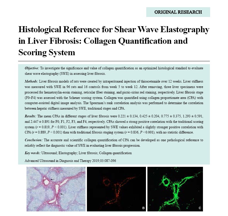
Objective: To investigate the significance and value of collagen quantification as an optimized histological standard to evaluate shear wave elastography (SWE) in assessing liver fibrosis.
Methods: Liver fibrosis models of rats were created by intraperitoneal injection of thioacetamide over 12 weeks. Liver stiffness was measured with SWE in 96 rats and 16 controls from week 5 to week 12. After removing, three liver specimens were processed for hematoxylin-eosin staining, reticular fiber staining, and picric-sirius red staining, respectively. Liver fibrosis stage (F0-F4) was assessed with the Scheuer scoring system. Collagen was quantified using collagen proportionate area (CPA) with computer-assisted digital image analysis. The Spearman’s rank correlation analysis was performed to determine the correlation between hepatic stiffness measured by SWE, traditional stages and CPA.
Results: The mean CPAs in different stages of liver fibrosis were 0.221 ± 0.134, 0.425 ± 0.204, 0.775 ± 0.375, 1.293 ± 0.591, and 2.447 ± 0.891 for F0, F1, F2, F3, and F4, respectively. CPAs showed a strong positive correlation with the traditional scoring system (r = 0.819, P < 0.001). Liver stiffness represented by SWE values exhibited a slightly stronger positive correlation with CPA (r = 0.889, P < 0.001) than with traditional fibrosis staging system (r = 0.836, P < 0.001), with no statistic difference.
Conclusion: The accurate and scientific collagen quantification of CPA can be developed as one pathological reference to reliably reflect the diagnostic value of SWE in evaluating liver fibrosis progression.
-
- The Role of Ultrasound Shear Wave Dispersion Imaging in Evaluating Carotid Viscoelasticity:A Preliminary Study
- Xianghong Luo, MD, Jianhui Zhang, MD, Sihui Shao, MD, Min Yan, MD, Rong Wu, MD, Lianfang Du, MD, Zhaojun Li, MD
- 2019, 3 (3): 97-102. DOI:10.37015/AUDT.2019.190816
- Abstract ( 636 ) HTML ( 36 ) PDF ( 1111KB ) ( 847 )
-
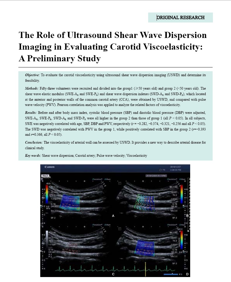
Objective: To evaluate the carotid viscoelasticity using ultrasound shear wave dispersion imaging (USWD) and determine its feasibility.
Methods: Forty-five volunteers were recruited and divided into the group1 (≥50 years old) and group 2 (<50 years old). The shear wave elastic modulus (SWE-AR and SWE-PR) and shear wave dispersion indexes (SWD-AR and SWD-PR), which located at the anterior and posterior walls of the common carotid artery (CCA), were obtained by USWD, and compared with pulse wave velocity (PWV). Pearson correlation analysis was applied to analyze the related factors of viscoelasticity.
Results: Before and after body mass index, systolic blood pressure (SBP) and diastolic blood pressure (DBP) were adjusted, SWE-AR, SWE-PR, SWD-AR and SWD-PR were all higher in the group 2 than those of group 1 (all P < 0.05). In all subjects, SWE was negatively correlated with age, SBP, DBP and PWV, respectively (r = -0.282, -0.374, -0.321, -0.256 and all P < 0.05). The SWD was negatively correlated with PWV in the group 1, while positively correlated with SBP in the group 2 (r=-0.393 and r=0.366, all P < 0.05).
Conclusion: The viscoelasticity of arterial wall can be assessed by USWD. It provides a new way to describe arterial disease for clinical study.
-
- The Significance of Heat Shock Protein 70 Expression in Benign Thyroid Nodules During Thermal Ablation
- Lei Yan, MM, Jianquan Zhang, MD, Jianguo Sheng, MM, Hang Zhang, MM, Zongping Diao, MM, Jianming Zheng, PhD
- 2019, 3 (3): 103-108. DOI:10.37015/AUDT.2019.190817
- Abstract ( 479 ) HTML ( 12 ) PDF ( 2020KB ) ( 935 )
-
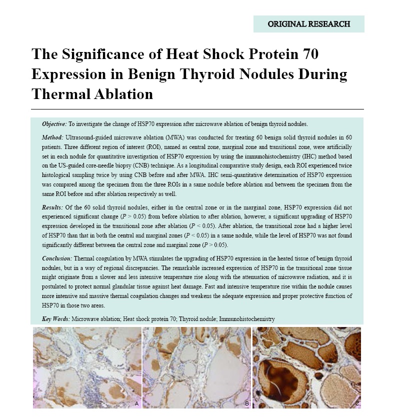
Objective: To investigate the change of HSP70 expression after microwave ablation of benign thyroid nodules.
Method: Ultrasound-guided microwave ablation (MWA) was conducted for treating 60 benign solid thyroid nodules in 60 patients. Three different region of interest (ROI), named as central zone, marginal zone and transitional zone, were artificially set in each nodule for quantitative investigation of HSP70 expression by using the immunohistochemistry (IHC) method based on the US-guided core-needle biopsy (CNB) technique. As a longitudinal comparative study design, each ROI experienced twice histological sampling twice by using CNB before and after MWA. IHC semi-quantitative determination of HSP70 expression was compared among the specimen from the three ROIs in a same nodule before ablation and between the specimen from the same ROI before and after ablation respectively as well.
Results: Of the 60 solid thyroid nodules, either in the central zone or in the marginal zone, HSP70 expression did not experienced significant change (P > 0.05) from before ablation to after ablation, however, a significant upgrading of HSP70 expression developed in the transitional zone after ablation (P < 0.05). After ablation, the transitional zone had a higher level of HSP70 than that in both the central and marginal zones (P < 0.05) in a same nodule, while the level of HSP70 was not found significantly different between the central zone and marginal zone (P > 0.05).
Conclusion: Thermal coagulation by MWA stimulates the upgrading of HSP70 expression in the heated tissue of benign thyroid nodules, but in a way of regional discrepancies. The remarkable increased expression of HSP70 in the transitional zone tissue might originate from a slower and less intensive temperature rise along with the attenuation of microwave radiation, and it is postulated to protect normal glandular tissue against heat damage. Fast and intensive temperature rise within the nodule causes more intensive and massive thermal coagulation changes and weakens the adequate expression and proper protective function of HSP70 in those two areas.
-
- Clinical Value of Contrast Enhanced Ultrasound in Differential Diagnosis of Early Hepatocellular Carcinoma and Dysplastic Nodules
- Yanhua Zhen, MD, Xuefeng Lu, MD, Chenyu Wang, MD, Huixia Li, MD, Ji-Bin Liu, MD
- 2019, 3 (3): 109-114. DOI:10.37015/AUDT.2019.190818
- Abstract ( 661 ) HTML ( 2 ) PDF ( 709KB ) ( 549 )
-
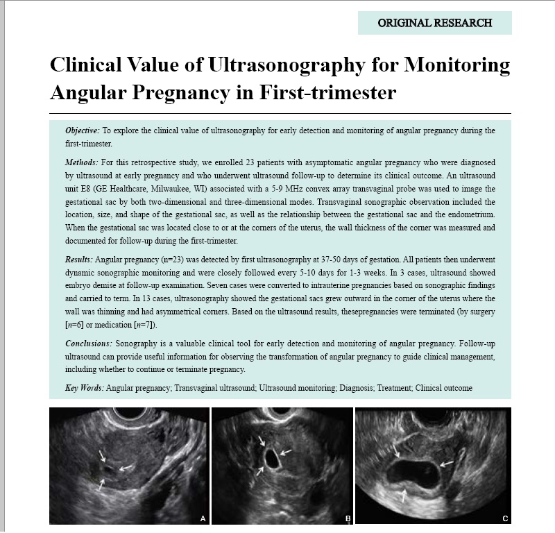
Objective: To explore the clinical value of ultrasonography for early detection and monitoring of angular pregnancy during the first-trimester.
Methods: For this retrospective study, we enrolled 23 patients with asymptomatic angular pregnancy who were diagnosed by ultrasound at early pregnancy and who underwent ultrasound follow-up to determine its clinical outcome. An ultrasound unit E8 (GE Healthcare, Milwaukee, WI) associated with a 5-9 MHz convex array transvaginal probe was used to image the gestational sac by both two-dimensional and three-dimensional modes. Transvaginal sonographic observation included the location, size, and shape of the gestational sac, as well as the relationship between the gestational sac and the endometrium. When the gestational sac was located close to or at the corners of the uterus, the wall thickness of the corner was measured and documented for follow-up during the first-trimester.
Results: Angular pregnancy (n=23) was detected by first ultrasonography at 37-50 days of gestation. All patients then underwent dynamic sonographic monitoring and were closely followed every 5-10 days for 1-3 weeks. In 3 cases, ultrasound showed embryo demise at follow-up examination. Seven cases were converted to intrauterine pregnancies based on sonographic findings and carried to term. In 13 cases, ultrasonography showed the gestational sacs grew outward in the corner of the uterus where the wall was thinning and had asymmetrical corners. Based on the ultrasound results, thesepregnancies were terminated (by surgery [n=6] or medication [n=7]).
Conclusions: Sonography is a valuable clinical tool for early detection and monitoring of angular pregnancy. Follow-up ultrasound can provide useful information for observing the transformation of angular pregnancy to guide clinical management, including whether to continue or terminate pregnancy.
-
Technical Development
- CMUT/CMOS-based Butterfly iQ - A Portable Personal Sonoscope
- Joyce Y Liu, BA, Jiajun Xu, MD, Flemming Forsberg, PhD, Ji-Bin Liu. MD, FAIUM
- 2019, 3 (3): 115-118. DOI:10.37015/AUDT.2019.190819
- Abstract ( 2863 ) HTML ( 2509 ) PDF ( 1393KB ) ( 4702 )
-
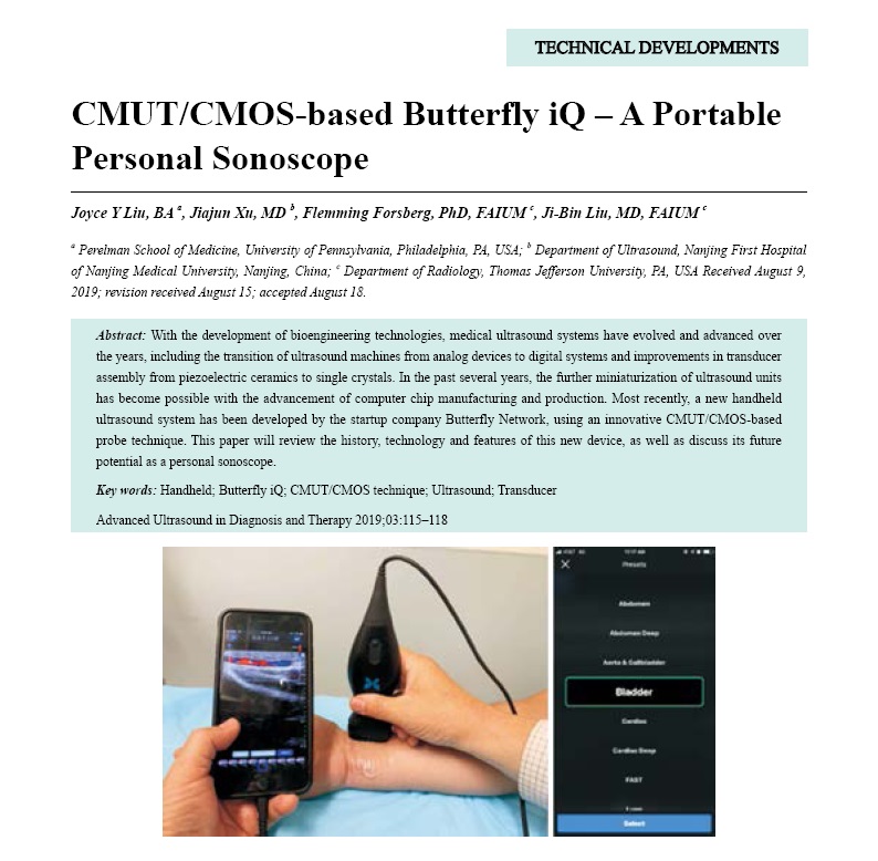
With the development of bioengineering technologies, medical ultrasound systems have evolved and advanced over the years, including the transition of ultrasound machines from analog devices to digital systems and improvements in transducer assembly from piezoelectric ceramics to single crystals. In the past several years, the further miniaturization of ultrasound units has become possible with the advancement of computer chip manufacturing and production. Most recently, a new handheld ultrasound system has been developed by the startup company Butterfly Network, using an innovative CMUT/CMOS-based probe technique. This paper will review the history, technology and features of this new device, as well as discuss its future potential as a personal sonoscope.
-
Educational Articles
- Review of Certification in Building a Successful Sonographer Program in China
- Hannah Mason, MA, Karen Caruth, MBA, Qiang Lu, MD, Dale R. Cyr, MBA, CAE
- 2019, 3 (3): 119-122. DOI:10.37015/AUDT.2019.190820
- Abstract ( 493 ) HTML ( 3 ) PDF ( 397KB ) ( 464 )
-
By working with local medical ultrasound experts, American Registry for Diagnostic Medical Sonography (ARDMS) of Inteleos, a non-profit certification organization, is striving to create global standards and international certifications in the field of diagnostic ultrasound that will improve clinical practice and patient safety. Through eligibility and application requirements, building a proper assessment, and determining continuing education requirements, standards are a critical part of Inteleos’ test development process. Since 2005, Inteleos/ARDMS has been working with Chinese physicians and professional societies to facilitate and promote certification and credentialing programs. Through the Registered Physician in Vascular Interpretation (RPVI-China) assessment, applicants can earn the internationally-recognized certificates in vascular ultrasound in China. Along with certification for Physicians, China’s growing population and the related burden on its healthcare system as well as the rapid increases in ultrasound use could contribute to the ideal situation for building a sonographer profession in China. Through a pilot education and certification program for Chinese sonographers, Inteleos will work with Chinese universities and collaborators to build and expand the sonographer profession in China in order to reduce workload on physicians, reduce wait times, decrease hospital costs, and provide consistently better care for their population.
-
Case Report
- Contrast-enhanced Ultrasonography: A New Strategy to Confirm Cervical Pregnancy
- Qingyun Song, MD, Sha Hu, Hong Luo, MD, Taizhu Yang, Qianqian Gao, MD, Fan Yang, MD
- 2019, 3 (3): 123-127. DOI:10.37015/AUDT.2019.190821
- Abstract ( 425 ) HTML ( 4 ) PDF ( 2680KB ) ( 696 )
-
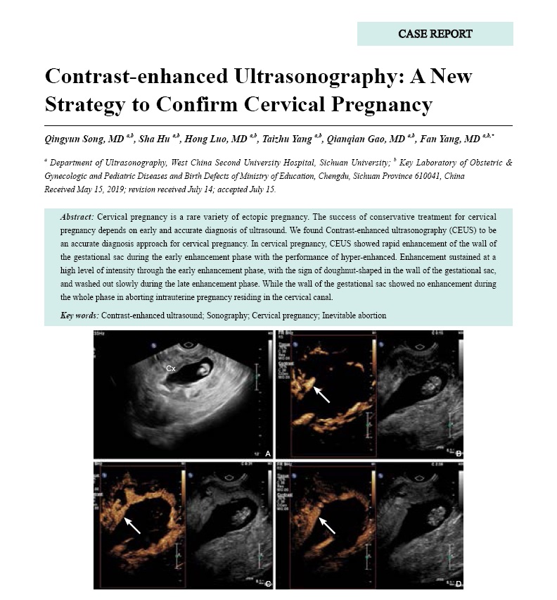
Cervical pregnancy is a rare variety of ectopic pregnancy. The success of conservative treatment for cervical pregnancy depends on early and accurate diagnosis of ultrasound. We found Contrast-enhanced ultrasonography (CEUS) to be an accurate diagnosis approach for cervical pregnancy. In cervical pregnancy, CEUS showed rapid enhancement of the wall of the gestational sac during the early enhancement phase with the performance of hyper-enhanced. Enhancement sustained at a high level of intensity through the early enhancement phase, with the sign of doughnut-shaped in the wall of the gestational sac, and washed out slowly during the late enhancement phase. While the wall of the gestational sac showed no enhancement during the whole phase in aborting intrauterine pregnancy residing in the cervical canal.
-
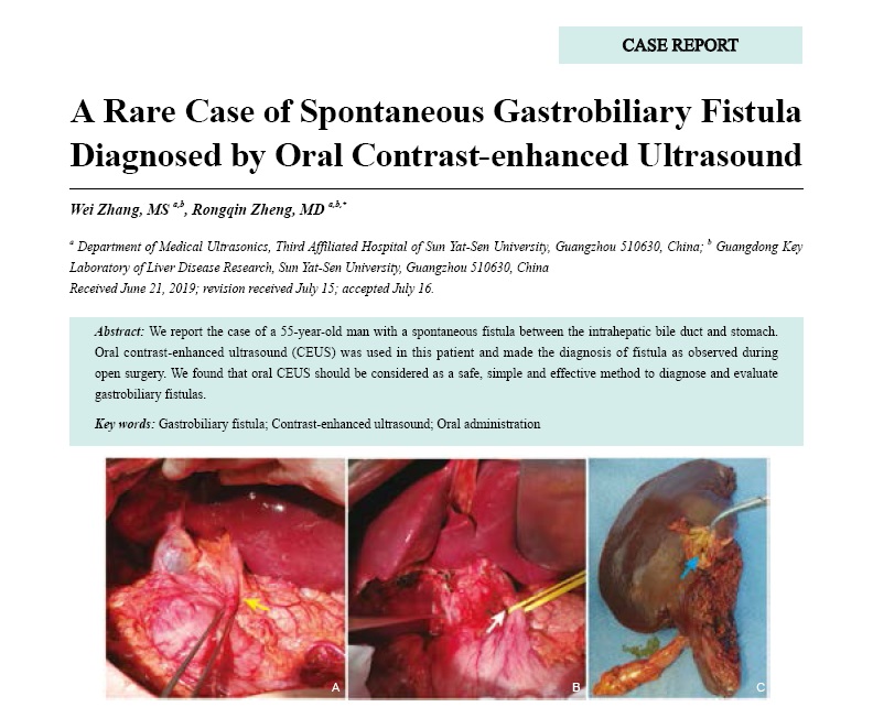
- A Rare Case of Spontaneous Gastrobiliary Fistula Diagnosed by Oral Contrast-enhanced Ultrasound
- Wei Zhang, MS, Rongqin Zheng, MD
- 2019, 3 (3): 128-131. DOI:10.37015/AUDT.2019.190822
- Abstract ( 543 ) HTML ( 5 ) PDF ( 1897KB ) ( 625 )
-
We report the case of a 55-year-old man with a spontaneous fistula between the intrahepatic bile duct and stomach. Oral contrast-enhanced ultrasound (CEUS) was used in this patient and made the diagnosis of fistula as observed during open surgery. We found that oral CEUS should be considered as a safe, simple and effective method to diagnose and evaluate gastrobiliary fistulas.
-
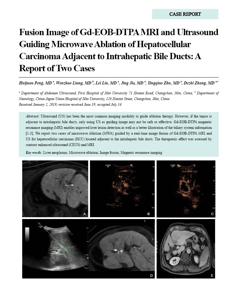
- Fusion Image of Gd-EOB-DTPA MRI and Ultrasound Guiding Microwave Ablation of Hepatocellular Carcinoma Adjacent to Intrahepatic Bile Ducts: A Report of Two Cases
- Huijuan Peng, MD, Wenzhao Liang, MD, Lei Liu, MD, Jing Jia, MD, Yingqiao Zhu, Dezhi Zhang, MD
- 2019, 3 (3): 132-135. DOI:10.37015/AUDT.2019.190823
- Abstract ( 365 ) HTML ( 2 ) PDF ( 1161KB ) ( 451 )
-
Ultrasound (US) has been the most common imaging modality to guide ablation therapy. However, if the tumor is adjacent to intrahepatic bile ducts, only using US as guiding image may not be safe or effective. Gd-EOB-DTPA magnetic resonance imaging (MRI) enables improved liver lesion detection as well as a better illustration of the biliary system information [1-3]. We report two cases of microwave ablation (MWA) guided by a real-time image fusion of Gd-EOB-DTPA MRI and US for hepatocellular carcinoma (HCC) located adjacent to the intrahepatic bile ducts. The therapeutic effect was assessed by contrast enhanced ultrasound (CEUS) and MRI.
-
- Microwave Ablation of An Autonomous Functioning Thyroid Nodule in A Pregnant Patient: A Case Report
- Shengnan Huo, MD, Lin Yin, Lili Peng, MD, Zhao Wang, MD, Ming-an Yu, MD
- 2019, 3 (3): 136-139. DOI:10.37015/AUDT.2019.190824
- Abstract ( 718 ) HTML ( 3 ) PDF ( 493KB ) ( 647 )
-
An autonomous functioning thyroid nodule (AFTN) is a benign disease. It can autonomously secrete excessive thyroid hormones without the need for TSH stimulation. It is not subject to the hypothalamic-pituitary-thyroid axis and is rare during pregnancy. Here we report a case of a 36-year-old pregnant woman with hyperthyroidism detected in early pregnancy, According to ultrasound and laboratory results, she was diagnosed with an AFTN. Ultrasound-guided (US-guided) microwave ablation (MWA) was used to treat AFTN during the second trimester of pregnancy. Hyperthyroidism crisis and other complications did not occur due to MWA. Thyroid function was normal at 2, 3 and 4 months after MWA. The volume reduction rate (VRR) was 83.67% at 4 months after MWA. The patient gave birth normally at 40 weeks gestation. All indices of thyroid function were normal during postpartum and lactation. US-guided MWA is a feasible and safe method for the treatment of AFTN during pregnancy, specifically in the second trimester.
-
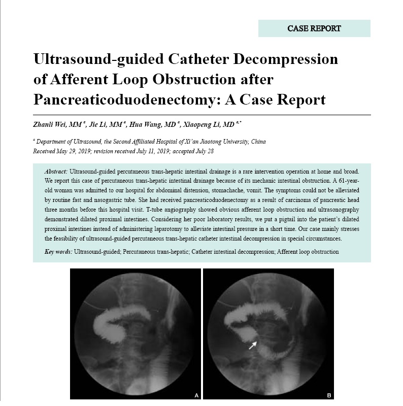
- Ultrasound-guided Catheter Decompression of Afferent Loop Obstruction after Pancreaticoduodenectomy: A Case Report
- Zhanli Wei, MM, Jie Li, MM, Hua Wang, MD, Xiaopeng Li, MD
- 2019, 3 (3): 140-143. DOI:10.37015/AUDT.2019.190825
- Abstract ( 420 ) HTML ( 4 ) PDF ( 933KB ) ( 489 )
-
Ultrasound-guided percutaneous trans-hepatic intestinal drainage is a rare intervention operation at home and broad.We report this case of percutaneous trans-hepatic intestinal drainage because of its mechanic intestinal obstruction. A 61-year-old woman was admitted to our hospital for abdominal distension, stomachache, vomit. The symptoms could not be alleviated by routine fast and nasogastric tube. She had received pancreaticoduodenectomy as a result of carcinoma of pancreatic head three months before this hospital visit. T-tube angiography showed obvious afferent loop obstruction and ultrasonography demonstrated dilated proximal intestines. Considering her poor laboratory results, we put a pigtail into the patient’s dilated proximal intestines instead of administering laparotomy to alleviate intestinal pressure in a short time. Our case mainly stresses the feasibility of ultrasound-guided percutaneous trans-hepatic catheter intestinal decompression in special circumstances.
-
Conference Paper
Technical Development
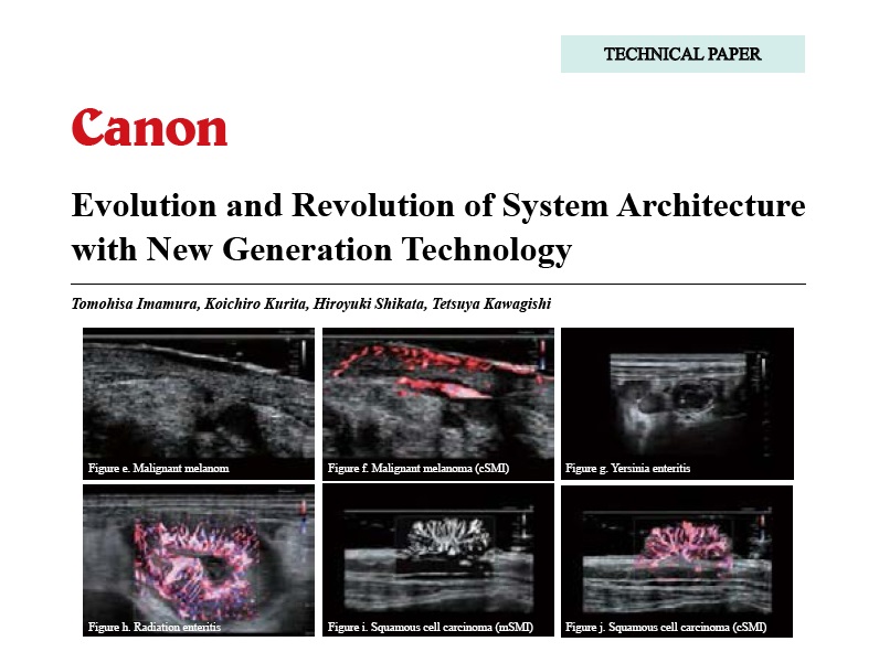
- Evolution and Revolution of System Architecture with New Generation Technology
- Tomohisa Imamura, Koichiro Kurita, Hiroyuki Shikata, Tetsuya Kawagishi
- 2019, 3 (3): 166-173.
- Abstract ( 527 ) PDF ( 2963KB ) ( 2254 )
-